
Lecture 8 The Cell Surface Part 1 Chapter
- Slides: 14
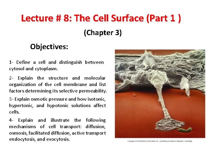
Lecture # 8: The Cell Surface (Part 1 ) (Chapter 3) Objectives: 1 - Define a cell and distinguish between cytosol and cytoplasm. 2 - Explain the structure and molecular organization of the cell membrane and list factors determining its selective permeability. 3 - Explain osmotic pressure and how isotonic, hypertonic, and hypotonic solutions affect cells. 4 - Explain and illustrate the following mechanisms of cell transport: diffusion, osmosis, facilitated diffusion, active transport endocytosis, and exocytosis.
Modern Cell Theory All organisms composed of cells and cell products. Cell is the simplest structural and functional unit of life (cells are alive) An organism’s structure and functions are due to the activities of its cells. In 1859, Louis Pasteur established beyond any reasonable doubt that ‘cells arise only from other cells Cells come only from preexisting cells, not from nonliving matter. Therefore, all life traces its ancestry to the same original cells Cells of all species have many fundamental similarities in their chemical composition and metabolic mechanisms.
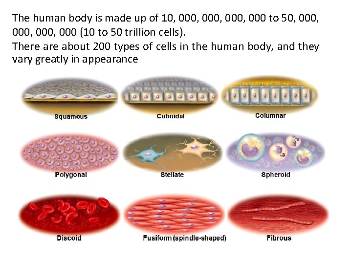
The human body is made up of 10, 000, 000 to 50, 000, 000 (10 to 50 trillion cells). There about 200 types of cells in the human body, and they vary greatly in appearance
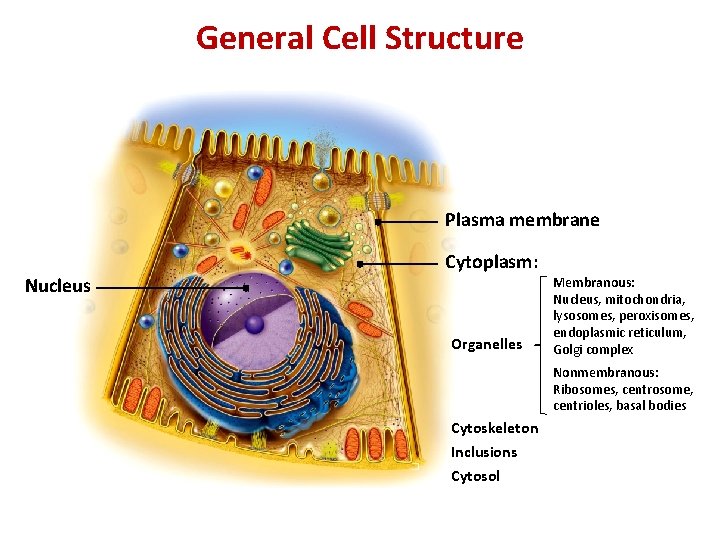
General Cell Structure Plasma membrane Nucleus Cytoplasm: Organelles Membranous: Nucleus, mitochondria, lysosomes, peroxisomes, endoplasmic reticulum, Golgi complex Nonmembranous: Ribosomes, centrosome, centrioles, basal bodies Cytoskeleton Inclusions Cytosol
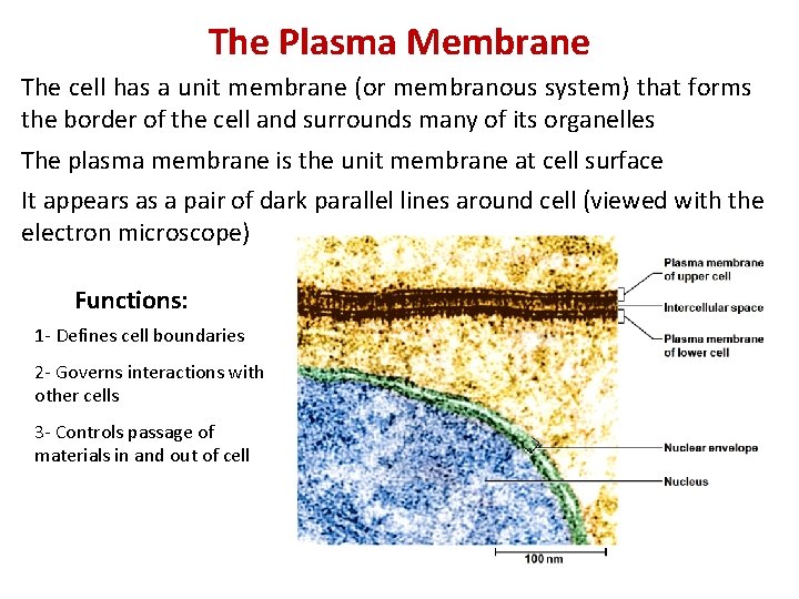
The Plasma Membrane The cell has a unit membrane (or membranous system) that forms the border of the cell and surrounds many of its organelles The plasma membrane is the unit membrane at cell surface It appears as a pair of dark parallel lines around cell (viewed with the electron microscope) Functions: 1 - Defines cell boundaries 2 - Governs interactions with other cells 3 - Controls passage of materials in and out of cell
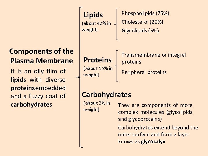
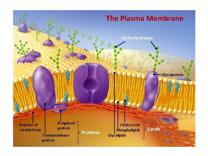
Lipids (about 42% in weight) Components of the Plasma Membrane It is an oily film of lipids with diverse proteins embedded and a fuzzy coat of carbohydrates Proteins (about 55% in weight) Phospholipids (75%) Cholesterol (20%) Glycolipids (5%) Transmembrane or integral proteins Peripheral proteins Carbohydrates (about 3% in weight) They are components of more complex molecules (glycolipids and glycoproteins) Carbohydrates extend beyond the outer surface and form a layer knows as glycocalyx
The Plasma Membrane G l y c o c a l y x Carbohydrates Glycoprotein Proteins of cytoskeleton Peripheral protein Transmembrane protein Proteins Cholesterol Phospholipids Glycolipids Lipids
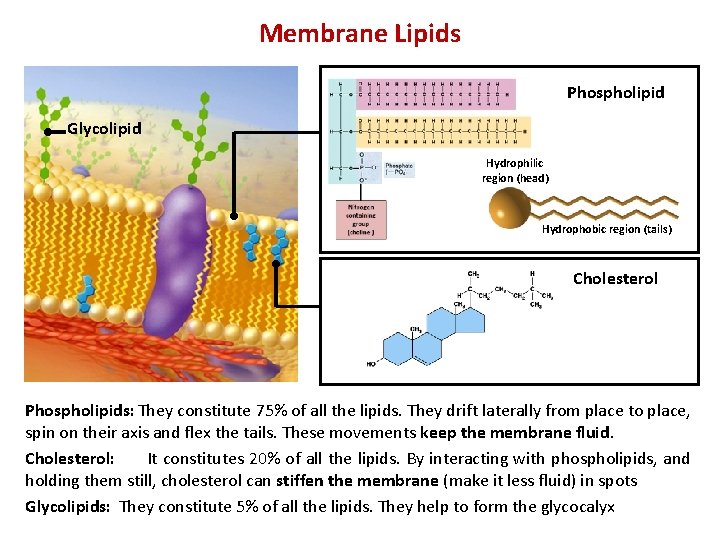
Membrane Lipids Phospholipid Glycolipid Hydrophilic region (head) Hydrophobic region (tails) Cholesterol Phospholipids: They constitute 75% of all the lipids. They drift laterally from place to place, spin on their axis and flex the tails. These movements keep the membrane fluid. Cholesterol: It constitutes 20% of all the lipids. By interacting with phospholipids, and holding them still, cholesterol can stiffen the membrane (make it less fluid) in spots Glycolipids: They constitute 5% of all the lipids. They help to form the glycocalyx
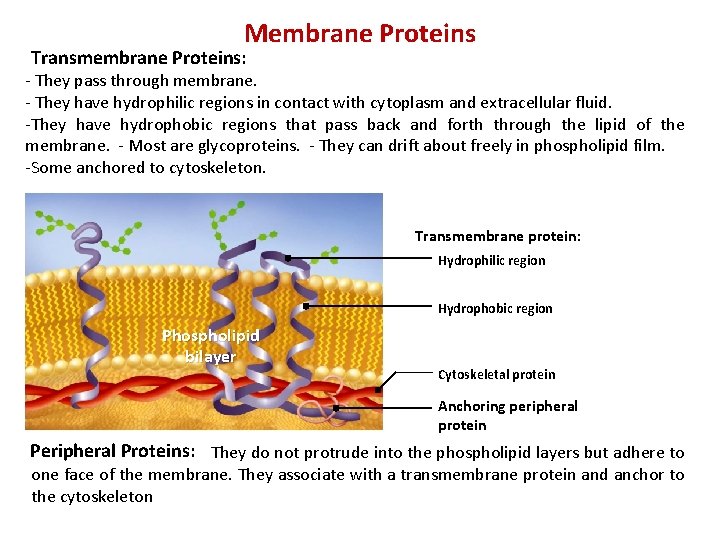
Membrane Proteins Transmembrane Proteins: - They pass through membrane. - They have hydrophilic regions in contact with cytoplasm and extracellular fluid. -They have hydrophobic regions that pass back and forth through the lipid of the membrane. - Most are glycoproteins. - They can drift about freely in phospholipid film. -Some anchored to cytoskeleton. Transmembrane protein: Hydrophilic region Hydrophobic region Phospholipid bilayer Cytoskeletal protein Anchoring peripheral protein Peripheral Proteins: They do not protrude into the phospholipid layers but adhere to one face of the membrane. They associate with a transmembrane protein and anchor to the cytoskeleton
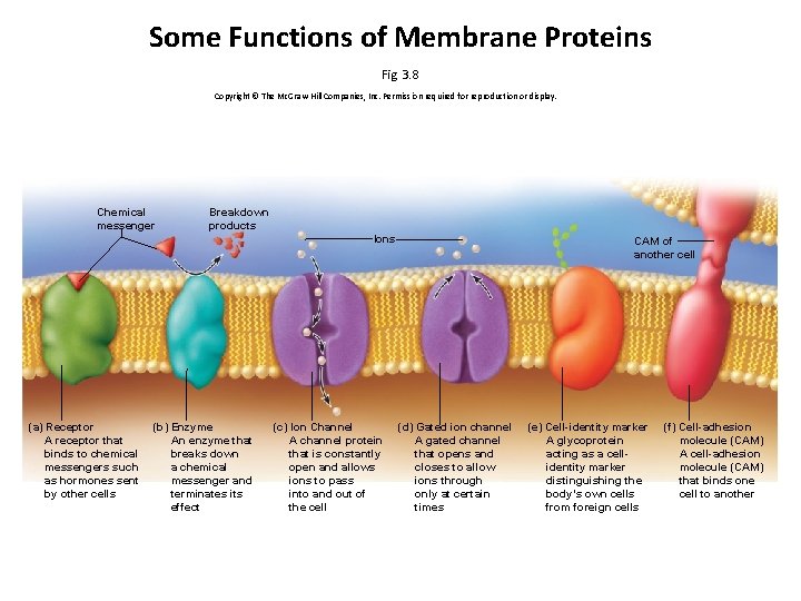
Some Functions of Membrane Proteins Fig. 3. 8 Copyright © The Mc. Graw-Hill Companies, Inc. Permission required for reproduction or display. Chemical messenger Breakdown products Ions (a) Receptor A receptor that binds to chemical messengers such as hormones sent by other cells (b) Enzyme An enzyme that breaks down a chemical messenger and terminates its effect (c) Ion Channel A channel protein that is constantly open and allows ions to pass into and out of the cell CAM of another cell (d) Gated ion channel A gated channel that opens and closes to allow ions through only at certain times (e) Cell-identity marker A glycoprotein acting as a cellidentity marker distinguishing the body’s own cells from foreign cells (f) Cell-adhesion molecule (CAM) A cell-adhesion molecule (CAM) that binds one cell to another
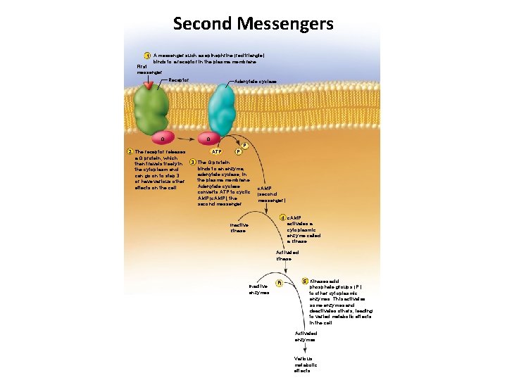
Second Messengers 1 A messenger such as epinephrine (red triangle) binds to a receptor in the plasma membrane. First messenger Receptor G 2 The receptor releases a G protein, which then travels freely in the cytoplasm and can go on to step 3 or have various other effects on the cell. Adenylate cyclase G ATP Pi Pi 3 The G protein binds to an enzyme, adenylate cyclase, in the plasma membrane. Adenylate cyclase converts ATP to cyclic AMP (c. AMP), the second messenger. c. AMP (second messenger) 4 c. AMP activates a cytoplasmic enzyme called a kinase. Inactive kinase Activated kinase Inactive enzymes Pi 5 Kinases add phosphate groups (Pi) to other cytoplasmic enzymes. This activates some enzymes and deactivates others, leading to varied metabolic effects in the cell. Activated enzymes Various metabolic effects
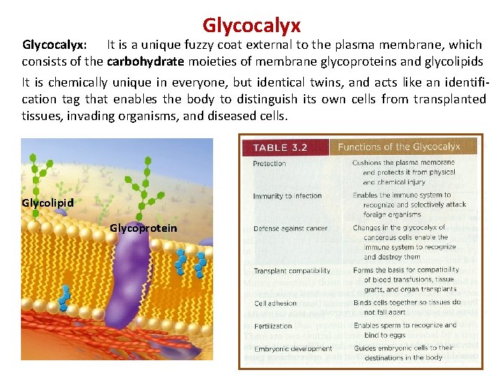
Glycocalyx: It is a unique fuzzy coat external to the plasma membrane, which consists of the carbohydrate moieties of membrane glycoproteins and glycolipids It is chemically unique in everyone, but identical twins, and acts like an identification tag that enables the body to distinguish its own cells from transplanted tissues, invading organisms, and diseased cells. Glycolipid Glycoprotein
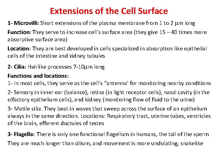
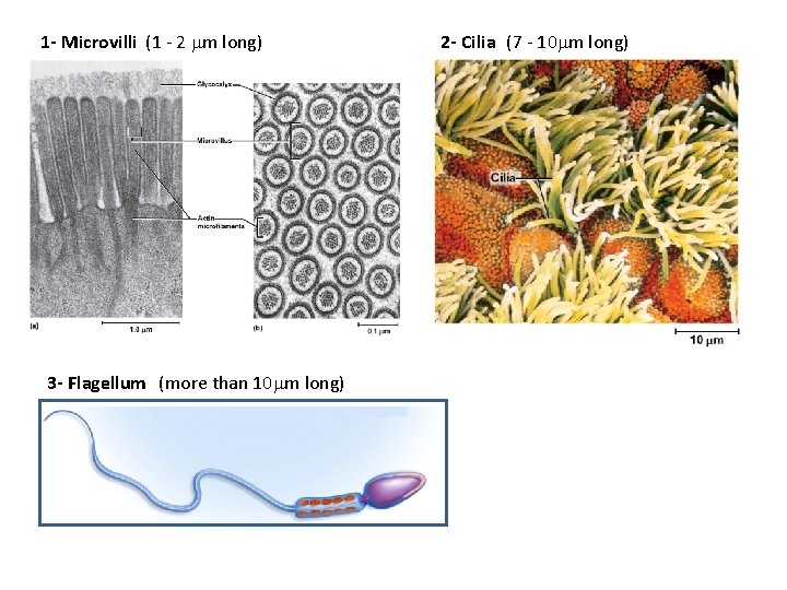
Extensions of the Cell Surface 1 - Microvilli: Short extensions of the plasma membrane from 1 to 2 m long Function: They serve to increase cell’s surface area (they give 15 – 40 times more absorptive surface area) Location: They are best developed in cells specialized in absorption like epithelial cells of the intestine and kidney tubules 2 - Cilia: Hairlike processes 7 -10 m long Functions and locations: 1 - In most cells, they serve as the cell’s “antenna’ for monitoring nearby conditions 2 - Sensory in inner ear (balance), retina (in light receptor cells), nasal cavity (in the olfactory epithelium cells), and kidney (monitoring flow of fluid to the urine) 3 - Motile cilia. They beat in waves that sweep across the surface of an epithelium always in the same direction. Locations: Respiratory tract, uterine tubes, ventricles of the brain, efferent ductules of testes 3 - Flagella: There is only one functional flagellum in humans, the tail of the sperm They are much longer than cilium, and movement is more undulating, snakelike
1 - Microvilli (1 - 2 m long) 3 - Flagellum (more than 10 m long) 2 - Cilia (7 - 10 m long)