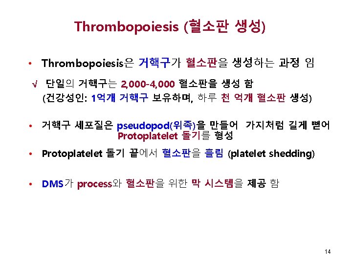
Lecture 5 Platelet Production Structure and Function Keohane


 • 분화중 또는 성숙한 거핵구는 내피세포를 경계로 정맥굴 반대편(abluminal) 표면가까이 위치 5")
 • BFU-Meg 와 CFU-Meg는 유사분열 가능 (증식계속 의미) • LD-CFU-Meg can undergo")


은 형태학적으로 다른계통")
 - 계속 Platelet을 흘리는 거핵구 Fig 13 -7, Rodak혈액학, 5 th")


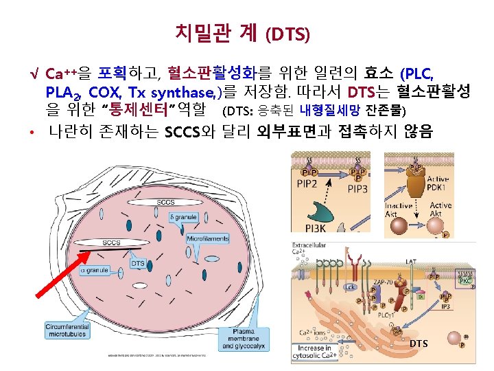
 DTS(Dense tubular system, 치밀관계, condensed RER)")


 Trauma Platelet Glycoprotein Ib-IX-V Endothelial Cell")


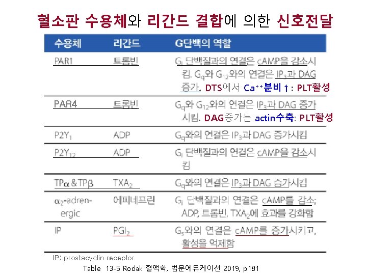
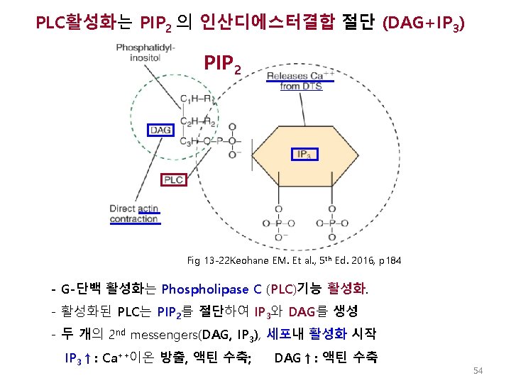
 √ 활성화 된 혈소판은 Thromboxane A 2를 합성하여 분비 함으로써")

 √ 섬유소원은 혈소판 막의 GP IIb-IIIa (Integrin IIb 3) 를 통해")
- Slides: 57
Lecture 5 Platelet Production , Structure and Function Keohane EM et al. Rodak Hematology 5 th Ed, 2016 Elaine M. Keohane, Ph. D, MLS(ASCP), SH keohanem@shrp. rutgers. edu Rutgers-The State University of New Jersey School of Health Related Professions and Kyung Jin Cho, Ph. D chokj@korea. ac. kr Korea University, College of Health Sciences Apr 5, 2019
Platelet Production Chapter author: George A. Fritsma
Fig 12 -1, p. 150 Keohane EM, et al. , Rodak Hematology, 5 th Ed, 2015 (CMP) (CLP) 4
Megakaryocyte Progenitors(거핵구선조세포) • 분화중 또는 성숙한 거핵구는 내피세포를 경계로 정맥굴 반대편(abluminal) 표면가까이 위치 5 Fig. 13 -1 Elaine EM. , Et al. , Rodak Hemaology 5 th Ed. , 2015, p 168
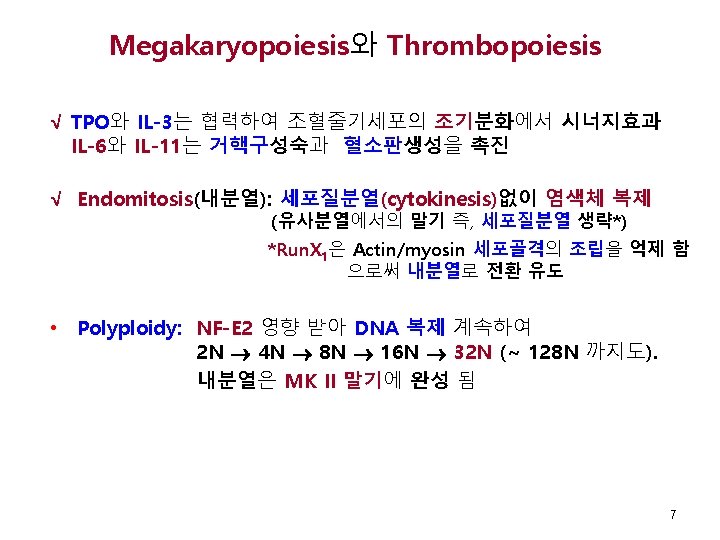
Megakaryocyte Progenitors(거핵구선조세포) • BFU-Meg 와 CFU-Meg는 유사분열 가능 (증식계속 의미) • LD-CFU-Meg can undergo endomitosis • BFU-Meg, CFU-Meg, LD-CFU-Meg형태: 림프구와 유사 (광학현미경 감별 불가) 2 N 2 N (RUNX 1) Fig 13 -2, Keohane EM, et al. , Rodak Hematology, 5 th Ed, 2015, p. 169
Megakaryopoiesis and Thrombopoiesis – cont’d 거핵모구 전거핵구 Fig 13 -3, Keohane EM, et al. , Rodak Hematology, 5 th Ed, 2015, p. 170 거핵구 8
Table 13 -1 Keohane EM, et al. , Rodak Hematology, 5 th Ed, 2015, p. 169
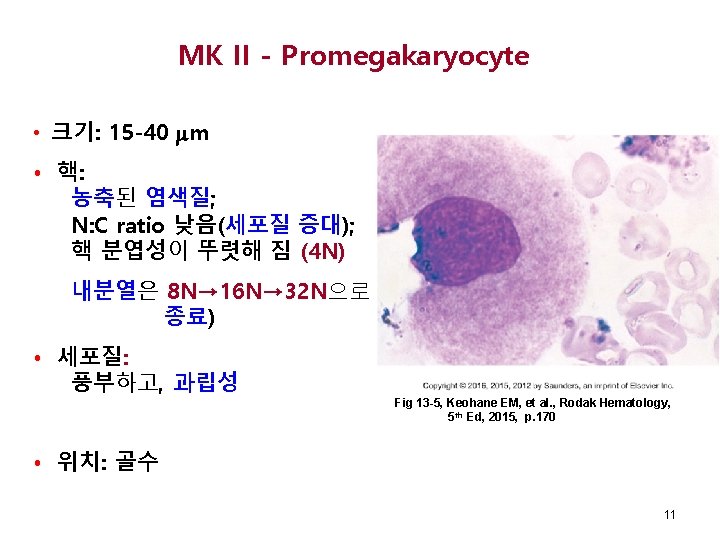
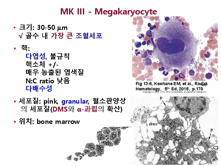
MK I - Megakaryoblast • 크기: 14 -18 m • 핵: 둥글고; 핵소체 2 -6개; 균등 구조의 염색질; N: C ratio 높음; 이배수성 (2 N) √ √ 세포질: 진한 푸른색, Cytoplasmic blebs (blunt projections) • 위치: 골수 Fig 13 -4, Keohane EM, et al. , Rodak Hematology, 5 th Ed, 2015, p. 170 MK I, MK III은 모두 granules (dense bodies) Demarcation system – DMS (원천은 혈소판막)가 나타남 10
거핵구의 동정 • 거핵구선조세포의 형태: 림프구와 유사 함 • MK I (megakaryoblast)은 형태학적으로 다른계통 blast와의 현미경감별 어려움 • BFU-Meg, CFU-Meg, LD-CFU-Meg: CD 34, HLA-DR, CD 41 • MK I, MK II의 감별표지자: Mpl (TPO receptor) CD 41/CD 61 (GPIIb/IIIa) CD 42 (GPIb) PF 4 Table 13 -2 Keohane EM, et al. , Rodak Hematology, 5 th Ed, 2015, p. 170 13
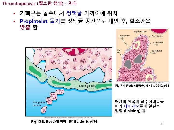
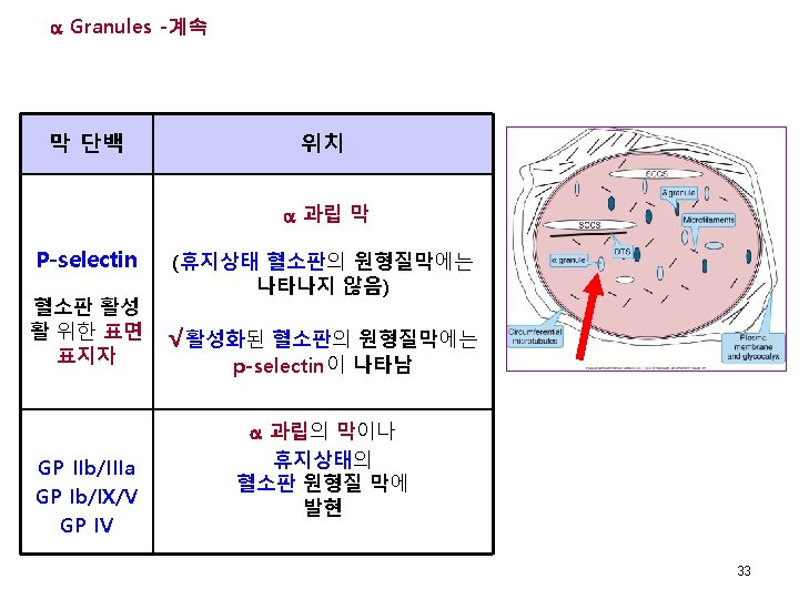
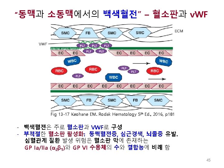
Thrombopoiesis (혈소판 생성) - 계속 Platelet을 흘리는 거핵구 Fig 13 -7, Rodak혈액학, 5 th Ed, 2019, p 179 15
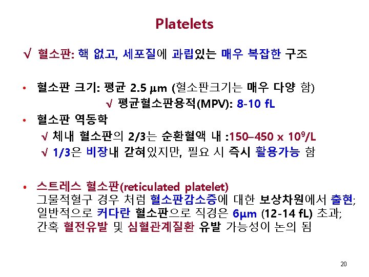
Platelet Structure
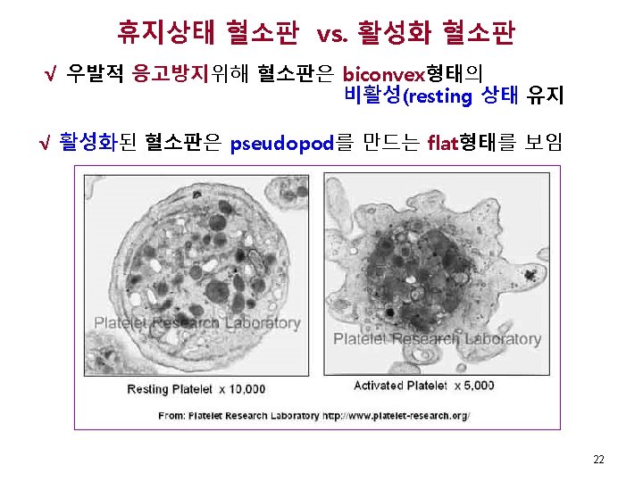
Stress Platelets 21 Fig 13 -8 Keohane EM. Hematology-Clinical Principles and Applications, Elsevier, 2015 p 173
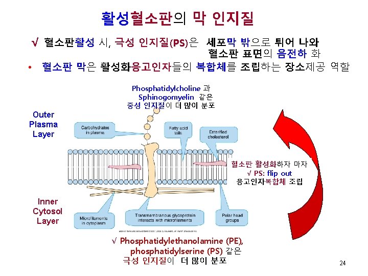
휴지상태 혈소판의 막 인지질 • 원형질막: 이중지질막은 혈소판활성화 위한 많은 수용체 함유 • Phospholipids – 비대칭적 분포(Asymmetric distribution) Phosphatidylcholine 과 Sphinogomyelin 더 많이 분포 Outer Plasma Layer (중성 인지질) Inner Cytosol Layer √ Phosphatidylethanolamine (PE), phosphatidylserine (PS) 더 많이분포 (극성 인지질) 23
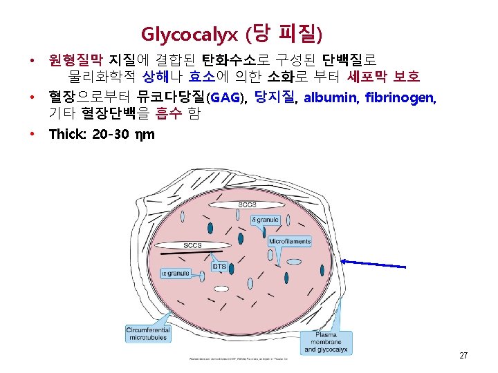
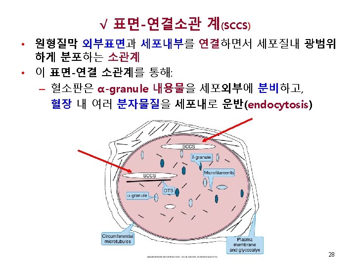
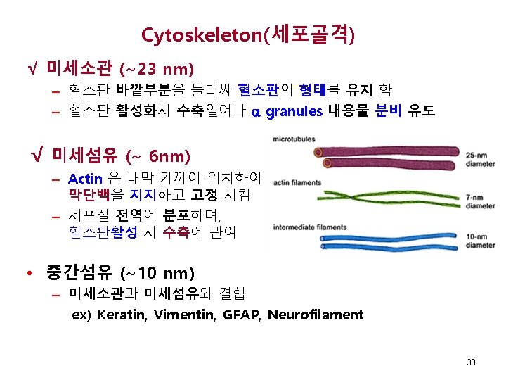
혈소판의 초미세구조 SCCS (Surface connected canalicular system, 표면연결소관계) DTS(Dense tubular system, 치밀관계, condensed RER) Microfilament (미세섬유: actin) Microtubule (미세소관: 튜뷸린이합체) Glycocalyx (당피질) α- 과립 β- 과립 25
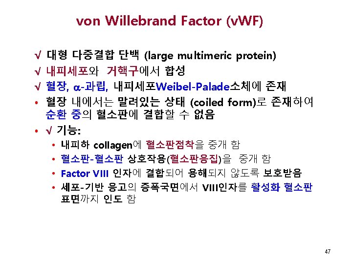
혈소판 막 수용체 • 혈소판 막에 50 종류 이상의 수용체 존재 • 당단백 수용체: 수용체 리간드 √ GP Ib/IX/V* √ VWF**, thrombin √ GP IIb/IIIa* (Integrin IIb 3) √ Fibrinogen, VWF GP Ia/IIa Collagen GP VI Collagen * : 가장 중요한 수용체들 ** : von Willebrand Factor 26
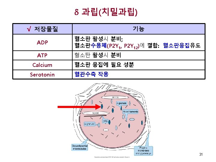
과립 저장물질 Fibrinogen v. WF* Fibronectin Thrombospondin* Vitronectin 기능 √ 접착단백 Platelet Derived Growth Factor (PDGF) Others 성장조절인자 Factor V* Fibrinogen Factor XI Platelet Factor 4* HMWK* Proteins C and S Protein C Inhibitor* Plasminogen* Others √응고촉진단백, 응고억제단백 Thromboglobulin* *: 거핵구에서 합성 32
혈소판수용체와 막 신호경로 GP Ia/IIa GT GP IIb/IIIa VWD BSS Fig 13 -11 Keohane EM. Hematology-Clinical Principles and Applications, Elsevier, 2015 p 175
IX Ibα Ibβ V GT: mutations in GPIIb/IIIa 35 Keohane EM. Hematology-Clinical Principles and Applications, Elsevier, 2015 p 176
Platelet Function
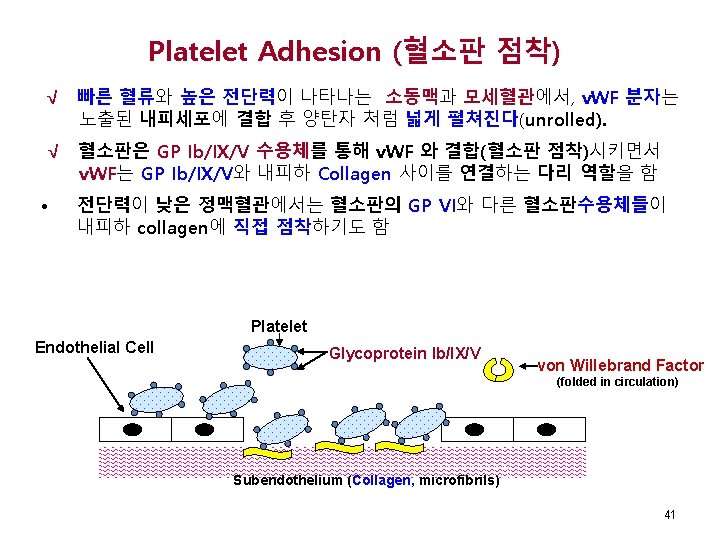
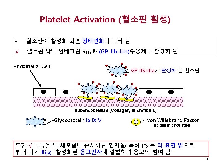
Blood Vessel Endothelial Cells Blood Flow Subendothelium (Collagen) Trauma Platelet Glycoprotein Ib-IX-V Endothelial Cell Subendothelium (Collagen) 내피세포의 노출 40
혈소판활성화의 혈소판점착 유발 Fig 13 -15 Keohane EM. Hematology-Clinical Principles and Applications, Elsevier, 2015 p 180 42
Platelet Activation leading to Platelet Adhesion – Cont’d 초기 혈소판활성화의 혈소판점착 유도 현상 GPIIb/IIIa 화 한 위 활성 ) 집을 위 응 체부 변 화 판 용 태 소 혈 D수 의 형 RG 용체 (수 Inside-Out reaction GPIa/IIa 착의 T점 L P 화 정 안 Outside-In reaction 도 은 합 착유 F 결판점 W V 소 혈 의 α Ib n 에 GP llage Co Fig 13 -14 Keohane EM. Hematology-Clinical Principles and Applications, Elsevier, 2015 p 179
추가 활성- COAT혈소판과 혈소판응집 응집촉진제: Col, Thr, ADP, Tx. A 2 α ules n -gra COAT 혈소판 (collagen과 thrombin에 의해 활성화 된 혈소판) (RGD receptor) (RGD) Fig 13 -16 Keohane EM. Hematology-Clinical Principles and Applications, Elsevier, 2015, p 180
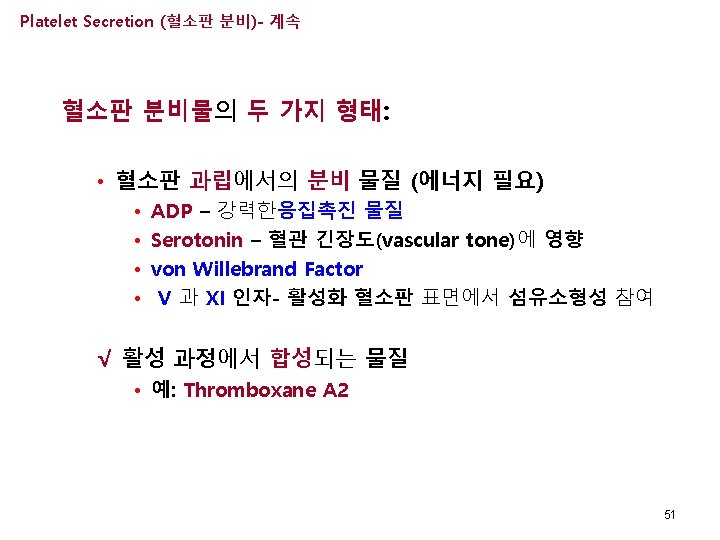
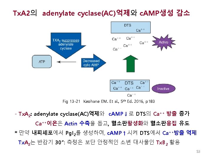
Platelet Secretion (혈소판 분비) √ 활성화 된 혈소판은 Thromboxane A 2를 합성하여 분비 함으로써 Adenylate cyclase (AC)억제하고, c. AMP감소로 DTS로부터 Ca++방출증가 시켜, Actin수축과 ADP방출증가로 혈소판활성화 및 혈소판응집 유도 함 Platelet with activated GP IIb-IIIa Endothelial Cell ADP Tx. A 2 Subendothelium (Collagen, microfibrils) von Willebrand Factor (folded in circulation) 50
Generation of Thromboxane A 2 Eicosanoid Pathway ates y l y t ce ibl a s r e n piri irrev * As -1 and it X CO tivates inac Pg I 2 Fig 13 -20 Keohane EM. Et al. , 5 th Ed. 2016, p 183
Platelet Aggregation (혈소판응집) √ 섬유소원은 혈소판 막의 GP IIb-IIIa (Integrin IIb 3) 를 통해 주변의 혈소판끼리 연결하여 일차 혈소판 마개를 형성 √ v. WF 또한 주변 혈소판들 GP IIb-IIIa(Integrin IIb 3) 분자를 연결 함 platelets Ca+2 required Endothelial Cell Fibrinogen von Willebrand Factor (folded in circulation) Subendothelium (Collagen, microfibrils) Glycoprotein Ib-IX-V Platelet with activated GP IIb-IIIa 55