
Lecture 5 Microscopy PHYS 430603 material Laszlo Takacs
- Slides: 16
Lecture 5: Microscopy PHYS 430/603 material Laszlo Takacs UMBC Department of Physics
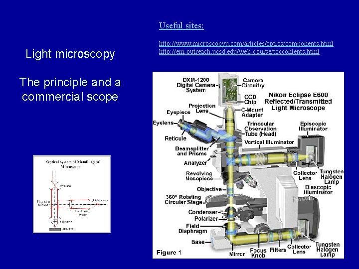
Useful sites: Light microscopy The principle and a commercial scope http: //www. microscopyu. com/articles/optics/components. html http: //em-outreach. ucsd. edu/web-course/toccontents. html
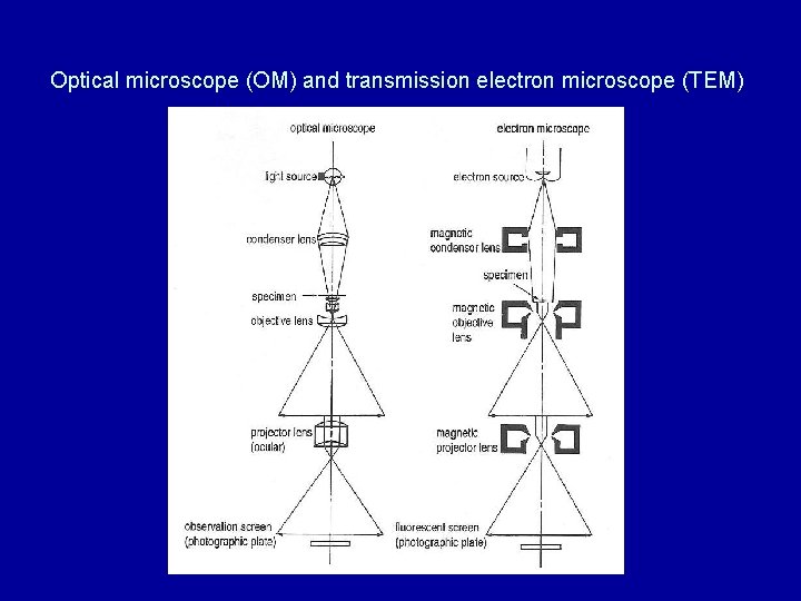
Optical microscope (OM) and transmission electron microscope (TEM)
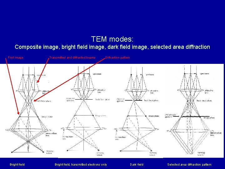
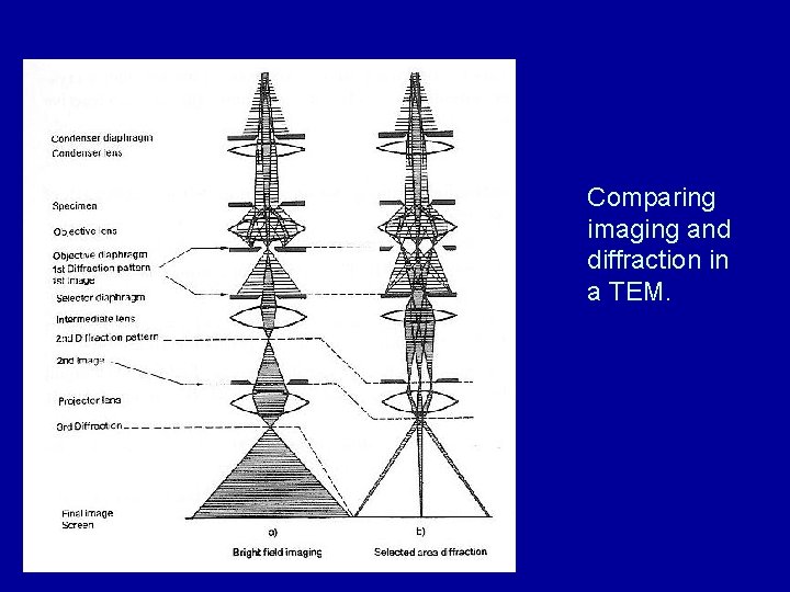
TEM modes: Composite image, bright field image, dark field image, selected area diffraction First image Bright field Transmitted and diffracted beams Diffraction pattern Bright field, transmitted electrons only Dark field Selected area diffraction pattern
Comparing imaging and diffraction in a TEM.
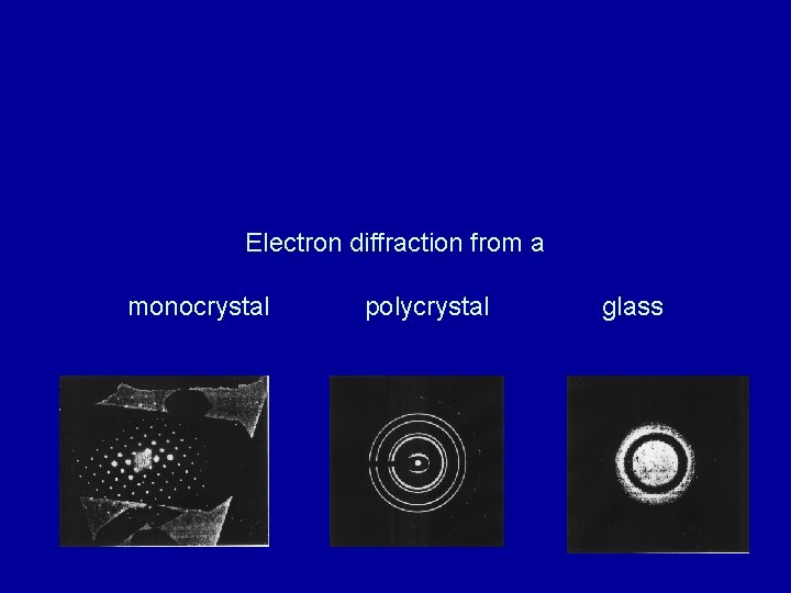
Electron diffraction from a monocrystal polycrystal glass
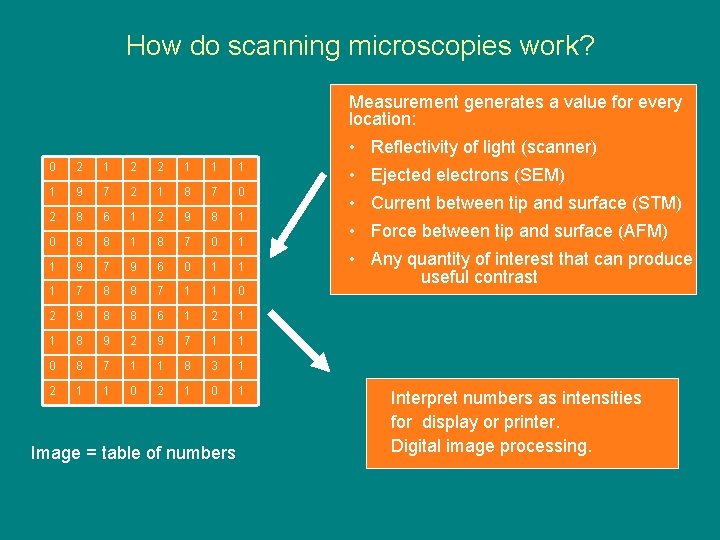
How do scanning microscopies work? Measurement generates a value for every location: • Reflectivity of light (scanner) 0 2 1 2 2 1 1 9 7 2 1 8 7 0 2 8 6 1 2 9 8 1 0 8 8 1 8 7 0 1 1 9 7 9 6 0 1 1 1 7 8 8 7 1 1 0 2 9 8 8 6 1 2 1 1 8 9 2 9 7 1 1 0 8 7 1 1 8 3 1 2 1 1 0 2 1 0 1 Image = table of numbers • Ejected electrons (SEM) • Current between tip and surface (STM) • Force between tip and surface (AFM) • Any quantity of interest that can produce useful contrast Interpret numbers as intensities for display or printer. Digital image processing.
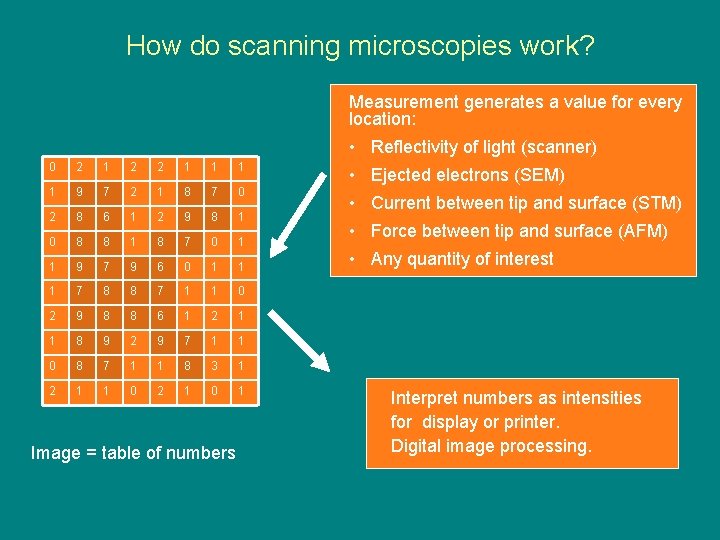
How do scanning microscopies work? Measurement generates a value for every location: • Reflectivity of light (scanner) 0 2 1 2 2 1 1 9 7 2 1 8 7 0 2 8 6 1 2 9 8 1 0 8 8 1 8 7 0 1 1 9 7 9 6 0 1 1 1 7 8 8 7 1 1 0 2 9 8 8 6 1 2 1 1 8 9 2 9 7 1 1 0 8 7 1 1 8 3 1 2 1 1 0 2 1 0 1 Image = table of numbers • Ejected electrons (SEM) • Current between tip and surface (STM) • Force between tip and surface (AFM) • Any quantity of interest Interpret numbers as intensities for display or printer. Digital image processing.
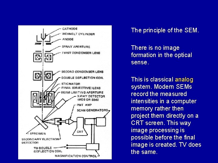
The principle of the SEM. There is no image formation in the optical sense. This is classical analog system. Modern SEMs record the measured intensities in a computer memory rather then project them directly on a CRT screen. This way image processing is possible before the final image is created. TV does the same.
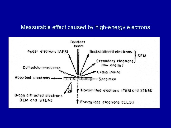
Measurable effect caused by high-energy electrons
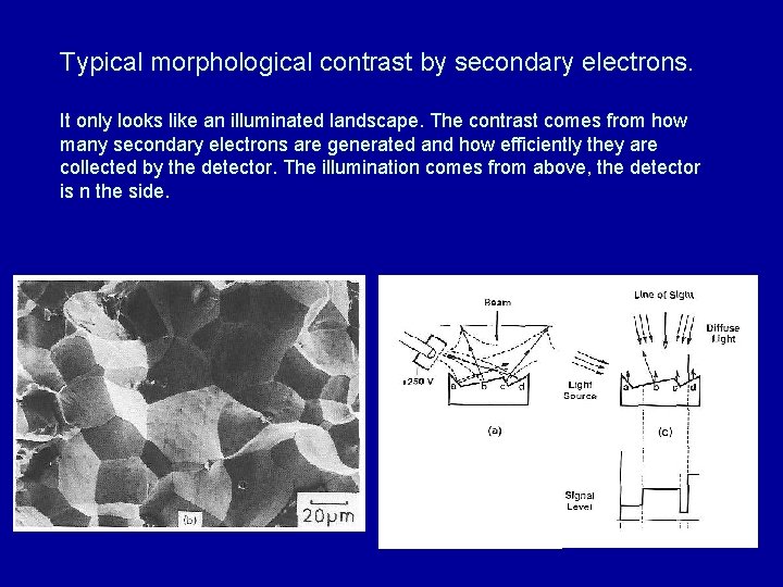
Typical morphological contrast by secondary electrons. It only looks like an illuminated landscape. The contrast comes from how many secondary electrons are generated and how efficiently they are collected by the detector. The illumination comes from above, the detector is n the side.
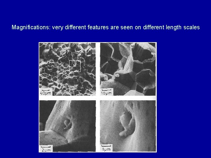
Magnifications: very different features are seen on different length scales
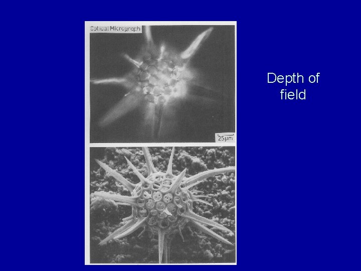
Depth of field
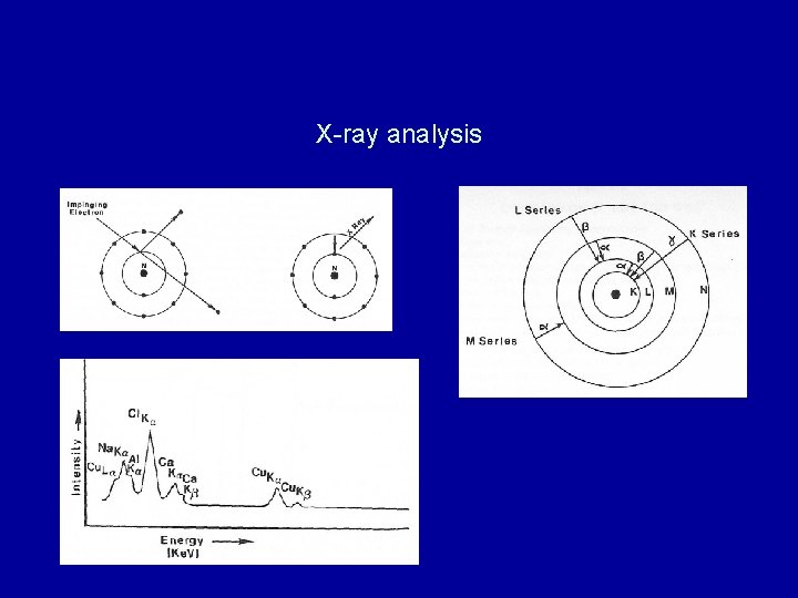
X-ray analysis
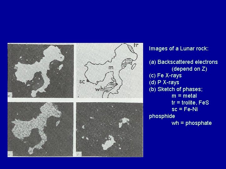
Images of a Lunar rock: (a) Backscattered electrons (depend on Z) (c) Fe X-rays (d) P X-rays (b) Sketch of phases; m = metal tr = trolite, Fe. S sc = Fe-Ni phosphide wh = phosphate
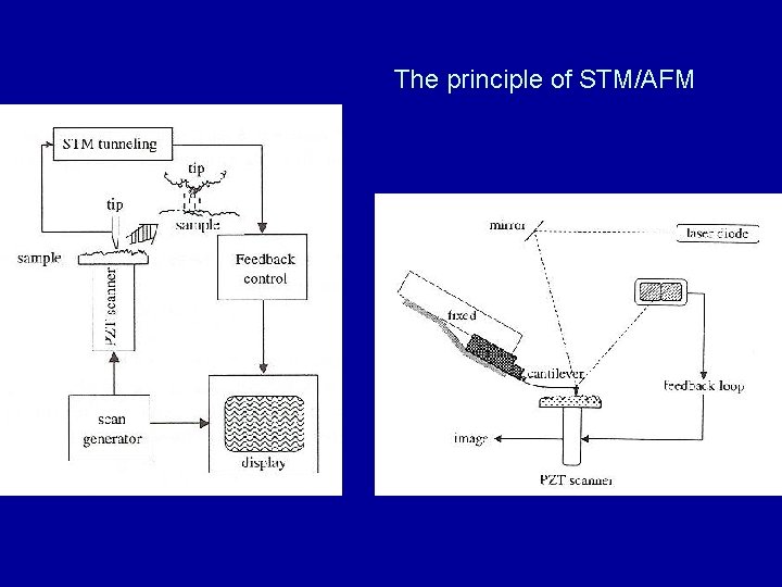
The principle of STM/AFM