Lecture 5 BONES and SKELETAL SYSTEM Skeletal system

Lecture 5 BONES and SKELETAL SYSTEM Skeletal system includes the bones, cartilage, ligaments, and connective tissues that hold all elements of the system together. Single bone is an organ and it has very complex construction. Most of bone constructed from a bone (osseous) tissue. Bones as ligaments are a special type of connective tissue.

Skeletal system has six functions: Protection. Bones create protective structures. Cranium protects brain; thoracic cage protects heart and lungs. Vertebral column protects spinal. Support. Skeleton forms body shape, helps to hold body weight, and provides surface for attachment of muscles and viscera. Movement. Bones anchor muscles. Together, they form system of levers for body movement. Mineral storage and acid-base homeostasis. Bones are major storage of minerals: calcium, phosphorus, magnesium, fluoride, and others. Minerals are stored as salts in ground matter. Salts are chemical compounds based on ionic bonds. Dissolved in water of body fluids, salt dissociates on ions. Accumulation and release of these minerals by bone tissue is a decisive factor in acid-base balance regulation. Formation of blood cells. Some bones harbor red bone marrow. Red bone marrow is the only hematopoietic tissue of adult animal. Fat storage. Yellow bone marrow in medullary cavity of long bones consists of adipocytes that store triglycerides - a source of energy.

Osteology Classification of bones by shape and size is the most convenient and is widely used by most anatomists. According this approach, bones are divided into 5 classes: – Long – Short – Flat – Irregular – Sesamoid

Bone tissue like, all other connective tissues, consists from three components Matrix of bone tissue Collagenous fibers • thick • composed of collagen • great tensile strength • abundant in dense CT • hold structures together • tendons, ligaments Ground substance is an intercellular space filled with solid depositions of calcium phosphate, Ca 3(PO 4), accounting for 2/3 of the bone’s weight. Calcium phosphate interacts with calcium hydroxide, Ca(OH)2, to form hydroxyapatite, Ca 10(PO 4)6(OH)2. As the crystals of hydroxyapatite form, they also incorporate other inorganic materials , such as calcium carbonate, sodium, magnesium, and fluoride. Cellular component of bone There are 3 types of cells in bone: Osteocytes are major type of cells that form and maintain bone body. Osteoblasts are young progenitor cells that very often divide and when maturate transforms into osteocytes. They are bone producing cells. Osteoclasts are multinuclear giant cells that originate from blood stem cells – monocytes. These cells produce very strong enzymes and hydrochloric acid (HCl), which destroy (reabsorb) bones.

Parts of a Long Bone Epiphysis • distal • proximal Diaphysis Medullary cavity Trabeculae Marrow • red • yellow Articular cartilage Periosteum Endosteum There are two types of bone tissue: compact and spongy bone. 5

Compact bone forms shaft of long bones and creates epiphysis coverage. Compact bone is strong and heavy. 85% of the bone is ECM. 65% of ECM consists of inorganic calcium. That makes compact bone strong able to hold body weight and pressure from outside. The structural unit of the compact bone is osteon or Haversian system. Osteon has a shape of a long tube with thick walls. Osteons are tightly packed side by side, like straws in broom. At the center of the osteon is a central or Haversian canal. Central canal contains blood vessels and nerve fibers. Bone tissue is organized around central canal in concentric rings called lamellae. Lacunae with osteocytes are located within lamellae. Lacunae are connected each-other and the central canal by a network of tiny canals called canaliculi. Central canals of osteons are bound perforating or Volkmann’s canals. 6

Microscopic Structure of Compact Bone tissue is very active and constantly in reconstruction by osteoclasts and osteoblasts. The remnants of old osteons called interstitial lamellae fill the space between new osteons 7
 bone Spongy or cancellous bone forms interior of long bone and most")
Cancellous (spongy) bone Spongy or cancellous bone forms interior of long bone and most body of epiphysis. Spongy bone has no heavy osteons. The ECM of spongy bone is organized in spicules or plates called trabeculae. Trabeculae organized in a network to leave a space for red bone marrow, which fills all available space inside spongy bone. Trabeculae are covered by periosteum and in cross section contain lamellae with osteocytes in lacunae. Lacunae of trabeculae are connected with red bone marrow for 8 blood supply by short canaliculi

Flat bone of the cranium on section looks as a sandwich. From both sides they are covered by compact bone. Inside bone contains a thick layer of spongy bone which got a special name diploe. Some bones have pneumatic diverticula (air sacs) (frontal, ethmoid, and maxillae). Pneumatic diverticula replace bone marrow by air sacs. The effect of the pneumatic diverticula reduses body weight. This reduction is an adaptation of pterosaurs and birds to fly, when the low body mass makes a big difference to keep animal aloft. Pneumatized vertebral column of sauropods reduces the weight and make it easier to support and move the massive neck

Bone Structure • periosteum: membrane covers the outer surfaces of bones – outer layer – fibrous tissue – inner layer – osteoblasts • endosteum: membrane lines hollow interior surfaces 10

Bone Cells • osteoblasts – cells that form bone • osteocytes – osteoblasts surrounded by bone • osteoclasts – remodel and remove bone From Patton KT, Thibodeau GA: Anatomy & physiology, ed 8, St Louis, 2013, Mosby. Copyright © 2016 by Elsevier, Inc. 11

Blood Supply to Bone • Volkmann’s canals – channels through bone matrix that contain blood vessels – join at right angles to haversian system Copyright © 2016 by Elsevier, Inc. 12

Bone Marrow • fills the spaces within bones • 2 basic types – red bone marrow • hematopoietic tissue • most common type in younger animals – yellow bone marrow • primarily adipose connective tissue • most common type in adult animals • can revert to red marrow if needed Copyright © 2016 by Elsevier, Inc. 13

Bone Features • articular surfaces – the joint surfaces • processes – lumps, bumps, and other projections on a bone • holes and depressed areas – foramen – fossa Copyright © 2016 by Elsevier, Inc. 14

Articular Surfaces • smooth areas of compact bone where bones come in contact with each other = joints • covered by smooth, thin layer of hyaline cartilage – articular cartilage • surfaces – condyle – head – facet Copyright © 2016 by Elsevier, Inc. 15

Bone Processes • projections on a bone where tendons attach • names depend on location: – spinous process of a vertebra – trochanter on the femur – tubercle on the humerus – tuberosity on the ischium – spine on the scapula – crest on the tibia – wing on the atlas Copyright © 2016 by Elsevier, Inc. 16

Bone Features foramen fossa – hole in a bone – something important passes through hole • blood vessel, nerve – depressed or sunken area on surface of bone – usually occupied by muscles or tendons Copyright © 2016 by Elsevier, Inc. 17

Bone Development Intramembranous Ossification • bones originate within sheet-like layers of connective tissues • broad, flat bones • skull bones (except mandible) • intramembranous bones Endochondral Ossification • bones begin as hyaline cartilage • form models for future bones • most bones of the skeleton • endochondral bones 18

Endochondral Ossification • hyaline cartilage model • primary ossification center • secondary ossification centers • epiphyseal plate • osteoblasts vs. osteoclasts 19

Growth at the Epiphyseal Plate First layer of cells • closest to the end of epiphysis • resting cells • anchors epiphyseal plate to epiphysis Second layer of cells • many rows of young cells • undergoing mitosis 20

Growth at the Epiphyseal Plate Third layer of cells • older cells • left behind when new cells appear • cells enlarging and becoming calcified Fourth layer of cells • thin • dead cells • calcified extracellular matrix 21

Homeostasis of Bone Tissue • Bone Resorption – action of osteoclasts and parathyroid hormone • Bone Deposition – action of osteoblasts and calcitonin 22
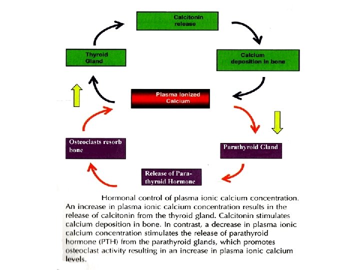

Homeostatic imbalances of bone can cause different deseases such as osteomalacia and rickets. Parturient paresis (milk fever) is result of low calcium in blood. The first milk produced by dairy cattle (called colostrum) contains high concentration of Ca 2+. Cow needs to produce 3 g of Ca 2+ per hour for colostrum. When she has not sufficient calcium supply from a food, she can develop milk fever within 72 hours following parturition. Symptoms include loss of appetite, muscle weakness, decrease of body temperature, labored breathing, and paralysis of hind legs. If left untreated, the cow can collapse into coma and die. To prevent milk fever, cows should be given sufficient vitamin D in the diet prior to parturition. If milk fever develops, cows are given an oral bolus of calcium carbonate.

Factors Affecting Bone Development, Growth, and Repair • Deficiency of Vitamin A – retards bone development • Deficiency of Vitamin C – results in fragile bones • Deficiency of Vitamin D – rickets, osteomalacia • Insufficient Growth Hormone – dwarfism • Excessive Growth Hormone – gigantism, acromegaly • Insufficient Thyroid Hormone – delays bone growth • Sex Hormones – promote bone formation; stimulate ossification of epiphyseal plates • Physical Stress – stimulates bone growth 25

Clinical Application • green stick • fissured • comminuted • transverse • oblique • spiral Types of Fractures 26

Repair • When a bone breaks, several things happen: – A hematoma forms; this is when you see signs of inflammation – A callus forms; this is an area where fibrocartilage (connective tissues, including osteoblasts) tries to bridge the gap – A bony callus results – Bone begins to remodel to re-establish its original size and shape

Cartilages Cartilage • rigid matrix • chondrocytes in lacunae • poor blood supply • three types • hyaline • elastic • fibrocartilage Hyaline cartilage • most abundant • ends of bones • nose, respiratory passages • embryonic skeleton Elastic cartilage • flexible • external ear, larynx Fibrocartilage • very tough • shock absorber • intervertebral discs • pads of knee and pelvic girdle 28

1. Cartilage has no nerves or blood vessels of its own 2. Cartilage is nourished by diffusion 3. Cartilage has no ossified structures 4. Bone is the reverse of all of the above Elastic Cartilage Hyaline Cartilage Fibrocartilage 29

Cartilage grows by two processes: 1. Appositional growth when new cartilage forms on the surfice of already existing cartilage. Cartilage increases in size by growing on its margins. 2. Interstitial growth when new cartilage growth from inside of the cartilage mass. In this case, chondrocytes that reside in the lacunae divide and secrete a new matrix. As a result cartilage expends from within.

Bones of the Skeleton • Axial skeleton – located along central axis of body – bones of the head and trunk • skull, hyoid bone, spinal column, ribs, sternum • Appendicular skeleton – bones of the limbs • Visceral skeleton – bones formed in soft organs (viscera) 31

HAVE QUESTIONS?
- Slides: 32