Lecture 4 The Cell The basic unit of

Lecture 4 The Cell The basic unit of life

Cells • Cells can be defined as fundamental to the living systems of biology as the atom is to chemistry or • Cell is the basic structural, functional, and biological unit of all known living organisms • Cell walls were first seen by Robert Hooke in 1665 as he looked through a microscope at dead cells from the bark of an oak tree

• However what Hooke actually saw was the dead cell walls of plant cells and gave no indication of the nucleus and other organelles found in most living cells • The first man to witness a live cell under a microscope was Anton van Leeuwenhoek in 1674

Cell Theory Developed in late 1800 s 1. All organisms are composed of one or more cells, and the life processes of metabolism and heredity occur within these cells. 2. Cells are the smallest living things, the basic units of organization of all organisms. 3. Cells arise only by division of a previously existing cell

Cell Diversity • Cells for organisms show Enormous Diversity in: – Size – Shape – Structure

1. Cell Size The size of Most cells are between 1 and 100 μm in diameter and are therefore visible only under a microscope.
 = 10– 2 meter (m) = 0. 4 inch 1 millimeter")
1 centimeter (cm) = 10– 2 meter (m) = 0. 4 inch 1 millimeter (mm) = 10– 3 m 1 micrometer (µm) = 10– 3 mm = 10– 6 m 1 nanometer (nm) = 10– 3 µm = 10– 9 m

Why Cells are small in Size? Home work

2 -Cell Shape The cells have different shaped , the cell shape is fit to its function

3 - Cell Types • There are of two distinct types of cells: prokaryotic and eukaryotic cells. • Organisms of the domains Bacteria and Archaea consist of prokaryotic cells. • Protists, fungi, animals, and plants all consist of eukaryotic cells.

Comparing Prokaryotic and Eukaryotic Cells All cells share certain basic features: 1. They are all bounded by a selective barrier, called the plasma membrane. 2. Inside all cells is a semifluid, jellylike substance called cytosol, in which subcellular components are suspended. 3. All cells contain chromosomes, which carry genes in the form of DNA. 4. And all cells have ribosomes, tiny complexes that make proteins according to instructions from the genes.

A major difference between prokaryotic and eukaryotic cells is the location of their DNA. • In a eukaryotic cell, most of the DNA is in an organelle called the nucleus, which is bounded by a double membrane • In a prokaryotic cell, the DNA is concentrated in a region that is not membrane-enclosed, called the nucleoid

• The word eukaryotic means “true nucleus” (from the Greek eu, true, and karyon, kernel, here referring to the nucleus) • The word prokaryotic means “before nucleus” (from the Greek pro, before), reflecting the fact that prokaryotic cells evolved before eukaryotic cells. • The interior of either type of cell is called the cytoplasm; • Within the cytoplasm of a eukaryotic cell, suspended in cytosol, are a variety of organelles of specialized form and function. • These membrane-bounded structures are absent in prokaryotic cells. • Thus, the presence or absence of a true nucleus is just one aspect of the disparity in structural complexity between the two types of cells.

Prokaryotic versus Eukaryotic Cells Feature Prokaryotic Organisms Bacteria Eukaryotic All others (animals, plants, fungi, and protozoa) Nucleus Absent DNA One chromosome Multiple chromosomes Size Small (1 -10 um) Large (10 or more um) Membrane Absent Bound Organelles Division Rapidprocess (Binary fission) Present (mitochondria, golgi, chloroplasts, etc. ) Complex process (Mitosis)

The Eukaryotic Cell

The Eukaryotic Cell • In addition to the plasma membrane at its outer surface, a eukaryotic cell has extensive and elaborately arranged internal membranes that divide the cell into compartments—the organelles • The cell’s compartments provide different local environments that facilitate specific metabolic functions, so incompatible processes can go on simultaneously inside a single cell • The plasma membrane and organelle membranes also participate directly in the cell’s metabolism, because many enzymes are built right into the membranes.

The Plasma Membranes The basic fabric of most biological membranes is a double layer of Phospholipids, which is called phospholipid biyaler. Embedded in this lipid bilayer or attached to its surfaces are diverse proteins Each type of membrane has a unique composition of lipids and proteins suited to that membrane’s specific functions. For example, enzymes embedded in the membranes of the organelles called mitochondria respiration. function in cellular

Membrane Components Phospholipids Proteins 19

Functions of Cell Membranes 1. Separate cell from nonliving environment. Form most organelles and partition cell into discrete compartments. 2. Regulate passage of materials in and out of the cell and organelles. Membrane is selectively permeable. 3. Receive information that permits cell to sense and respond to environmental changes. • Hormones • Growth factors • Neurotransmitters 4. Communication with other cells and the organism as a whole. Surface proteins allow cells to recognize each other, adhere, and exchange materials.

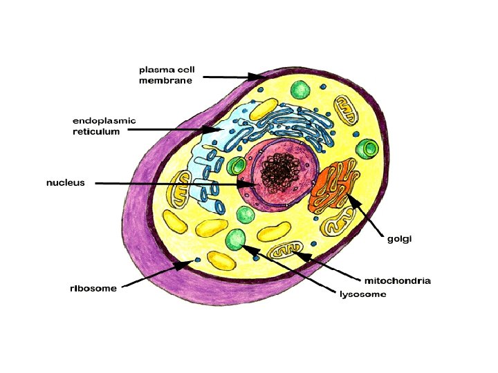
A eukaryotic cell has internal membranes that partition the cell into organelles. Plant and animal cells have most of the same organelles Membrane-Bound Organelles of Eukaryotic Cells • Nucleus • Rough Endoplasmic Reticulum (RER) • Smooth Endoplasmic Reticulum (SER) • Golgi Apparatus • Lysosomes • Vacuoles • Chloroplasts • Mitochondria

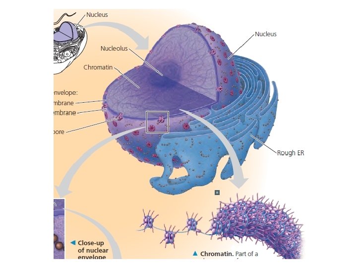
The Nucleus • nucleus contains most of the genes in the It is generally the most conspicuous organelle in a eukaryotic cell, averaging about 5 μm in diameter • The eukaryotic cell. (Some genes are located in mitochondria and chloroplasts. ) • The nuclear envelope encloses the nucleus separating its contents from the cytoplasm.

• The nuclear envelope is a double membrane. The two membranes, each a lipid bilayer with associated proteins, are separated by a space of 20– 40 nm • The envelope is perforated by pore structures that are about 100 nm in diameter • At the lip of each pore, the inner and outer membranes of the nuclear envelope are continuous. • An intricate protein structure called a pore complex lines each pore and plays an important role in the cell by regulating the entry and exit of proteins and RNAs
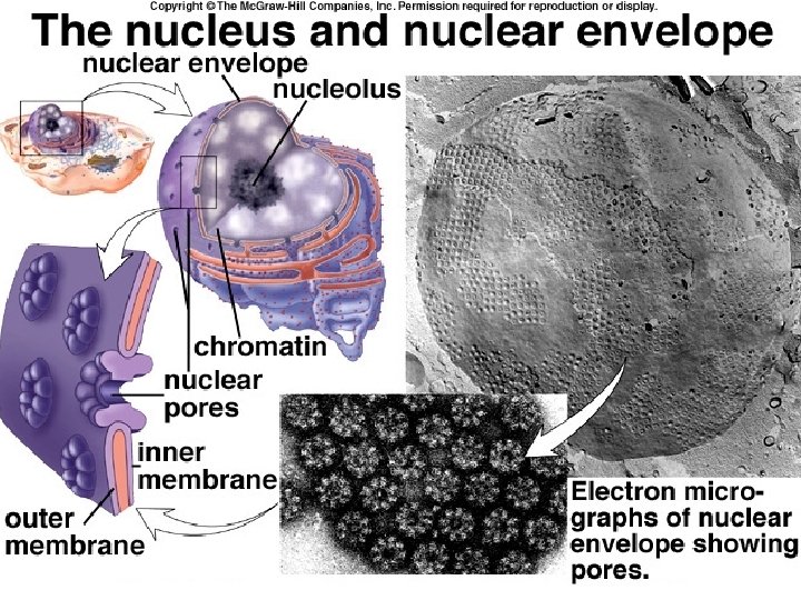

• Within the nucleus, the DNA is organized into discrete units called chromosomes, structures that carry the genetic information. • Each chromosome contains one long DNA molecule associated with many proteins. • Some of the proteins help coil the DNA molecule of each chromosome, reducing its length and allowing it to fit into the nucleus. • The nucleolus is located within the nucleus and is the site of ribosomal RNA (r. RNA) synthesis

nucleolus • A prominent structure within the non-dividing nucleus is the nucleolus • Here a type of RNA called ribosomal RNA (r. RNA) is synthesized from instructions in the DNA. • It is primarily serves as the site of ribosome synthesis and assembly • The proteins imported from the cytoplasm are assembled with r. RNA into large and small subunits of ribosomes. • These subunits then exit the nucleus through the nuclear pores to the cytoplasm, where a large and a small subunit can assemble into a ribosome.

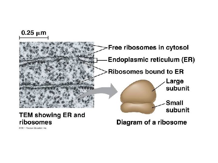
Ribosomes: Protein Factories – Smallest organelle – NOT surrounded by a membrane – Ribosomes, which are complexes made of ribosomal RNA and protein, – Makes proteins according to DNA instructions.

Cells that have high rates of protein synthesis have particularly large numbers of ribosomes. For example, a human pancreas cell has a few million ribosomes. Not surprisingly, cells active in protein synthesis also have prominent nucleoli. Two Types of ribosomes: • • Free ribosomes: float free in cytosol Bound ribosomes: attached to rough ER and nuclear membrane

The endomembrane system regulates protein traffic and performs metabolic functions in the cell • Components of the endomembrane system – Nuclear envelope – Endoplasmic reticulum – Golgi apparatus – Lysosomes – Vacuoles – Plasma membrane • These components are either continuous or connected via transfer by vesicles © 2011 Pearson Education, Inc.
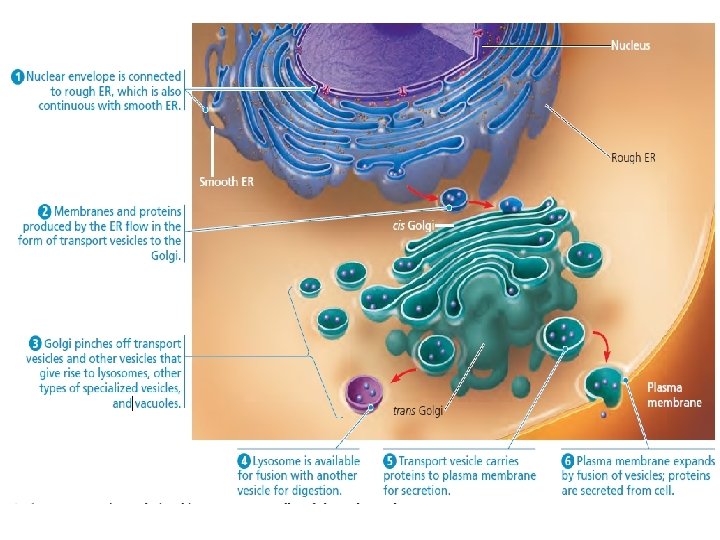

Endoplasmic Reticulum • is such an extensive network of membranes that it accounts for more than half the total membrane in many eukaryotic cells. • The ER consists of a network of membranous tubules and sacs called cisternae (from the Latin cisterna, a reservoir for a liquid). • The ER membrane separates the internal compartment of the ER, called the ER lumen (cavity) or cisternal space, from the cytosol.

The ER membrane is continuous with the nuclear envelope

• There are two distinct, though connected, regions of the ER that differ in structure and function: smooth ER and rough ER. • Smooth ER is so named because its outer surface lacks ribosomes. • Rough ER is studded with ribosomes on the outer surface of the membrane and thus appears rough the electron microscope

Functions of Smooth ER The smooth ER functions in diverse metabolic processes, which vary with cell type. These processes include 1. Synthesis of lipids 2. Metabolism of carbohydrates, 3. Detoxification of Drugs and poisons 4. Storage of calcium ions.

• Enzymes of the smooth ER are important in the synthesis of lipids, including oils, phospholipids, and steroids. • Among the steroids produced by the smooth ER in animal cells are the sex hormones of vertebrates and the various steroid hormones secreted by the adrenal glands • The cells that synthesize and secrete these hormones—in the testes and ovaries, for example—are rich in smooth ER, a structural feature that fits the function of these cells.

• Other enzymes of the smooth ER help detoxify drugs and poisons, especially in liver cells. • Detoxification usually involves adding hydroxyl groups to drug molecules, making them more soluble and easier to flush from the body. • The sedative phenobarbital and other barbiturates are examples of drugs metabolized in this manner by smooth ER in liver cells. • In fact, barbiturates, alcohol, and many other drugs induce the proliferation of smooth ER and its associated detoxification enzymes, thus increasing the rate of detoxification. • This, in turn, increases tolerance to the drugs, meaning that higher doses are required to achieve a particular effect.

• The smooth ER also stores calcium ions. In muscle cells, for example, the smooth ER membrane pumps calcium ions from the cytosol into the ER lumen. • When a muscle cell is stimulated by a nerve impulse, calcium ions rush back across the ER membrane into the cytosol and trigger contraction of the muscle cell. • In other cell types, calcium ion release from the smooth ER triggers different responses, such as secretion of vesicles carrying newly synthesized proteins

Functions of Rough ER : • Synthesis and modification of proteins(Has bound ribosomes, which secrete glycoproteins ) • Synthesis of cell and organelle membranes(Is a membrane factory for the cell) • Packaging, and transport of proteins that are secreted from the cell.

Protein Synthesis • Many types of cells secrete proteins produced by ribosomes attached to rough ER. For example, certain pancreatic cells synthesize the protein insulin in the ER and secrete this hormone into the bloodstream • As a polypeptide chain grows from a bound ribosome, the chain is threaded into the ER lumen through a pore formed by a protein complex in the ER membrane • As the new polypeptide enters the ER lumen, it folds into its native shape. Most secretory proteins are glycoproteins, proteins that have carbohydrates covalently bonded to them.

• After secretory proteins are formed, the ER membrane keeps them separate from proteins that are produced by free ribosomes and that will remain in the cytosol. • Secretory proteins depart from the ER wrapped in the membranes of vesicles that bud like bubbles from a specialized region called transitional ER

• In addition to making secretory proteins, rough ER is a membrane factory for the cell; it grows in place by adding membrane proteins and phospholipids to its own membrane. • As polypeptides destined to be membrane proteins grow from the ribosomes, they are inserted into the ER membrane itself • Like the smooth ER, the rough ER also make membrane phospholipids; enzymes built into the ER membrane assemble phospholipids from precursors in the cytosol. • The ER membrane expands and portions of it are transferred in the form of transport vesicles to other components of the endomembrane system.
- Slides: 43