Lecture 4 Structures of plant cells 1 Cell


 at")


- Slides: 11
Lecture – 4 Structures of plant cells
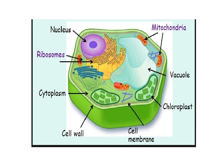
1 - Cell wall: The cell wall around plant and fungal cells, and some single-celled eukaryotes, consists mostly of cellulose fibers and adds strength and rigidity to the cell. Spaces between the cellulose fibers allow molecules to pass to and from the cell plant. Cells that form part of a supporting structure in a plant have a secondary stronger, cell wall, contains lignin, inside the primary cell. Unlike the cell membrane, the cell wall doesn't control the materials that can pass through it. However, the cell wall does help the cell deal with hypotonic or hypertonic environments. It prevents the cell from bursting in hypotonic environments. In hypertonic environments, it provides structural support when the rest of the cell contents shrink.
2 - Central Vacuole: Plant cells usually have a large, fluid-filled central vacuole. This presses outwards on the cell wall to help support it. It also provides storage space for water and other substances. These may include toxins that make the cell, and thus the plant, taste bad to animals. 3 - Plastids: The green chloroplast is one of a group of organelles in plant cells called plastids. All plastids contain stacked internal membrane sacs these sacs are enclosed within a double membrane and have the ability to perform photosynthesis.
Functions of plastids 1 - Plastids exposed to light develop pigments and participate ( share ) in collecting energy from light. 2 -Plastids also act as storage containers for starches, lipids, and proteins. 3 - The chlorophyll, other pigments, and enzymes necessary for photosynthesis are contained within a special membrane system.
Lysosomal Digestion : Only eukaryotic cells contain lysosomes. In plant cells, the specialized vesicles that recycle cellular material (the way that a lysosome does) are often called vacuoles. Each lysosome (or vacuole) contains over 40 different digestive enzymes. After the lysosome leaves the Golgi apparatus, its membrane actively pumps in hydrogen ions to make its interior environment more acidic. This acidic environment activates the lysosomal enzymes. When active, these enzymes can break apart macromolecules in a step-by-step process. This lysosomal digestion can occur in two ways: A-By breaking down material ingested through endocytosis B- By recycling the cell’s own organic material.
Endocytosis: After a cell engulfs material by endocytosis, a lysosome fuses with the food vacuole formed to fill the vacuole with digestive enzymes and break down the captured material. Recycling : A lysosome fuses with and digests organelles no longer useful to the cell. It then recycles their components back to the cytoplasm. You can follow The steps involved in this process in Figure below
Cells and energy photosyntheses and energy
Enzyme Function : Enzymes make possible reactions that would otherwise not proceed( Forward) at temperatures low enough for life to be possible. An enzyme is a protein that functions as an organic catalyst. A catalyst helps a particular reaction go forward without being used up in the process. The cell makes a different enzyme for each reaction it requires. An enzymatic reaction may either combine(join) molecules to produce a new product or break a molecule into smaller parts. Chemical bonds within a substrate can be broken or bonds between substrates can be formed. Then the enzyme releases the product(s) and can start the process again.
Figure 2. 22 illustrates a model of the way an enzyme breaks apart a molecule, proteins haven three-dimensional shapes. An enzyme’s shape allows its substrate(s) to attach at a spot called the active site. Here, chemical bonds within a substrate can be broken or bonds between substrates can be formed. Then the enzyme releases the product(s) and can start the process again.
THANK YOU FOR ATTENTION