Lecture 4 Iron Kinetics Leukocyte Development Kinetics Functions
 Keohane EM et")

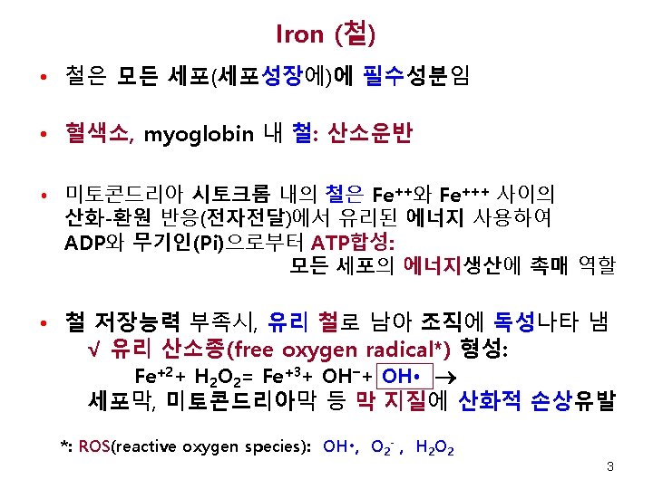
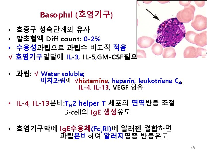
 기능 철 운반 철 저장 철 √")

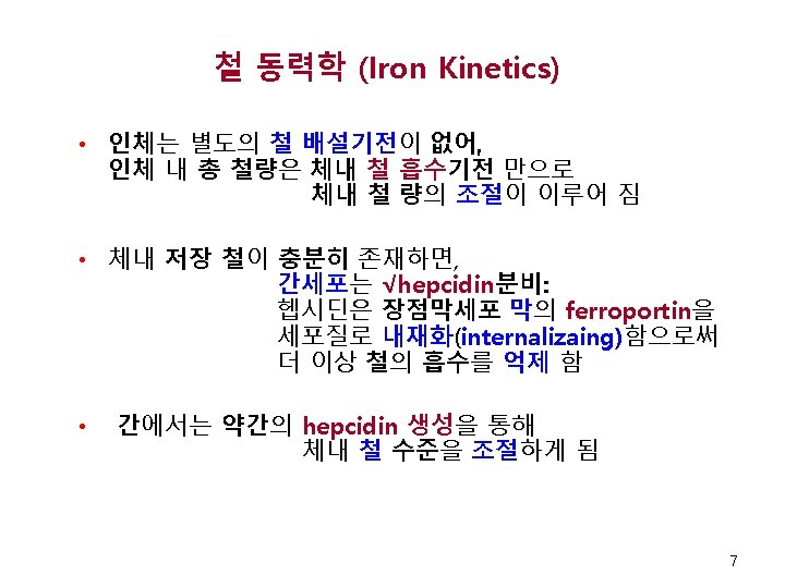

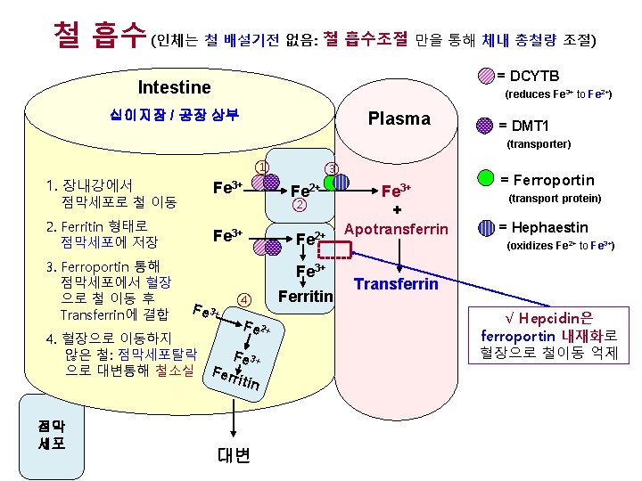
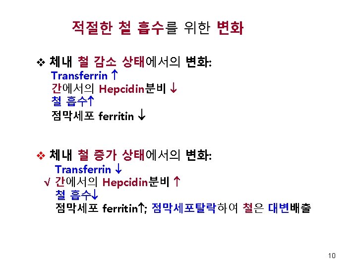
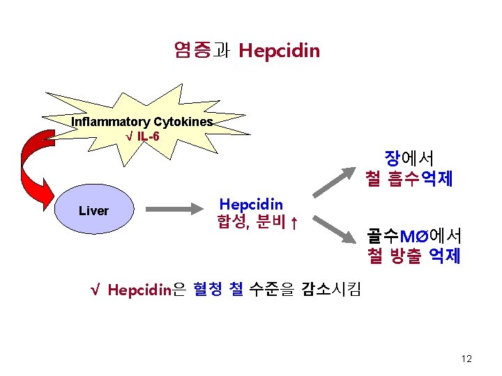
 Fig 11 -4. Hepatic")
 Apotransferrin (No Bound Iron) Transferrin (Tf) Diferric Transferrin Receptor")
 • 그물적혈구에서의")


 Bone Marrow Large cell (14")
 과립내용물 Myeloperoxidase, cathepsins,")
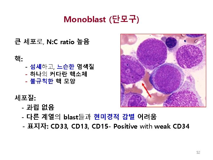
 골수유핵세포의 1 -2 % 차지 형태학적으로 어떤 계통의 blast인지 감별이 어려움 감별을")


 with classic red-orange globular granules in the cytoplasm √ √ Maslak, P.")

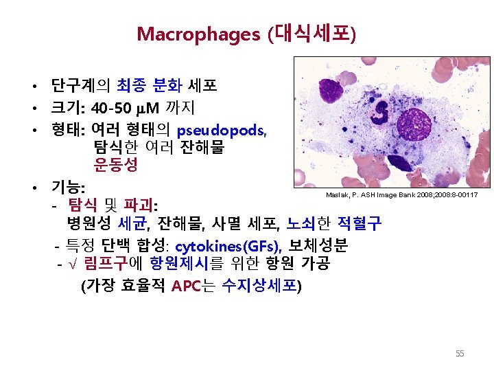
 • 선조세포 : GMP • 성숙단계: Monoblast Promonocyte blood monocyte √macrophage •")
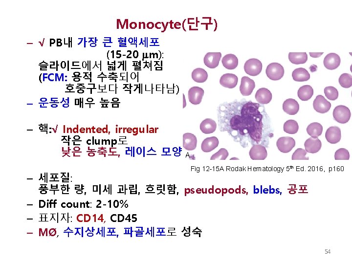
 • 운동성 있음 • 탐식기능 있음 • 핵: √ folded, twisted, indented")

 크기: 직경 10 -18 M N: C ratio 높음 – 핵: 섬세,")
 – 림프모구와 유사한 형태 – 핵: - 염색질은 약간 뭉쳐짐 - 핵소체는 덜")
 – 분열(증식) 없음 – 크기: 소형, 중형, 대형으로 다양 함 – 핵:")
 • • • Dark blue cytoplasm 편재성 핵 핵 주변의")
- Slides: 67
Lecture 4 Iron Kinetics; Leukocyte Development, Kinetics, Functions (Chapters 11, 12) Keohane EM et al. , Rodak’s Hematology 5 th Ed. , 2016 Elaine M. Keohane, Ph. D, MLS(ASCP), SH keohanem@shrp. rutgers. edu Rutgers-The State University of New Jersey School of Health Related Professions and Kyung Jin Cho, Ph. D chokj@korea. ac. kr Korea University, College of Health Sciences Mar 29, 2018 1
철의 이동 및 검사실 평가 Chapter 11 Chapter author: Doig K 2
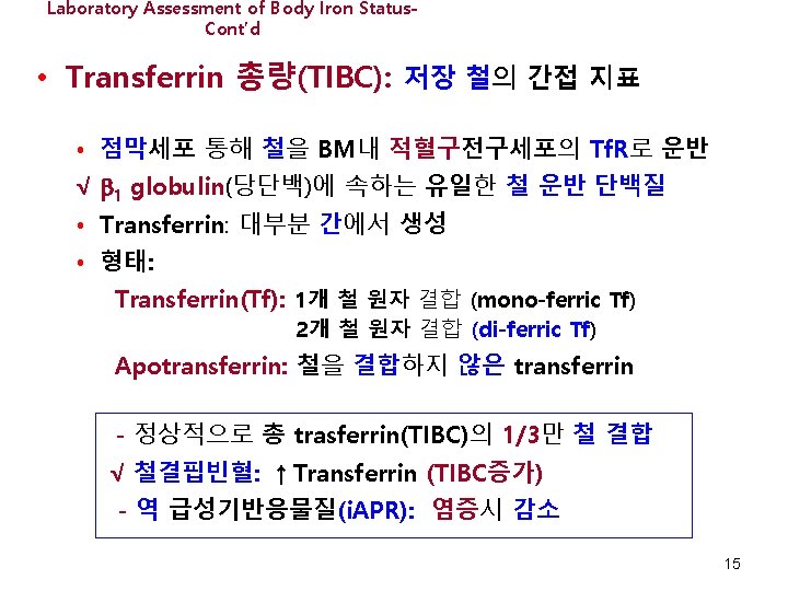
성인에서 철의 분포 구획 별 철 분포도(%) 기능 철 운반 철 저장 철 √ Hemoglobin Myoglobin Heme Enzymes Nonheme Enzymes ~ 70% ~ 5% < 1% Transferrin Ferritin ~25% Hemosiderin Total body Iron: Men 50 mg/kg Women 40 mg/kg 4
성인에서 철의 분포 간 세포 골수 내 적혈구 전구세포 √ Intestines ~3. 5 mg Fe/day ( 최대량) Tf 배설 ~ 1 mg/day 대변, 피부세포, 점막세포 순환 중의 적혈구 Tf = Transferrin (transport iron) = Functional iron 근육 및 기타 세포 Macrophages = Storage iron Adapted from: Brittenham GM. Disorders of Iron Metabolism: Iron deficiency and Overload. In Hoffman et al, eds. Hematology Basic Principles and Practice, 3 rd ed. Churchill Livingstone, 2000. 5
체내 철 조절 DCYTB Hephaestin DCYTB: duodenal cytochrome B DMT 1: divalent metal transporter 1 Fig 11 -2, 11 -3. Body Iron Regulation. Rodak’s Hematology 5 th Ed. 2016. p 140 -141
간세포의 철-감지시스템에 의한 hepcidin생성 HFE: hemochromatosis protein HJV: hemojuvelin (coreceptor) Fig 11 -4. Hepatic Iron-Sensing and helpcidin production. Rodak’s Hematology 2015. p 142
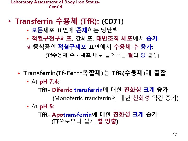
Intracellular Transport of Iron (Fe) Apotransferrin (No Bound Iron) Transferrin (Tf) Diferric Transferrin Receptor (Tf. R) 2 Transferrins bound to Tf. R Fig 11 -5. Cellular Iron Regulation. Rodak’s Hematology 2016. p 143
Laboratory Assessment of Body Iron Status. Cont’d • 그물적혈구혈색소량 (Advia CHr/Sysmex Ret-He) • 그물적혈구에서의 MCH 측정과 같은 의미. • 그물적혈구: 24시간 이전의 적혈구조혈 상태나타내므로, CHr/Ret-He*는 Hb생성위해 활용가능한 철 수준을 실시간으로 제공 • CHr/Ret-He는 적혈구조혈에 사용할 철성분제한시 감소 *: CHr: cellular hemoglobin content for reticulocyte Ret-He: Reticulocyte hemoglobin equivalent 20
Leukocyte Development, Kinetics, and Functions Chapter 12 Roquiz W, Diffalha SA, Kini AR 22
Rodak Hemaology, 5 th E. 2016. Fig 12 -1 p 150 23
Stem Cell Theory Purple = HSC Red = progenitor cell Blue = mature cell 다능조혈줄기세포 (HSC) √ 공통림프구계 선조세포 (CLP) 공통 골수구계 선조세포 (CMP) 과립구-단구계 선조세포 (GMP) 호산구-호염기구계 선조세포 T 선조세포 거핵구-적혈구계 선조세포 Dendritic cell Neut Mono Eos Baso Platele ts B 선조세포 NK cell T Lymph B Lymph Erythrocytes Mast 24 Cell Rodak Hematology, 5 th Ed. , 2016, Fig 7 -13 p. 86
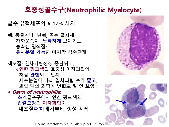
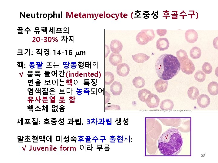
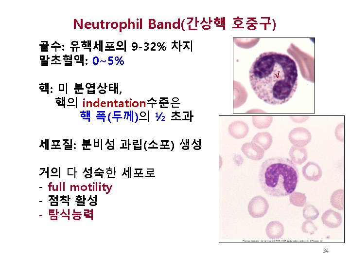
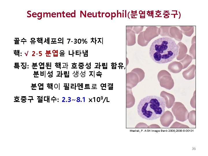
과립구생성 과정 MYELOBLAST (1 -2% of nucleated BM cells) Bone Marrow Large cell (14 -20 m); high N: C ratio; smooth, open nuclear chromatin; 1 -4 nucleoli; blue cytoplasm PROMYELOCYTE (1 -6% of nucleated BM cells) Large cell (16 -25 m), high N: C ratio, slightly clumped chromatin, nucleoli, nonspecific, primary granules (dark red) NEUTROPHILIC MYELOCYTE (6 -17% of nucleated BM cells) Smaller cell, lower N: C ratio, nucleus round or flat on one side, slightly clumped chromatin, blue-pink cytoplasm with neutrophilic (lavender) granules (specific or secondary granules) NEUTROPHILIC METAMYELOCYTE (20 -30% of nucleated BM cells) Lower N: C ratio, nucleus indented less than half of width, coarse chromatin, pink cytoplasm with neutrophilic granules Peripheral blood NEUTROPHILIC BAND (9 -32% of nucleated BM cells) Nucleus indented more than half of width, coarse chromatin, pink cytoplasm with neutrophilic granules SEGMENTED NEUTROPHIL (Seg, Poly) (50 -70% of cells in PB) 2 -5 nuclear lobes connected by a filament, coarse chromatin, pink cytoplasm with neutrophilic granules Image From: Diggs, Sturm, Bell: The Morphology of Human Blood Cells, 5 th ed. Abbott Laboratories, 1985, p. 7 27 Other Images: http: //www. med-ed. virginia. edu/courses/path/innes/nh/wcbmaturation. cfm
과립구계 과립의 종류 과립 형성시기 √ 일차과립 (Azurophilic 전골수구 단계 ) 과립내용물 Myeloperoxidase, cathepsins, defensins, elastase √이차과립 (Specific) 골수구 와 후골수 collegenase, gelatinase, lactoferrin, 구 단계 gelatinase-관련 lipocalin 삼차과립 후골수구와 간상 핵과립구 단계 Gelatinase, collagenase, lysozyme, acetyltransferase 간상핵 과 초기분엽핵 단계 CD 11 b/CD 18, alkaline phosphatase, 소 포-관련막-2, CD 10, CD 13, CD 14, CD 16, cytochrome b 558, complement receptor 1 분비 소포 28
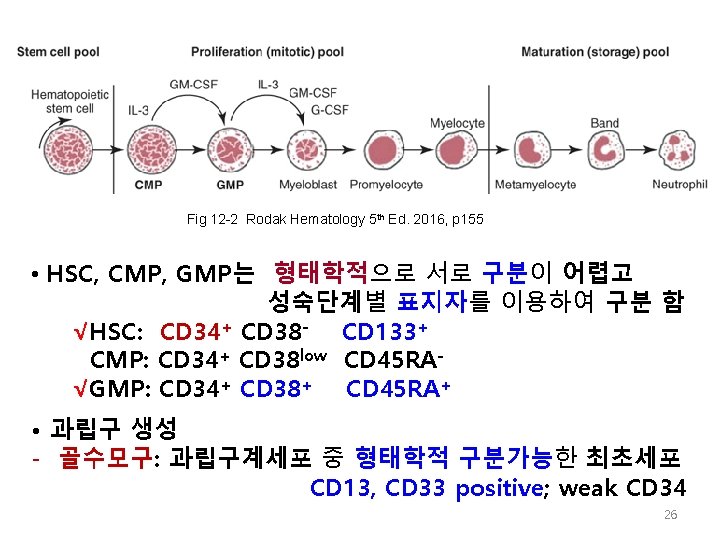
Myeloblast (골수모구) 골수유핵세포의 1 -2 % 차지 형태학적으로 어떤 계통의 blast인지 감별이 어려움 감별을 위해서표지자 활용해야: Weak CD 34 Positive: CD 33, CD 13 크기: 직경 14 -20 μm, √N: C ratio 높음 (8: 1~4: 1) Nucleolus http: //www. med핵: 등글거나 난형이며, ed. virginia. edu/courses/path/innes/nh/wcbmaturation. cfm √섬세하고, open chromatin)과 1 -4의 핵소체 보유 Type I: N: C ratio 8: 1~4: 1, 핵소체 2 -4개, 과립 없음 Type II and Type III: azurophilic granules 보임 29
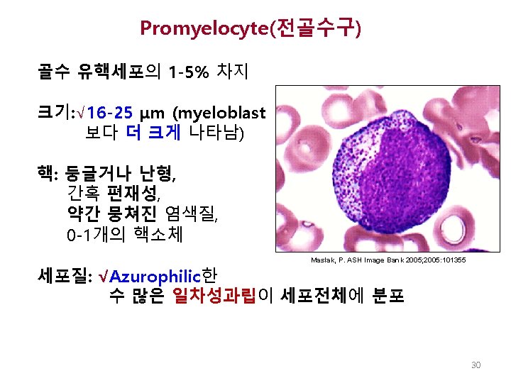
Myeloblast http: //www. meded. virginia. edu/courses/path/innes/nh/wcbmaturation. cfm √ Promyelocyte Maslak, P. ASH Image Bank 2005; 2005: 101355 31
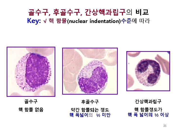
Figure 1. A cluster of myeloid cells in a bone marrow aspirate 골수구 후골수구 간상핵과립구 Maslak, P. ASH Image Bank 2009; 2009: 9 -00027 http: //ashimagebank. hematologylibrary. org/ Copyright © 2009 American Society of Hematology. Copyright restrictions may apply. 37
Fig 12 -2 Rodak BF, Fritsma GA, Keohane EM. Hematology-Clinical Principles and Applications, Elsevier, 2012, p 136. Copyright © 2012, 2007, 2002, 1995 by Saunders, an imprint of Elsevier Inc. Blast 골수구 (dawn of neutrophilia) 후골수구 → 간상핵과립구 38
Myelocytes NRBC √ √ √ Promyelocyte √ Metamyelocyte Fig 12 -4 A Keohane EM et al. , Rodak Hematology, 5 th Ed. , 2016, p 153. Copyright © 2012, 2007, 2002, 1995 by Saunders, an imprint of Elsevier Inc.
Dawn of Neutrophilia 초기골수구 Rodak Hematology 5 th Ed. 2016, p 153 Fig 12 -5 40
골수구 후골수구 간상핵과립구 Rodak Hematology 5 th Ed. 2016, p 154 Fig 12 -6 41
분엽핵호중구 간상핵과립구 Rodak Hematology 5 th Ed. 2016, p 153 Fig 12 -5 42
1° 과립: Cathepsins, Elastase 분비과립: Cytochrome b www. google. co. kr/url? sa=i&rc
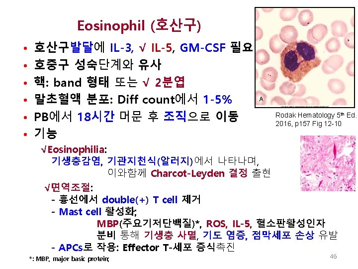
Eosinophil (top) with classic red-orange globular granules in the cytoplasm √ √ Maslak, P. ASH Image Bank 2008; 2008: 8 -00127 http: //ashimagebank. hematologylibrary. org/ Copyright © 2008 American Society of Hematology. Copyright restrictions may apply.
Fig 19 -1. Abbas AK. Et al, Cellular and Molecular Immunology, 7 th Ed. 2016, p 426 “allergic reaction”
Monopoiesis (단구생성) • 선조세포 : GMP • 성숙단계: Monoblast Promonocyte blood monocyte √macrophage • 단구의 순환: 3일동안 순환계 체류 • 조직: 조직의 미세환경에 따라 다양하게 분화 (Box 12 -5) Keohane EM, et al. , 2015, p 161 Copyright © 2012, 2007, 2002, 1995 by Saunders, an imprint of Elsevier Inc.
Promonocyte (전단구) • 운동성 있음 • 탐식기능 있음 • 핵: √ folded, twisted, indented • 세포질: irregular, some granules, vacuoles Fig 12 -14 Rodak Hematology 5 th Ed. 2016, p 159 53
Fig 12 -1, p. 150 57
Lymphoblast (림프모구) 크기: 직경 10 -18 M N: C ratio 높음 – 핵: 섬세, 느슨한 염색질 1개 정도의 핵소체 둥글거나 난형 √세포질: 매우 적음, 과립없음 Maslak, P. 8/29/2003 ASH Image Bank. This image was originally published in ASH Image Bank. © the American Society of Hematology. " – 감별은 표지자(CD antigens)로 59
Prolymphocyte(전림프구) – 림프모구와 유사한 형태 – 핵: - 염색질은 약간 뭉쳐짐 - 핵소체는 덜 뚜렷함 -세포질: 약간 증가 함 (N: C ratio 감소) Maslak, P. 6/10/2010. ASH Image Bank. This image was originally published in ASH Image Bank. © the American Society of Hematology. " 60
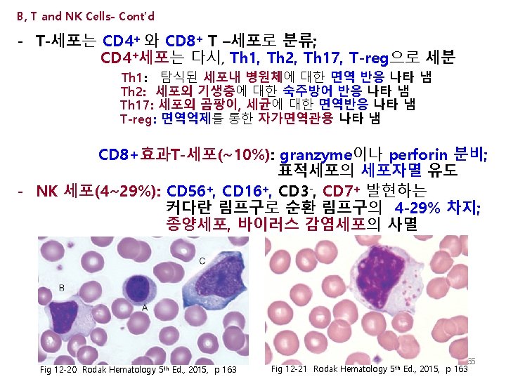
Lymphocyte (림프구) – 분열(증식) 없음 – 크기: 소형, 중형, 대형으로 다양 함 – 핵: √ 뭉쳐져 block양상 염색질 √ 둥글거나 난형, 때로는 indented, stretched형태 – 세포질 - Blue color - 적은 수의√azurophilic 과립 존재 Maslak, P. 10/1/08. This image was originally published in ASH Image Bank. © the American Society of Hematology. " 61
√ Plasma Cell (형질세포) • • • Dark blue cytoplasm 편재성 핵 핵 주변의 halo Fig 12 -18 Keohane EM. , et al. , Rodak Hematology 5 th Ed. , 2015, p 162. Copyright © 2012, 2007, 2002, 1995 by Saunders, an imprint of Elsevier Inc. Saito, T. et al. ASH Image Bank 2004; 2004: 101098 67