Lecture 3 Tissues and Membranes A 4 types

Lecture 3 Tissues and Membranes

A. 4 types of tissues n n Epithelial tissue Connective tissue Nervous tissue Muscular tissue

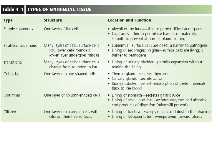
Classification epithelial of tissues Alveoli and capillaries Glandular epithelium Salivary and thyroid glands Skin (keratin) and oral cavity, esophagus in women b. Transitional epithelium Urinary bladder c. Ciliated epithelium Stomach, goblet cells (intestine and trachea
 n n n n Covers the body - “sheets”")
I. Epithelial Tissue (p. 33) n n n n Covers the body - “sheets” (except for glands) Lines organs and body cavities No blood supply Apical Surface – free surface Basement Membrane – acts as a anchor to cells Function: protects, absorbs, secretes, filters Examples: skin, glands, lining of the digestive and respiratory tracts

Types of Epithelial Tissue

Cuboidal Epithelium n Cells are cube-like
")
3. Columnar Epithelium n Cells are tall and narrow (kidney)

Classified also by layers Simple – one layer Stratified – multi layered

Ciliated Pseudostratified columnar

B-Epithelial Tissue
 § Multicellular glands")
Glands § Unicellular glands (goblet cells in respiratory and digestive tracts) § Multicellular glands (a) Exocrine glands (duct glands, Salivary, sweat and stomach glands) (b) Endocrine glands (ductless glands, hormones) Pancreas- secrete digestive enzyme-duodenum of small intestine Pancreatic islets or islets of langerhans , secrete hormone insulin and glucagon directly into the blood

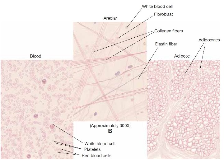
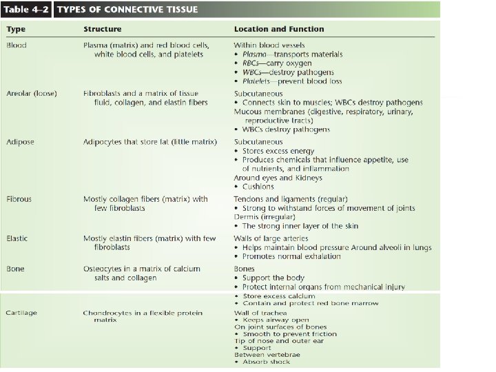
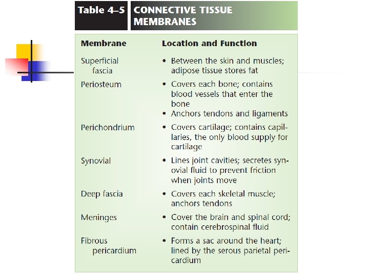
II. Connective Tissue n n Most abundant Supports and joins various part of the body Good blood supply except tendons and ligaments Examples: cartilage, bone, blood, ligaments, tendons
 able")
Connective Tissues n n n Intercellular matrix – fills area between cells (non-living) able to bear weight may be liquid, semi-solid, gel, hard able to bear weight, stretch, withstand abuse Blood (plasma) Bone (salts)
 a.")
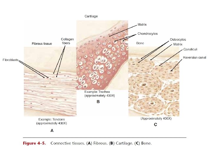
Connective Tissue n n B. Classification of Connective Tissue 1. Bone (Osseous tissue) a. Bone cells are in tiny cavities called lacunae surrounded by layers of calcified matrix b. Protect and Support

Connective Tissue - Cartilage n n n 1. Hyaline Cartilage: rubbery & smooth; somewhat hard found in larynx, attaches ribs to sternum, covers ends of bone

2. Fibrocartilage n Discs in the spinal column

3. Elastic Cartilage a. found in external ear and nose

Dense Fibrous Tissue n Makes up tendons and ligaments

Loose Connective Tissue n Areoler tissue n n Anchors body parts Surround organs

Connective – Adipose Tissue n n Stores Fat Protection from extreme temperatures

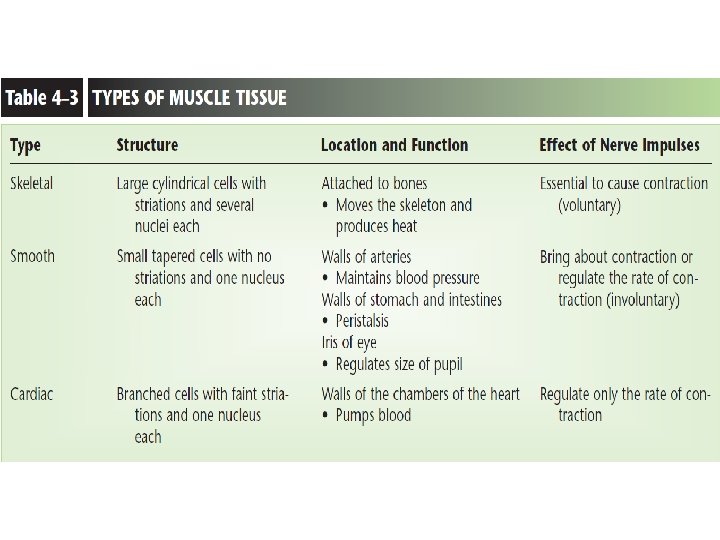
III. Muscle Tissue n n Made of specialized cells that can contract. 3 types of : Skeletal muscle (tendon) Smooth muscle Cardiac muscle

a. Skeletal Muscle n n Voluntary Striated Movement Multi-nucleated

2. Smooth muscle n n n n Not voluntary Found in various organs Non- striated Single nucleus Stomach and intestine Arteries iris

3. Cardiac muscle n n n Heart Striated Involuntary One nuclei per cell Intercalated discs

Smooth muscle

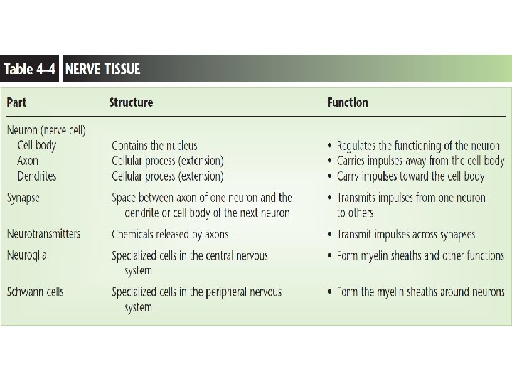
III. Nervous Tissue n n Carry an impulse Two types of cells: n n Neurons: Neuroglia: support the neurons

Nervous Tissue n n Receive and conduct impulses Neuron – nerve cells

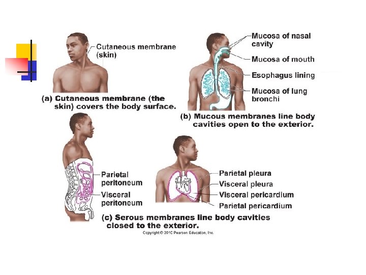
Membranes

Membranes: 3 Types: n n 1. Epithelial Membranes Cutaneous membranes Serous membranes Mucous membranes 2. Connective tissue Membranes

Membranes: 3 Types: n 1. Cutaneous Membranes n n n Skin Epidermis: Stratified epithelium tissue Dermis: connective tissue
 n n - Simple epithelium tissue Line cavities that are")
2. Mucous Membrane (muscosa) n n - Simple epithelium tissue Line cavities that are open to the external environment Many produce mucous Lining of mouth, digestive tract, reproductive tract, urinary tract, respiratory tract
 n n n Named according to location Line cavities that")
3. Serous Membrane (Serosa) n n n Named according to location Line cavities that are not open to the external environment n Visceral layer: the layer against the internal organs (organs=viscera) n Parietal layer: the layer against the inside wall of the cavity

Integumentary System Chapter 5

Functions • Protection against abrasion and ultraviolet light • Protection from entry of microorganisms and dehydration • Sensation receptors for heat, cold, touch, pressure, and pain • Production of precursors to Vitamin D when exposed to ultraviolet light • Regulation of temperature by controlling blood flow to the skin and activity of sweat glands • Excretion of waste products through the skin and the gland secretions

Facts • • • The integumentary system consists of the skin and the structures derived from it including hair, nails, and glands. The skin is the largest organ of the body, covering approximately 2 square meters and having a mass of about 5 kilograms. The study of the integument is called dermatology.

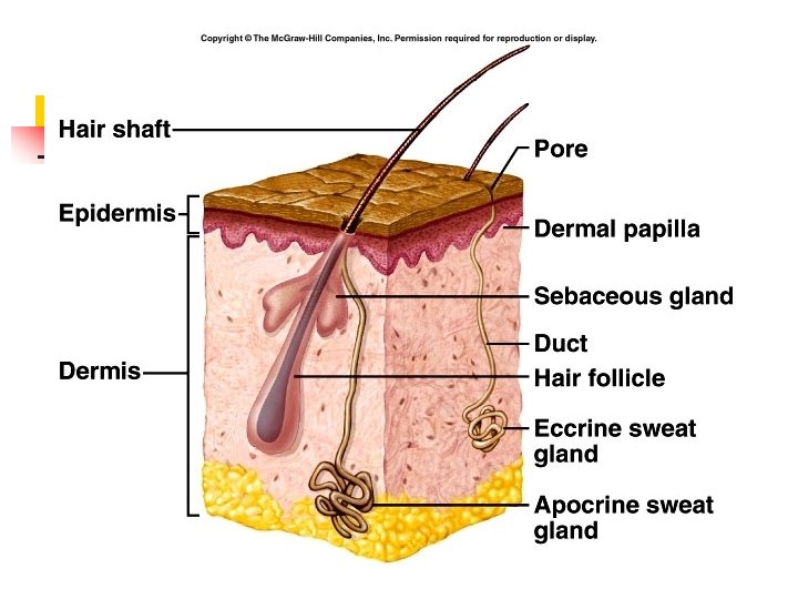
Anatomy of Skin • • • Epidermis - the five outermost thin layers, mitosis in the deepest layer produces new cells as the older cells move towards the most superficial layer where they are sloughed off Dermis - the thicker layer below the epidermis containing blood vessels, nerve endings, hair follicles, smooth muscles, lymphatic vessels, sweat glands, and sebaceous glands Hypodermis - the layer of loose connective tissue under the dermis and not considered part of the skin, it contains half of the body’s stored fat which acts as padding and insulation, it attaches to underlying bones and muscles

Structure of Skin

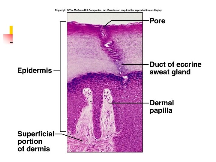
This is a micrograph of actual skin sliced thinly and stained. Note the epidermis, and hypodermis. The outer cells of the epidermis are sloughing off. The dermal papillae are projections of the dermis which extend into the epidermis. They contain tiny blood vessels that supply the epidermis and aid in regulation of body temperature. Fingerprints are projections of the dermal papillae into the epidermis of the fingertips. They increase friction and help improve grip.

This image shows the five layers of the epidermis. The outermost layer is called the stratum corneum: -25 layers of dead skin cells -Keratinized Keratin is a protein that builds up in cells of the epidermis as they move from the deepest layer, the stratum basale, towards the skin’s surface. This process is called keratinization and takes 2 -4 weeks.

Epidermal Layers Composed primarily of keratinized stratified squamous epithelial cells -- consisting of 4 -5 layers: 1. 2. 3. 4. 5. stratum corneum: outermost layer, keratinized dead epithelium stratum lucidum: layer found in thickest areas of skin stratum granulosum: 3 -5 layers of flattened granular cells, developing keratin fibers stratum spinosum: multiple layers of cells stratum basale: deepest layer of single cuboidal or columnar cells, also contains melanocytes - Blisters, Calluses &Psoriasis

Skin Color - Genetics, Environment & Physiology: 1. 2. 3. 4. 5. # of melanocytes - difference in kind, amount & size sunlight, UV light, etc. blood in dermis, blood vessels, freckles & moles other pigments - carotinoids jaundice – liver disorder

Skin, hair, and eye color are all due to a molecule called melanin. This provides protection from the sun’s ultraviolet rays. Melanin is made by special cells in the stratum basale called melanocytes which have extensions reaching towards the skin’s surface. Note the pigment granules in these cell extensions.

Variations in skin color are due to the color, amount, and distribution of melanin, not to the number of melanocytes present in the epidermis.

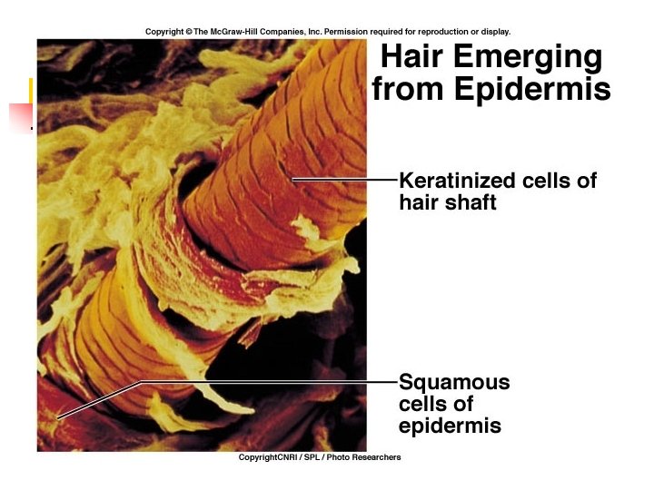
Special Features of the Dermis • • Hair follicles - hair grows from hair bulbs in the follicle, the follicles are actually extensions of the epidermis deep into the dermis Arrector pili (muscles) - an arrector pili muscle is associated with each hair follicle, when the muscle contracts the hair stands on end, contraction of these muscles also causes goose bumps Sweat glands - sweat is produced in these coiled glands and is secreted on the skin’s surface through pores, sweat is sometimes produced due to emotional stress but is usually produced in order to decrease body temperature by evaporative cooling Sebaceous glands - most sebaceous glands are connected to hair follicles and produce an oily substance called sebum, the sebum protects against drying of the hair and skin and against

Skin Glands Ø Sebaceous glands: - assoc. w/ hair follicles, secrete sebum - oils hair, lubricates skin & prevent water loss Ø Sweat glands: § § Eccrine: deep in dermis or subcutaneous layer, odorless secretions, function in thermoregulation Apocrine: found near hair follicles, in axillary regions, released during pain, fear & stress or sexual arousal - Ceruminous & mammary glands

Note the extension of the epidermis far into the dermis forming the hair follicle. Also, note the coiled sweat gland the sebaceous gland connected to the hair follicle.

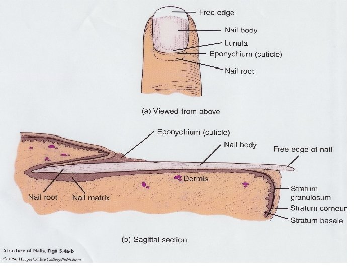
Nails

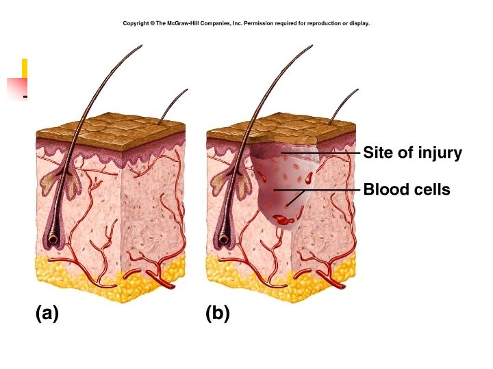
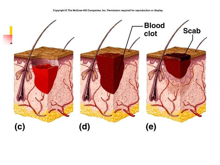
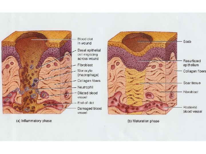
Wound healing
- Slides: 63