Lecture 3 Amino Acids Peptides and Proteins 3


 R Groups • The most hydrophilic R groups are those that")
 R Groups • The two amino acids having R groups with")


 9")


 are first ionized in")


- Slides: 86
Lecture 3 Amino Acids, Peptides, and Proteins
3. 1 Structure of Amino Acids
Importance of Amino Acids • Proteins are the most abundant biological macromolecules, occurring in all cells and all parts of cells. • Proteins also occur in great variety. • Proteins are the molecular instruments through which genetic information is expressed. • Using amino acids as building blocks, different organisms can make extremely diverse products, such as enzymes, hormones, antibodies, transporters, muscle fibers, the lens protein of the eye, feathers, spider webs with distinct biological activities.
Some functions of proteins fireflies erythrocytes rhinoceros l The light produced by fireflies is the result of a reaction involving the protein luciferin and ATP, catalyzed by the enzyme luciferase. l Erythrocytes contain large amounts of the oxygen-transporting protein hemoglobin. l The protein keratin, formed by all vertebrates, is the chief structural component of hair, scales, horn, wool, nails, and feathers.
Amino Acids • Proteins are polymers of amino acids, with each amino acid residue joined to its neighbor by a peptide bond. • Twenty different amino acids are commonly found in proteins. • Asparagine was the first amino acid discovered (in 1806) and the last to be discovered was threonine, in 1938.
Amino Acids Share Common Structural Features • All 20 of the common amino acids are α-amino acids, and except proline, all have the following structure. • For all the common amino acids except glycine, the α carbon is bonded to four different groups: a carboxyl group, an amino group, an R group, and a hydrogen atom. • The α-carbon atom is thus a chiral center, thus amino acids have two stereoisomers called enantiomers.
Stereoisomerism in α-amino acids The absolute configurations of simple sugars and amino acids are specified by the D, L system, which refers only to the absolute configuration of the four substituents around the chiral carbon, not to optical properties of the molecule.
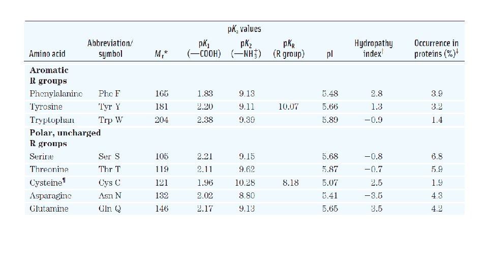
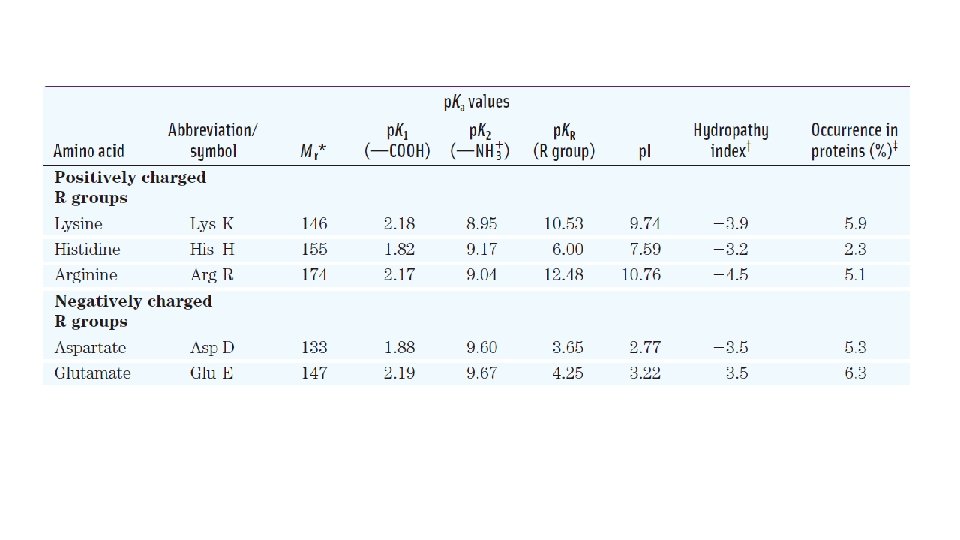
Properties and Conventions Associated with the Common Amino Acids Found in Proteins
The Amino Acid Residues in Proteins Are L Stereoisomers • The amino acid residues in protein molecules are exclusively L-stereoisomers. D-Amino acid residues have been found only in a few, generally small peptides, including some peptides of bacterial cell walls and certain peptide antibiotics. • Cells are able to specifically synthesize the L isomers of amino acids because the active sites of enzymes are asymmetric, causing the reactions they catalyze to be stereospecific.
Amino Acids Can Be Classified by R Group Nonpolar, Aliphatic R Groups • The side chains of alanine, valine, leucine, and isoleucine tend to cluster together within proteins, stabilizing protein structure by means of hydrophobic interactions. • Glycine has the simplest structure. • Proline has an aliphatic side chain with a distinctive cyclic structure.
Aromatic R Groups • Phenylalanine, tyrosine, and tryptophan, with their aromatic side chains, are relatively nonpolar. All can contribute to the hydrophobic effect. • Tyrosine and tryptophan are significantly more polar than phenylalanine. • Tryptophan and tyrosine, and to a much lesser extent phenylalanine, absorb ultraviolet light. Absorption of ultraviolet light by aromatic amino acids
Polar, Uncharged R Groups • This class of amino acids includes serine, threonine, cysteine, asparagine, and glutamine. • Asparagine and glutamine are the amides of aspartate and glutamate, respectively. They are easily hydrolyzed by acid or base. • Cysteine is readily oxidized to form a covalently linked dimeric amino acid called cystine, in which two cysteine molecules or residues are joined by a disulfide bond.
Reversible formation of a disulfide bond by the oxidation of two molecules of cysteine Disulfide bonds between Cys residues stabilize the structures of many protein.
Positively Charged (Basic) R Groups • The most hydrophilic R groups are those that are either positively or negatively charged. • R groups of lysine, arginine and histidine have significant positive charge at p. H 7. 0. • Histidine has an ionizable side chain with a p. Ka near neutrality. • His residue facilitates many enzyme-catalyzed reactions by serving as a proton donor/ acceptor. guanidino group imidazole group
Negatively Charged (Acidic) R Groups • The two amino acids having R groups with a net negative charge at p. H 7. 0 are aspartate and glutamate, each of which has a second carboxyl group.
Uncommon Amino Acids Also Have Important Functions • Proteins may contain residues created by modification of common residues already incorporated into a polypeptide. • Among these are: 4 -hydroxyproline - a derivative of proline that is found in plant cell walls and collagen. 5 -hydroxylysine - derived from lysine, and is found in both plant cell walls and collagen.
• 6 -N-Methyllysine - a constituent of myosin, a contractile protein of muscle. • g-carboxyglutamate - found in the blood-clotting protein prothrombin and in certain other proteins that bind Ca 2+ as part of their biological function. • desmosine, a derivative of four Lys residues, which is found in the fibrous protein elastin.
• Selenocysteine and pyrrolysine are not created through a post synthetic modification, but are introduced during protein synthesis through an unusual adaptation of the genetic code. • Pyrrolysine is found in a few proteins in several methanogenic (methane-producing) archaea and in one known bacterium; it plays a role in methane biosynthesis.
• Some amino acid residues in a protein may be modified transiently to alter the protein’s function. • The addition of phosphoryl, methyl, acetyl, adenylyl, ADP-ribosyl, or other groups to particular amino acid residues can increase or decrease a protein’s activity.
• Some 300 additional amino acids have been found in cells. They have a variety of functions, but not all are constituents of proteins. • Ornithine and citrulline deserve special note because they are key intermediates (metabolites) in the biosynthesis of arginine and in the urea cycle.
Amino Acids Can Act as Acids and Bases • The amino and carboxyl groups of amino acids, along with the ionizable R groups of some amino acids, function as weak acids and bases. • When an amino acid is dissolved in water, it exists in solution as the dipolar ion, or zwitterion. • A zwitterion can act as either an acid (proton donor) or a base: it predominates at neutral p. H.
• The additional carbons in an R group are commonly designated β, γ, δ, ε, and so forth, proceeding out from the α carbon.
• Substances having acid-base nature amphoteric and are often called ampholytes. • A simple monoamino monocarboxylic a-amino acid, such as alanine, is a diprotic acid when fully protonated—it has two groups, the COOH group and the NH 3 group, that can yield protons:
Amino Acids Have Characteristic Titration Curves Titration curve of 0. 1 M glycine at 25 o. C. • The p. Ka values for the ionizable groups in glycine are lower than those for simple, methylsubstituted amino and carboxyl groups. • These downward perturbations of p. Ka are due to intramolecular interactions.
Effect of the chemical environment on p. Ka
Titration Curves Predict the Electric Charge of Amino Acids • At p. H 5. 97, the point of inflection between the two stages in its titration curve, glycine is present predominantly as its dipolar form, fully ionized but with no net electric charge. This is called the isoelectric point (p. I). • Glycine has no ionizable group in its side chain, so its p. I is the sum of two p. Ka values. 1 1 p. I = (p. K 1 + p. K 2) = (2. 34 + 9. 60) = 5. 97 2 2
Amino Acids Differ in Their Acid-Base Properties • All amino acids with a single a-amino group, a single acarboxyl group, and an R group that does not ionize have titration curves resembling that of glycine. • These amino acids have very similar, although not identical, p. Ka values: p. Ka of the COOH group in the range of 1. 8 to 2. 4, and p. Ka of the NH 3 group in the range of 8. 8 to 11. 0.
• Amino acids with an ionizable R group have more complex titration curves, with three stages corresponding to the three possible ionization steps; thus they have three p. Ka values.
Peptides Are Chains of Amino Acids • Biologically occurring polypeptides range in size from small to very large, consisting of two or three to thousands of linked amino acid residues. • Two amino acid molecules can be covalently joined by a peptide bond, to yield a dipeptide, with the elimination of water molecules (dehydration) from the a-carboxyl group of one amino acid and the a-amino group of another.
• Oligopeptide consists of a chain of few amino acids, whereas polypeptide consists of a long chain of amino acids. • Polypeptides usually have molecular weights of below 10 000, while proteins have higher molecular weights. The pentapeptide serylglycyltyrosylalanylle ucine, Ser–Gly–Tyr–Ala– Leu, or SGYAL. • The peptide bonds in proteins are quite stable, with an average halflife (t 1/2) of about 7 years under most intracellular conditions.
Molecular Data on Some Proteins The vast majority of naturally occurring proteins are much smaller than this, containing fewer than 2, 000 amino acid residues.
Peptides Can Be Distinguished by Their Ionization Behavior • The acid-base behavior of a peptide can be predicted from its free αamino and α-carboxyl groups combined with the nature and number of its ionizable R groups. • Peptides also have characteristic titration curves and a characteristic isoelectric p. H (p. I) at which they do not move in an electric field. Alanylglutamylglycyllysine The groups ionized at p. H 7. 0 are in red
• In a protein, the p. Ka value for an ionizable R group can be affected by the loss in charge of the α-carboxyl and α-amino groups, the interactions with other peptide R groups, and other environmental factors.
Biologically Active Peptides and Polypeptides Occur in a Vast Range of Sizes and Compositions • Cannot tell the function of a protein from its molecular weight. • Naturally occurring peptides range in length from two to many thousands of amino acid residues. • The artificial sweetener aspartame or Nutra. Sweet is a dipeptide. • Many vertebrate hormones are small peptides.
Examples of vertebrate peptide hormones Hormone Number of amino acids Function oxytocin (CYIQNCPLG) 9 Secreted by posterior pituitary & stimulates uterine contractions bradykinin (RPPGFSPFR) 9 Inhibits inflammation of tissues thyrotropin-releasing 3 Formed in the hypothalamus & factor (EHP) stimulates the release of thyrotropin Some extremely toxic mushroom poisons, such as amanitin, are also small peptides, as are many antibiotics.
• The individual polypeptide chains in a multi-subunit protein may be identical or different. • The protein is said to be oligomeric if at least two subunits are identical. The identical subunits are called protomers. Example: hemoglobin is either four polypeptide subunits or a dimer of ab protomers. insulin • Polypeptide chains in a protein may be linked by covalent bonds. Example, insulin consists of two chains linked by disulfide bonds.
Some Proteins Contain Chemical Groups Other Than Amino Acids • Simple proteins contain only amino acid residues and no other chemical constituents, e. g. , ribonuclease A & chymotrypsin. • Conjugated proteins contain permanently associated chemical components in addition to amino acids. The non– amino acid part of a conjugated protein is usually called its prosthetic group. • Examples of conjugated proteins: lipoproteins contain lipids, glycoproteins contain sugar groups, and metalloproteins contain a specific metal.
Conjugated proteins
3. 2 Working with Proteins
Protein can be Separated and Purified • To determine the properties and activities of a protein, we need get the protein in a pure form. • Classical methods for separating proteins take advantage of properties that vary from one protein to the next, including size, charge, and binding properties. • DNA cloning and genome sequencing have now been used to simplify the process of protein purification.
Breaking up tissue/cell General protocol for purifying proteins to release proteins At this stage, differential centrifugation can be used to prepare subcellular fractions or to isolate Crude extract specific organelles Fractionation based (NH 4)2 SO 4 is particularly effective and is often used on size or charge to salt out proteins。 Partially purified protein Dialysis Column chromatography Pure protein This step remove (NH 4)2 SO 4 the from the protein preparation This step takes advantage of differences in protein charge, size, binding affinity, and other properties
Chromatography • Different types of chromatography can be used to purify proteins. 1. Ion-exchange chromatography 2. Size exclusion chromatography 3. Affinity chromatography
Ion-exchange chromatography • Separate proteins on the basis of charge differences. • Synthetic polymer (resin) containing bound charged groups are used. Two types: cation exchangers have bound anionic groups, whereas anion exchangers (have bound cationic groups). • The affinity of each protein for the charged groups to the resin is affected by the p. H (which determines the ionization state of the molecule) and the concentration of competing free salt ions in the buffer.
In ion-exchange columns, the expansion of the protein band in the mobile phase (the protein solution) is caused both by separation of proteins with different properties and by diffusional spreading. Column chromatography
Ion-exchange chromatography exploits differences in the sign and magnitude of the net electric charges of proteins at a given p. H.
Worked Example 3 -1 - Ion Exchange of Peptides • A biochemist wants to separate two peptides by ionexchange chromatography. At the p. H of the mobile phase to be used on the column, one peptide (A) has a net charge of − 3 due to the presence of more Glu and Asp residues than Arg, Lys, and His residues. Peptide B has a net charge of +1. Which peptide would elute first from a cation-exchange resin? Which would elute first from an anion-exchange resin?
Solution: A cation-exchange resin has negative charges and binds positively charged molecules, retarding their progress through the column. Peptide B, with its net positive charge, will interact more strongly than peptide A with the cationexchange resin, and thus peptide A will elute first. On the anion-exchange resin, peptide B will elute first. Peptide A, being negatively charged, will be retarded by its interaction with the positively charged resin.
Size-exclusion chromatography, also called gel filtration, separates proteins according to size. The solid phase consists of cross-linked polymer beads with engineered pores or cavities of a particular size.
Affinity chromatography separates proteins by their binding specificities.
• Chromatographic methods are typically enhanced by the use of HPLC, or high-performance liquid chromatography. • By reducing the transit time on the column, HPLC can limit diffusional spreading of protein bands and thus greatly improve resolution. A Purification Table for a Hypothetical Enzyme
Proteins Can Be Separated and Characterized by Electrophoresis • Electrophoresis is the separation of proteins based on the migration of charged proteins in an electric field. • Electrophoresis allows both protein separation and visualization. It can also determine the molecular weight and p. I of a protein. • Electrophoresis of proteins is generally carried out in gels made up of the cross-linked polymer polyacrylamide. • The polyacrylamide gel acts as a molecular sieve, slowing the migration of proteins approximately in proportion to their charge-tomass ratio.
Polymerization of acrylamide
Electrophoresis Proteins can be visualized after electrophoresis by treating the gel with a stain such as Coomassie blue, which binds to the proteins but not to the gel itself.
• An electrophoretic method commonly employed for estimation of purity and molecular weight makes use of the detergent sodium dodecyl sulfate (SDS) (“dodecyl” denoting a 12 -carbon chain). • One molecule of SDS for each amino acid residue. • SDS binding partially unfolds proteins, such that most SDS-bound proteins assume a similar rod-like shape.
• Isoelectric focusing is a procedure used to determine the isoelectric point (p. I) of a protein. • A p. H gradient is established by allowing a mixture of low molecular weight organic acids and bases (ampholytes) to distribute themselves in an electric field generated across the gel. • When a protein mixture is applied, each protein migrates until it reaches the p. H that matches its p. I. • Combining isoelectric focusing and SDS electrophoresis sequentially in a process called two-dimensional electrophoresis permits the resolution of complex mixtures of proteins.
Two-dimensional electrophoresis
• Estimating the molecular weight of a protein. The electrophoretic mobility of a protein on an SDS polyacrylamide gel is related to its molecular weight, Mr.
Unseparated Proteins Can Be Quantified • For proteins that are enzymes, the amount in a given solution or tissue extract can be measured, or assayed, in terms of the catalytic effect the enzyme produces. • Enzymes are usually assayed at their optimum p. H and at some convenient temperature within the range 25 to 38 °C. • 1. 0 unit of enzyme activity for most enzymes is defined as the amount of enzyme causing transformation of 1. 0 μmol of substrate to product per minute at 25 °C under optimal conditions of measurement. • The term activity refers to the total units of enzyme in a solution. The specific activity is the number of enzyme units per milligram of total protein.
3. 3 The Structure of Proteins: Primary Structure
Levels of Structure in Proteins
• Primary structure – the amino acid sequence of the polypeptide. • Secondary structure – the stable arrangements of amino acid residues giving rise to recurring structural patterns. • Tertiary structure - describes all aspects of the threedimensional folding of a polypeptide. • Quaternary structure – the arrangement of two or more polypeptide subunits in space.
The Function of a Protein Depends on Its Amino Acid Sequence • Proteins with different functions always have different amino acid sequences. • Thousands of human genetic diseases have been traced to the production of defective proteins. Example: Sickle cell disease is caused by a single change in the amino acid sequence of hemoglobin. Duchenne muscular dystrophy is caused by deletion of a larger portion of the polypeptide chain of dystrophin. • For some proteins, the amino acid sequences can be polymorphic. In human, an estimated 20% to 30% of the proteins are polymorphic, having amino acid sequence variants in the human population.
• Most proteins contain crucial regions that are essential to their function and thus have sequences that are conserved.
The Amino Acid Sequences of Millions of Proteins Have Been Determined • Following the elucidation of the double-helical structure of DNA by James D. Watson and Francis Crick in 1953, Frederick Sanger worked out the sequence of amino acid residues in the polypeptide chains of the hormone insulin. Amino acid sequence of bovine insulin
Direct protein sequencing Sequence each polypeptide Agents used to label the amino-terminal α-amino group of a protein.
The protein sequencing chemistry devised by Pehr Edman The process is repeated until, typically, as many as 40 sequential amino acid residues are identified.
Breaking disulfide bonds in proteins 2 -Mercaptoethanol Performic acid or 2 - merc apto e than ol
The Specificity of Some Common Methods for Fragmenting Polypeptide Chains
Reagents used to modify the sulfhydryl groups of Cys residues • The sulfhydryl group on Cys residues can be modified with iodoacetamides, maleimides, benzyl halides, and bromomethyl ketones
Mass Spectrometry Offers an Alternative Method to Determine Amino Acid Sequences • The Edman degradation procedure is relatively slow and requires a larger sample than does mass spectrometry. • Mass spectrometry can be used for small amounts of sample and for mixed samples. It provides sequence information, but the fragmentation processes can leave unpredictable sequence gaps. • Mass spectrometry is the method of choice to identify proteins that are present in small amounts.
Concept of Mass Spectrometry • Molecules to be analyzed (analytes) are first ionized in a vacuum, and then passed through an electric and/or magnetic field, where their paths through the field are a function of their mass-to-charge ratio, m/z, which can be used to determine the mass (m) of the analyte with very high precision. • Two methods for determining molecular mass of a protein by mass spectrometry. 1. Matrix-assisted laser desorption/ionization mass spectrometry (MALDI-MS) 2. Electrospray ionization mass spectrometry (ESI-MS)
Electrospray ionization mass spectrometry of a protein A protein solution is dispersed into highly charged droplets by passage through a needle under the influence of a high-voltage electric field. The spectrum generated is a family of peaks, with each successive peak
Obtaining protein sequence information with tandem MS Concept of tandem MS A typical spectrum with peaks representing the peptide fragments generated from a sample of one small peptide (21 residues).
Small Peptides and Proteins Can Be Chemically Synthesized • There are three ways to obtain a peptide: 1. purification from tissue, 2. genetic engineering 3. direct chemical synthesis • In 1962, a major breakthrough in peptide synthesis technology was provided by R. Bruce Merrifield, which involves synthesizing the peptide while keeping it attached at one end to a solid support. • The peptide is built up on this support one amino acid at a time, through a standard set of reactions in a repeating cycle.
Chemical synthesis of a peptide on an insoluble polymer support. Reactions 1 through 4 are necessary for the formation of each peptide bond. Chemical synthesis proceeds from the carboxyl terminus to the amino terminus.
Effect of Stepwise Yield on Overall Yield in Peptide Synthesis
Amino Acid Sequences Provide Important Biochemical Information • The amino acid sequence of a protein can offer insights into its three-dimensional structure and its function, cellular location, and evolution. • Exactly how the amino acid sequence determines the threedimensional structure is not understood in detail, nor can we always predict function from sequence. • Protein families that have some shared structural or functional features can be readily identified on the basis of amino acid sequence similarities.
• Evolutionary relationships can also be inferred from the structural and functional similarities within protein families. • Certain amino acid sequences serve as signals that determine the cellular location, chemical modification, and half-life of a protein. • A consensus sequence is the one that reflects the most common base or amino acid at each position.
Protein Sequences Help Elucidate the History of Life on Earth • The biochemical information conveyed by a protein sequence is limited only by our understanding of structural and functional principles. • Functional segments in new proteins can be identified using bioinformatics tools, allowing both their sequence and their structural relationships to proteins already in the databases to be established. • On a different level of inquiry, protein sequences are beginning to tell us how the proteins evolved and, ultimately, how life evolved on this planet.
• Members of a family of proteins that share a common ancestor are called homologous proteins, or homologs. • If two proteins in a family (that is, two homologs) are present in the same species, they are referred to as paralogs. • Homologs from different species are called orthologs. Partial sequence alignment of a short section of the Hsp 70 proteins.
• Certain segments of a protein sequence may be found in the organisms of one taxonomic group but not in other groups; these segments can be used as signature sequences for the group in which they are found. A signature sequence in the EF-1αEF-Tu protein family.
Evolutionary tree • By considering the entire sequence of a protein, researchers can now construct more elaborate evolutionary trees with many species in each taxonomic group. Evolutionary tree derived from amino acid sequence comparisons. The free end points of lines are called “external nodes”; each represents an extant species. The points where two lines come together, the “internal nodes, ” represent extinct ancestor species.
Summary of Major Concepts • Amino acids share a common structure. • The different chemical properties of amino acids are the result of the different properties of their R groups, which are the basis for categorizing amino acids as nonpolar, aromatic, polar, positively charged, or negatively charged. • Amino acids ionize in aqueous solution. • Polymers of amino acids are polypeptides or proteins. • Proteins can be studied using a variety of techniques.
• A protein’s function depends on its primary structure, which can be determined experimentally. • Proteins that have similar functions in different species have similar amino acid sequences.