Lecture 2 Structure of DNA Discoveries Hereditary material


")


 provides more evidence that DNA = genetic")


")
- Slides: 30
Lecture 2 Structure of DNA
Discoveries • Hereditary material exists – Mendel’s plant genetics • Chemical nature of DNA • Physical nature of DNA • Structure of DNA
Discoveries: The Chemical Nature of DNA • 1869—Fredrich Miescher named the chemical nuclei contained nuclein. Other chemists discovered it was acidic and named it nucleic acid. • It was soon realized that there were two types of nucleic acids: DNA and RNA. • Early in the 20 th century, 4 types of nucleotides were discovered.
Physical Nature of DNA – A Physical Unit • You can see it – experiments demonstrated that inheritance is on chromosomes • You can move it from one place to another – experiments demonstrated that DNA can carry new traits into an organism • You can change it with other physical processes – traits can be changed by physical processes
The Search for Genetic Material Leads to DNA • Once Morgan showed that genes are located on chromosomes, proteins and DNA were the candidates for the genetic material. • Until the 1940 s, the specificity of function of proteins seemed to indicate that they were the genetic material. • However, this was not consistent with experiments with microorganisms, like bacteria and viruses. Copyright © 2002 Pearson Education, Inc. , publishing as Benjamin Cummings
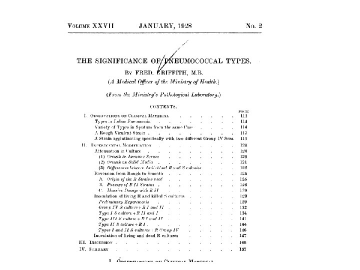
I. Bacterial Transformation is Mediated by DNA • Experiment by Frederick Griffith – 1928 – Demonstrated first evidence that genes are molecules – Two different strains of Streptococcus pneumoniae • Non-pathogenic = Avirulent = ROUGH cells (R) • Pathogenic = virulent = SMOOTH (S) – Smooth outer covering = capsule – Capsule = slimy, polysaccharide – Encapsulated strains escape phagocytosis
Continued. – The capsule alone did not cause pneumonia • Heat-killed S strain was avirulent • Ability to escape immune detection and multiply – When heat-killed S strain was mixed with living R strain the mouse dies of pneumoniae • Encapsulated strain (S) recovedred from dead mouse Now a live strain • The R strain had somehow acquired the ability to produce the polysaccharide capsule – Transformation – Ability to produce coat was an inherited trait Daughter cells also produced capsule
The Experiment
First Demonstration of Transformation – Uptake of genetic material from an external source resulting in the acquisition of new traits (phenotype is changed) – Griffith’s expriment was the earliest document evidence of transformation
What was this transforming agent? • Avery, Mac. Leod and Mc. Carty defined the transforming agent of Griffith’s experiment as DNA (1944) – Chemical components of heat-killed S strain bacteria were purified and co-injected with live R strain • Polysaccharide/Carbohydrate • Lipids • Protein • Nucleic acids – DNA – RNA
• Oswald Avery, Maclyn Mc. Carty and Colin Mac. Leod announced that they found that only DNA transformed the cells. • To replicate, a virus infects a host cell and takes over the cell’s metabolic machinery. • Viruses that specifically attack bacteria are called bacteriophages or just phages. Copyright © 2002 Pearson Education, Inc. , publishing as Benjamin Cummings
II. Viral DNA is Transferred into Cells During Infection – The Hershey-Chase Experiment (1952) • T 2 Bacteriophage studies – Bacteriophage = viruses that infect bacteria – Major chemical components = DNA and protein – Escherichia coli infected with T 2 produce thousands of new viruses in the host cell • Host cell lyses and phage are released
Hershey Chase Experiment • Determination of whether DNA or protein was directing synthesis of new phage particles – Viral proteins were radioactively labeled with: • 35 S by growing T 2 -infected bacteria in 35 S-methionine = 1 st Batch – Amino acid labeling – DNA does not contain any sulfur atoms • 32 P by growing T 2 -infected bacteria in 32 -P – Nucleic acid labeling – Amino acids do not contain phosphorous
Continued. . – Radioactively labeled viruses were isolated from the culture and used to REINFECT new host cells • Batch 1 = protein labeled • Batch 2 = DNA labeled – Blender used to disrupt phage on surface of bacteria from cells and their cytoplasmic components then centrifuged • Supernatant? ? (Protein never entered the cell) • Pellet? ? (DNA injected into the cell)
III. Chargaff’s Rules • Erwin Chargaff (1947) provides more evidence that DNA = genetic material – Analysis of base composition of DNA compared between different organisms • Nitrogenous bases – Adenine (A) – Thymine (T) – Guanine (G) – Cytosine (C) – Conclusions of Chargaff • DNA composition is species specific • The amounts of A, G, C and T are not the same between species – Ratios of nitrogenous bases vary between species
– This diversity strengthened argument that DNA is the molecular basis of inheritance – Chargaff’s Rules • Amount of A = T • Amount of G = C
IV. X-Ray Crystallography Data Provides James Watson and Francis Crick with Insight into DNA Structure • The Race is On – Linus Pauling – Maurice Wilkins and Rosalind Franklin – Watson and Crick • X-ray Crystallography defined – Diffracted X-rays as they pass through a crystallized substance – Patterns of spots are translated by mathematical equations to define 3 -D shape
Watson and Crick Discovered the Structure of DNA by Building Models of X-ray Data • Maurice Wilkins and Rosalind Franklin used X-ray crystallography to study the structure of DNA. – X-rays are diffracted as they passed through purified, crystallized DNA. – The diffraction pattern can be used to deduce threedimensional shape of molecules. • James Watson learned from their research that DNA was helical in shape.
X-ray Chrystallograph of DNA • The diffraction pattern obtained by Franklin and Wilkins showed a X pattern which hinted of a 2 stranded helical form
• The helical turn of DNA correlates to the horizontal lines in the picture which measures to 34 Angstroms. • They also calculated that the gap between based pairs was 3. 4 A as measured on the distance from the center of the X to the ends. • Simple math deduced that there are 10 nucleotides per turn.
• Franklin and Wilkins also showed that the sugar phosphate backbones were found to be on the outside of the helix. • The hydrated and dry forms of DNA showed that water could easily come in and bind to DNA, a fact that could only happen if the feature showed sugar phosphate backbones being on the outside.
• Rosalind Franklin’s data provide clues about DNA’s 3 -D shape – Helix – Width = 2 nm probably two strands (DOUBLE HELIX) – Nitrogenous bases = 0. 34 n. M apart – One turn every 3. 4 n. M (10 base pairs per turn)
• The arrangement of the three major components in nucleic acid polymers was already well known – but the 3 -D shape was still unclear – Sugar phosphate backbone – Bases
• Putting the hydrophobic nitrogenous bases on the inside, and the sugar-phosphate groups on the outside was a stable arrangement
• Base pairing was worked out by trial and error – The distance between the sugar-phosphate backbone groups is constant • Therefore purine-purine or pyrimidine were not allowed because spacing would be in inconsistent with data – Purines = A and G (two organic rings) – Pyrimidines – C and T ( one organic ring)
• Purine-pyrimidine base pairing would be consistent with X-ray data
• Hydrogen bonding between purines and pyrimidines established the appropriate pairs and reinforced Chargaff’s Rules – 2 hydrogen bonds between A and T – 3 hydrogen bonds between G and C
Nature 171: 737 -738 – April 1953 • Watson JD and Crick FC (1953) Molecular Structure of Nucleic Acids: A Structure for Deoxyribose Nucleic Acid. • 1962 – Nobel Prize awarded to three men – Watson, Crick and Wilkins