Lecture 13 Hemostasis and Coagulation Keohane EM et


: Vascular Intima in Hemostasis 5")

는 혈소판수용체 GP Ib/IX/V의 결함에")


: Coagulation 14")
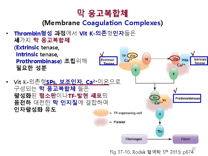
, 보조인자, 조절단백, 기질 Zymogen 활성형: Serine protease Fig 37 -7 Keohane EM et")
: Plasma-Based Model XII PK HMWK XI TF Extrinsic IX VIII Common")
 √ Serine")


 Ca 2+ VIIa TF-VIIa Ca")
")


 V IIa VIII IIa Va XI Fibrinogen (I)")
 Fig 37")

 응고 혈관외 TF-발현세포에서의 개시 단계: 소량(3 -5%) thrombin생성 이루어짐 √ √")


 √ Antithrombin (AT) • 트롬빈(IIa), IXa, XIa, XIIa, Kallikrein 등의 serine")

 Fig 37 -17. Rodak 혈액학 5 th 2019, p 681 44")
 경로: 과다응고 억제 √ √ 염증 시 C 4 b. BP증가:")
 • Vitamin K antagonist (slide#22 참조) •")
의 AT-Thrombin반응 활성화 - AT는 UFH의 오탄당 특정서열에 결합 함. UFH에 의해")


- Slides: 50
Lecture 13 Hemostasis and Coagulation Keohane EM et al, 2016, Chapter 37 Elaine M. Keohane, Ph. D, MLS(ASCP), SH keohanem@shrp. rutgers. edu Rutgers-The State University of New Jersey School of Health Related Professions and Kyung Jin Cho, Ph. D chokj@korea. ac. kr Korea University, College of Health Sciences Jun 7, 2019 1
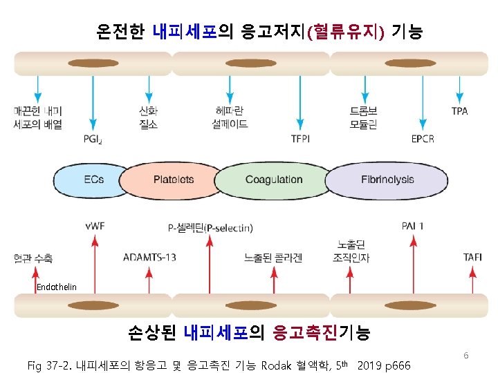
모세혈관, 동맥과 정맥의 구조 Capillary Dreamstime. com √ √ √ https: //www 2. highlands. edu/academics/divisions/scipe/biology/faculty/harn den/2122/notes/cvbv. htm
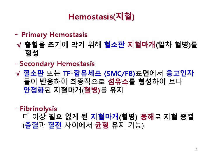
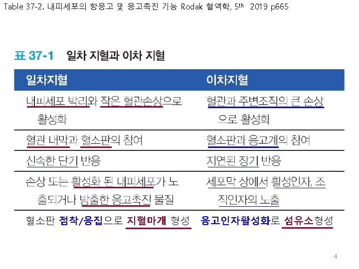
지혈과 혈관내막 (일차적지혈): Vascular Intima in Hemostasis 5
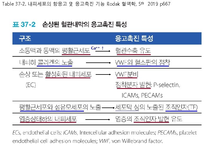
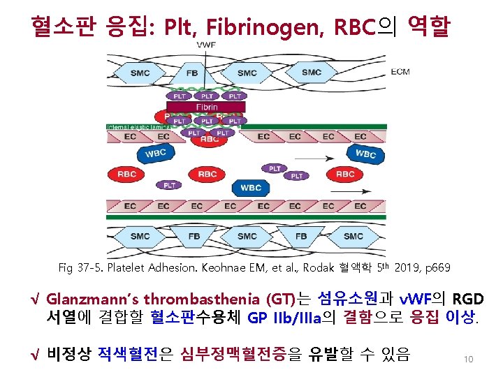
손상된 혈관벽에 혈소판과 v. WF 의 점착 노출된 SMC/FB: TF발현 Fig 37 -3. Keohnae EM, et al. , Rodak 혈액학 5 th 2019 p 668 8
혈소판 점착: PLT, VWF, Collagen의 역 할 √ Bernard-Soulier syndrome(BSS)는 혈소판수용체 GP Ib/IX/V의 결함에 의한 혈소판 점착 이상 √비정상적 백색혈전(PLTs, v. WFs)은 AMI, stroke의 유발 가능 Fig 37 -4. Keohnae EM, et al. , Rodak 혈액학 5 th 2019, p 668 9
√ √ √ Keohane EM et al. , Rodak 혈액학, Elsevier, 5 th Ed, 2019, p 668 11
√ √ √ √ Keohane EM et al. , Rodak 혈액학, Elsevier, 5 th Ed, 2019, p 669 12
Arachidonic acid와 Aspirin 효과 그림 13 -20 Eicosanoid합성 참조 √ PL P 2 Y ADP P 2 Y: ADP수용체 √ √ √ Fig 37 -6. Keohnae EM, et al. , Rodak 혈액학 5 th 2019, p 670 13
Secondary Hemostasis (이차적지혈): Coagulation 14
응고인자(효소원), 보조인자, 조절단백, 기질 Zymogen 활성형: Serine protease Fig 37 -7 Keohane EM et al. , Rodak Hematology, Elsevier, 5 th Ed, 2016, p 649
응고기전의 단순모형(Cascade Reaction): Plasma-Based Model XII PK HMWK XI TF Extrinsic IX VIII Common X V II I Clot Intrinsic
혈장내 응고인자 √ √ IIa, VIIa, IXa, XIa, XIIa, Kallikrein, (APC, AT-inhibitors) √ Serine Protease: √ Vit K-dependant facors: II, VII, IX, X, PC, PS, PZ Cofactors: √ Control proteins: Substrate: Transglutaminase: TF(III), V, VIII, HMWK, Protein S, Protein Z, TM PC, AT, ZPI, TFPI, Heparin Cof II, α 1 -anti-trypsin, α 2 -macrogl. Fibrinogen XIIIa Keohane EM et al. , Rodak Hematology, Elsevier, 5 th Ed, 2016, p 649 17
혈장내 응고인자 - 계속 Cont’d Keohane EM et al. , Rodak Hematology, Elsevier, 5 th Ed, 2016, p 649 18
√ √ Feed-back !! √ √ Table Fig 37 -6 Keohane EM et al. , Rodak Hematology, Elsevier, 5 th Ed, 2016, p 65020
Vit K에 의한 번역후-변형 γ-카르복실화 √ Vit K √ VII, IX, X, PC, PS, PZ • Gla(γ-carboxy glutamic acid)도메인은 Ca 2+과 높은 친화성 결합에 결정적 Fig 37 -9 Keohane EM et al. , Rodak 혈액학, Elsevier, 5 th Ed, 2019, p 673
√ Table Fig 37 -7 Keohane EM et al. , Rodak Hematology, Elsevier, 5 th Ed, 2016, p 65023
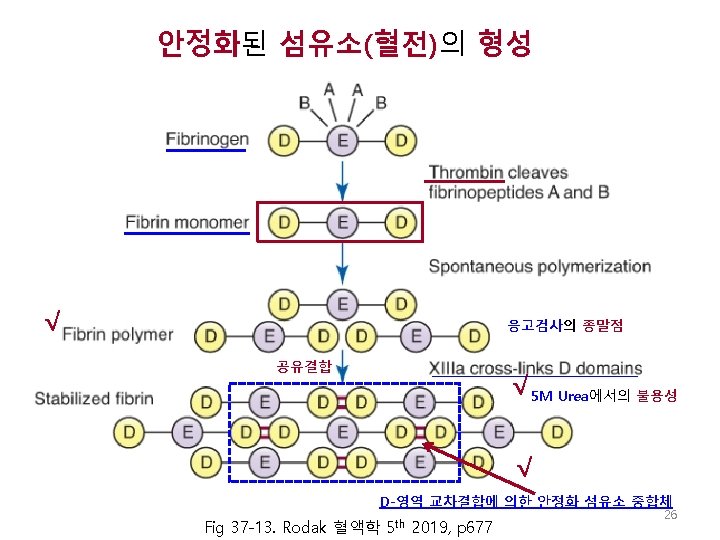
섬유소원 구조 D E D Fig 37 -12. Keohnae EM, et al. , Rodak Hematology 5 th p 654 24
섬유소원 도메인과 Thrombin의 절단 √ Fig 37 -12. Keohnae EM, et al. , Rodak Hematology 5 th p 654 25
In Vitro Blood Coagulation: Plasma-Based Model XII PK HMWK XI TF Extrinsic IX VIII Common X V II I Clot Intrinsic
In-vitro 혈장-기반 응고기전 Extrinsic √ PT Intrinsic √ 표면접촉 √ Tissue Thromboplastin, PL, Ca++ √ PTT PF 3 (Partial thromboplastin) Neg charged surface (activator) Fig 37 -14. Rodak 혈액학, 5 th 2019, p 678
in-vitro 혈장-기반 응고기전 - 계속 Extrinsic Tissue Factor (TF) Ca 2+ VIIa TF-VIIa Ca 2+ Extrinsic Tenase complex X Xa 외인계기전-관련 인자 : VII 29
In Vitro Blood Coagulation- Plasma Based Model – Cont’d Intrinsic Neg charged Surface (Kaolin) XII Prekallikrein HMWK XIIa HMWK XI XIa Ca 2+ Phospholipid IX IXa VIIIa- IXa Ca 2+ X Xa Intrinsic tenase complex 내인계-관련 인자: XII, XI, IX, VIII 30
In Vitro Blood Coagulation- Plasma Based Model – Cont’d 공통계-관련 인자: X, V, II, I Common Prothrombinase complex Xa X Ca 2+ Va-Xa XIII Prothrombin (II) Fibrinogen (I) Thrombin (IIa) Fibrin Monomer Fibrin Polymer (Gel) XIIIa Cross-Linked Fibrin 31
In Vitro Blood Coagulation- Plasma Based Model – Cont’d Intrinsic Extrinsic Neg Charged Surface XII Prekallikrein Tissue Factor (TF) HMWK Ca 2+ VII XIIa HMWK VIIa XI Common TF-VIIa Ca 2+ XIa Ca 2+ Phospholipid VIIIa- IXa IX IXa Ca 2+ X Xa Va-Xa Ca 2+ Prothrombin (II) Fibrinogen (I) XIII Thrombin (IIa) Fibrin Monomer Fibrin Polymer (Gel) XIIIa Cross-Linked Fibrin 32
지혈에 있어서의 Thrombin 역할 Thrombin (IIa) V IIa VIII IIa Va XI Fibrinogen (I) Fibrin IIa VIIIa XIa XIIIa Cross-Linked Fibrin XIII IIa 33
응고경로 단순화 모형 Contact group √ Thrombin의 Feed-back (V, VIII, XI 활성화) Fig 37 -8 Keohane EM et al. , Rodak Hematology, Elsevier, 5 th Ed, 2016, p 650 34
Table 37 -8 Rodak 혈액학, Elsevier, 5 th Ed, 2019, p 674 36
세포-기반 in-vivo (생리학적) 응고 혈관외 TF-발현세포에서의 개시 단계: 소량(3 -5%) thrombin생성 이루어짐 √ √ ac b d fee TF-발현세포의 개시: 소량의 X, IX 활성화 Extrinsic tenase Intrinsic tenase X or 50배 t c Fa 화 활성 √ √ k 혈관내 혈소판상에서의 전파 단계: Thrombin생성의 95% cover √ Prothrombinase Thrombin 생성의 95% Burst COAT(collagen and thrombin activated): 노출된 Coll에 혈소판점착과 Thrombin에 의해 활성화 Fig 37 -15. Rodak 혈액학 5 th 2019, p 679
Table 37 -9 Keohane EM et al. , Rodak 혈액학, Elsevier, 5 th Ed, 2019, p 674 √ √ √ 38
v. WF-VIII 복합체 √ √ Fig 37 -11. Rodak 혈액학 5 th 2019, p 675 39
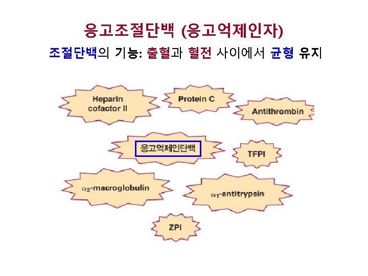
Table 37 -10 Keohane EM et al. , Rodak 혈액학, Elsevier, 5 th Ed, 2019, p 680 응고조절 단백 (응고억제인자) TF: VIIa +Xa복합체에 TFPI결합 41
주요 응고억제인자 (SERPIN) √ Antithrombin (AT) • 트롬빈(IIa), IXa, XIa, XIIa, Kallikrein 등의 serine protease를 결합하여 중화시키는 SERPIN임 √ Activated Protein C (APC); Protein S (APC 보조인자) • 억제대상: Va, VIIIa 42
SERPIN의 응고조절 부위 √ √ √ Fig 37 -16. Rodak 혈액학 5 th 2019, p 680
조직인자경로억제인자 (TFPI) Fig 37 -17. Rodak 혈액학 5 th 2019, p 681 44
Protein C (APC) 경로: 과다응고 억제 √ √ 염증 시 C 4 b. BP증가: bound form 증가로 상대적 free form 감소 이는 혈전 위험도 증가 C 4 b. BP(C 4 b binding protein): Acute phase reactant(APR) Fig 37 -18. Rodak 혈액학 5 th 2019, p 681 45
Major Anticoagulants √ Warfarin, Coumadin (경구용 항응고제) • Vitamin K antagonist (slide#22 참조) • 쿠마딘 투여는 간에서 합성 후 번역후 변형 실패로 γ-carboxylaton 이 이뤄지지 않아 기능 없는 II, VII, IX, X (PIVKA) 로 남아 PT, a. PTT 연장 • Warfarin 치료 투여량은 monitoring 되어야: Ø Prothrombin Time (PT) and INR (>3. 0) √ Unfractionated Heparin (정맥 내 투여) • Anti-Thrombin(AT) 과 Thrombin을 결합 하여 – IIa (thrombin)과 Xa 를 중화 – IXa, Xia, XIIa도 기능 억제 • 헤파린 투여량도 monitoring 되어야: Ø Activated Partial Thromboplastin Time (APTT)
√ 미분획 헤파린(UFH)의 AT-Thrombin반응 활성화 - AT는 UFH의 오탄당 특정서열에 결합 함. UFH에 의해 AT의 thrombin억제활성이 2000배 이상강화 Fig 37 -20. Keohane EM et al. , Rodak Hematology, Elsevier, 5 th Ed, 2016, p 659 47
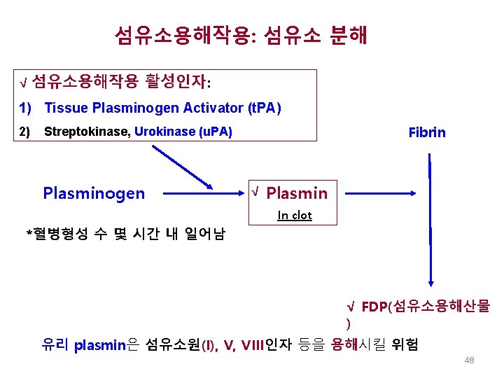
섬유소용해작용 - 요약 √ √ Fig 37 -23. Keohane EM et al. , Rodak Hematology, Elsevier, 5 th Ed, 2016, p 662 49
섬유소 용해경로와 억제인자 • • 염증이나 응고가 발생하면, 그 반응으로 내피세피와 상피세포/대식세포에서 각각 t. PA나 u. PA가 방출 됨 섬유소용해작용억제인자: TAFI, PAI-1, α 2 -antiplasmin FDPs Digest I, VIII and fibronectin: fatal fibrinolysis Fig 37 -21. Keohane EM et al. , Rodak Hematology, Elsevier, 5 th Ed, 2016, p 660 50