Lecture 11 Automated Cell Counting Instrumentation Keohane EM

 구성 • • • • White Blood Cell Count (WBC) Red Blood")
 – 계속 혈액장비의 주요 측정 기술 √ Electrical Impedance(전기저항) • 저")
 • 1950년대, W. Coulter에 의해 개발 • 전도성액체에 부유된")

 (Size) 세포 내부 복잡성 세포 크기(용적) Figure 15")
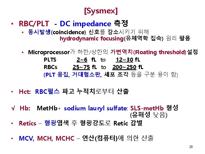
![[Beckman Coulter] • 직접측정 항목 • √ • • • RBC, PLT - DC](https://slidetodoc.com/presentation_image_h/010f93e9fde7ecc021a405c0c88c133b/image-16.jpg "[Beckman Coulter] • 직접측정 항목 • √ • • • RBC, PLT - DC")


 R 1 √ √ √ 24")
 – Cont’d • 3차원 분석 - 3 D Scatterplot •")


 FSC IRF, immature reticulocyte fraction;")
 기술 • Polarized Helium")

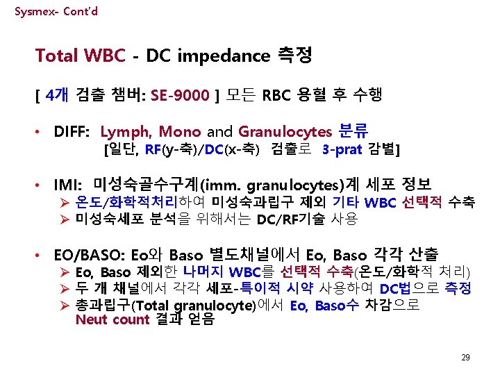
![Cell-Dyn – Cont’d 다형핵구 – 단핵구 감별 [산점도 1] 900 (분엽성) • 분엽성(900) vs.](https://slidetodoc.com/presentation_image_h/010f93e9fde7ecc021a405c0c88c133b/image-36.jpg "Cell-Dyn – Cont’d 다형핵구 – 단핵구 감별 [산점도 1] 900 (분엽성) • 분엽성(900) vs.")
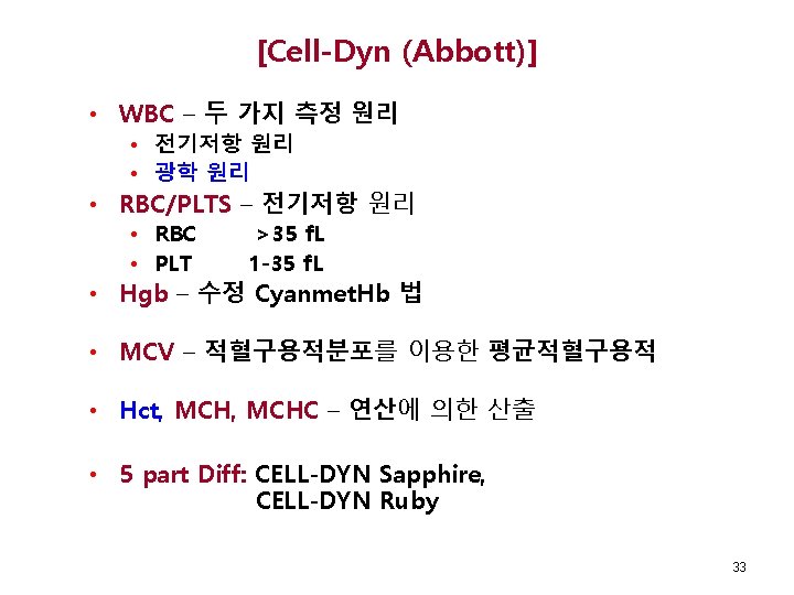
![Cell-Dyn – Cont’d 호산구 – 호중구 감별 [산점도 2] 900 D (과립성) • Plot](https://slidetodoc.com/presentation_image_h/010f93e9fde7ecc021a405c0c88c133b/image-37.jpg "Cell-Dyn – Cont’d 호산구 – 호중구 감별 [산점도 2] 900 D (과립성) • Plot")
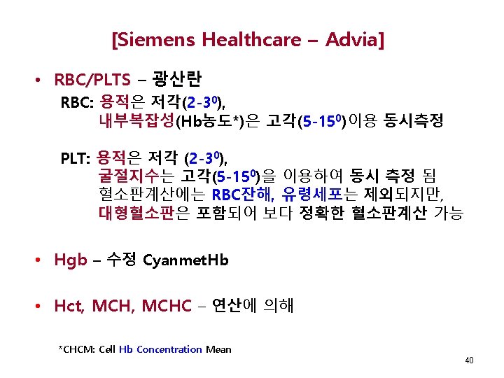
![Cell-Dyn – Cont’d [산점도 3] 백혈구 감별 00 (용적) • 단핵구 분석 • 용적](https://slidetodoc.com/presentation_image_h/010f93e9fde7ecc021a405c0c88c133b/image-38.jpg "Cell-Dyn – Cont’d [산점도 3] 백혈구 감별 00 (용적) • 단핵구 분석 • 용적")

![[Siemens Healthcare – Advia]- Cont’d • WBC/Differential - 광학(Tungsten Lamp), 세포화학 • 2 채널](https://slidetodoc.com/presentation_image_h/010f93e9fde7ecc021a405c0c88c133b/image-41.jpg "[Siemens Healthcare – Advia]- Cont’d • WBC/Differential - 광학(Tungsten Lamp), 세포화학 • 2 채널")
 FSC Scatter Neut LUC")

- Slides: 46
Lecture 11 Automated Cell Counting Instrumentation Keohane EM, Smith LJ, Walenga JM Rodak Hematology 5 th Ed, 2016, Chapter 15 Elaine M. Keohane, Ph. D, MLS(ASCP), SH keohanem@shrp. rutgers. edu Rutgers-The State University of New Jersey School of Health Related Professions and Kyung Jin Cho, Ph. D chokj@korea. ac. kr Korea University, College of Health Sciences May 24, 2019 1
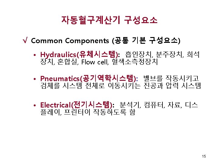
CBC (전혈구계산) 구성 • • • • White Blood Cell Count (WBC) Red Blood Cell Count (RBC) Hemoglobin (HGB) Hematocrit (HCT) Platelet Count (PLT) Mean Cell Volume (MCV) Mean Cell Hemoglobin (MCH) Mean Cell Hemoglobin Concentration (MCHC) Red Cell Distribution Width (RDW) WBC Differential Count (Relative and Absolute) NRBC Reticulocyte Count (RETIC), IRF Other parameters 2
혈액분석장비(Hematology CBC Analyzer) – 계속 혈액장비의 주요 측정 기술 √ Electrical Impedance(전기저항) • 저 전압 직류 저항 (DC Resistance) • 고 전압 교류 저항 [AC(RF) Resistance] √ Optical Scatter (광 산란) • Laser Light • Non Laser Light 4
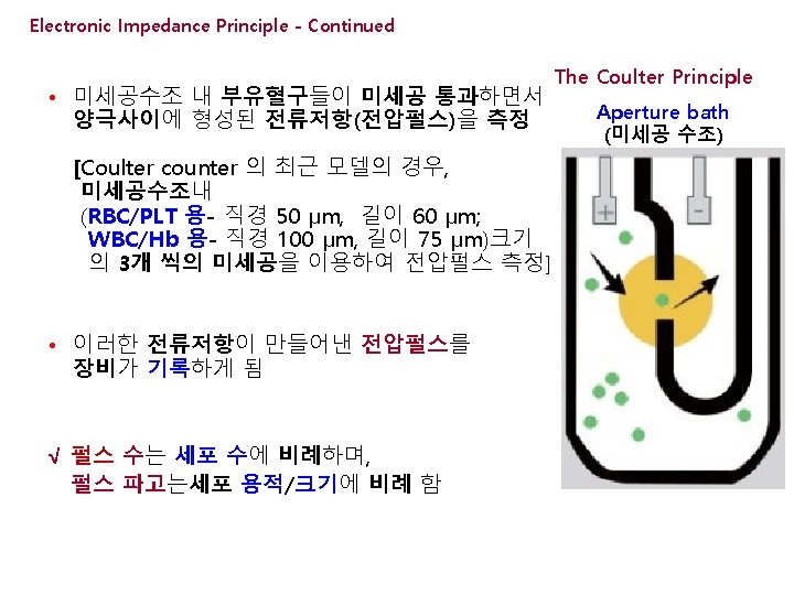
Electronic Impedance Principle (전기저항 원리) • 1950년대, W. Coulter에 의해 개발 • 전도성액체에 부유된 세포를 음압펌프를 이용하여 aperture tube의 세공을 통해 흡인하면서 전기저항이 발생 • 미세공 수조내 외부전극과 aperture tube내 내부전극 사이 저전압 직류전기 연결 됨 Fig 15 -1. Coulter Principle of Cell Counting. Keohane EM et al. Rodak Hematology, Elsevier 2016, p 209
Electronic Impedance Principle - Continued • 파고분석기는 일정간격으로 설정된 size thresholds에 의해 모든 펄스를 각 펄스크기(혈구크기)별로 분류 함 • X-축: 용적, Y-축: 혈구수로 표시되는 크기분포히스토그램(size distribution histogram)상에 측정값을 플롯 함 Fig 39 -2: Modified from Coulter Electronics, Inc. From Rodak, Fritsma, Keohane, 2012, p. 599. 7
DC vs RF 산점도 (Internal complexity) (Size) 세포 내부 복잡성 세포 크기(용적) Figure 15 -4: Rodak Hematology Keohane EM, 2016, p. 210 11
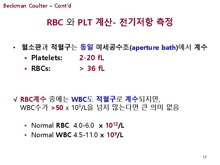
[Beckman Coulter] • 직접측정 항목 • √ • • • RBC, PLT - DC impedance 측정 MCV – RBC용적분포를 이용한 평균RBC용적 Hgb – 수정 Cyanmet. Hb법 WBC – VCS technology MPV - Plt용적분포를 이용한 평균PLT용적 √ 연산 산출: HCT*, MCHC, RDW *: Hct = (MCV x RBC) / 10 16
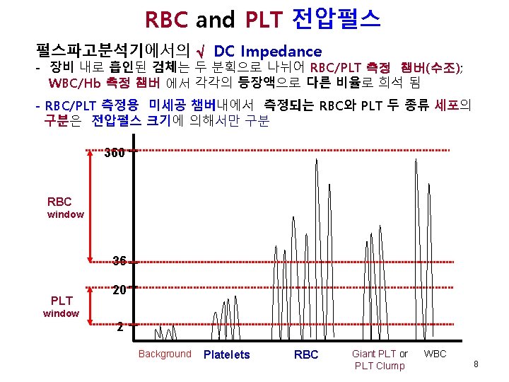
Beckman Coulter – Cont’d RBC 와 PLT 전압펄스 비교 360 RBC window 36 PLT 20 window 2 Background Platelets RBC √ Giant PLT, PLT Clump √ WBC 18
Beckman Coulter – Cont’d √ RDW 와 RBC Histogram √ Normal MCV # cells Normal RDW 70 f. L 90 f. L √ Normal MCV = 80 -100 f. L √ Normal RDW = 11. 5 -14. 5% 110 f. L # cells Normal MCV Increased RDW 70 f. L 90 f. L 110 f. L 19
Beckman Coulter – Cont’d √ Platelet Histograms 혈구수 2 20 f. L 정상분포 작은 입자들의 간섭현상 커다란 혈소판의 간섭으로 곡선이 기준선에 닿지 못함 혈구수 2 20 f. L √ Noise, Debris, 세균 2 20 f. L Giant PLTs, Clumped PLTs, Micro RBCs
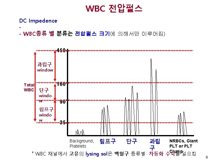
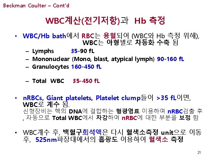
Beckman Coulter – Cont’d WBC Voltage Pulses 450 Gran window Total WBC Mono 160 window 90 Lymph window 35 Background, Platelets Lymph Gran Mono Blast, Atyp, Lymh Imm. Granulo NRBCs, Giant PLT or PLT Clump 22
Beckman Coulter – Cont’d √ WBC Histogram • Coulter: 3 Part Differential Ø Lymphs 35 - 90 f. L Ø Mononuclear 90 -160 f. L* Ø Granulocytes 160 -450 f. L • Total WBC 35 -450 f. L Lymph # cells Mononuclear 50 100 (35 -90) Granulocytes 200 (90 -160) 400 (160 -450) f. L * Mononuclear영역에서는 monocytes외에도 reactive lymphs, 일부 blasts, 미성숙 과립구도 포함 됨
Beckman Coulter – Cont’d Coulter STKR (구형 model) R 1 √ √ √ 24 Figure 15 -5: Printout from Coulter STKR , Rodak Hematology Keohane EM, 2016, p. 213
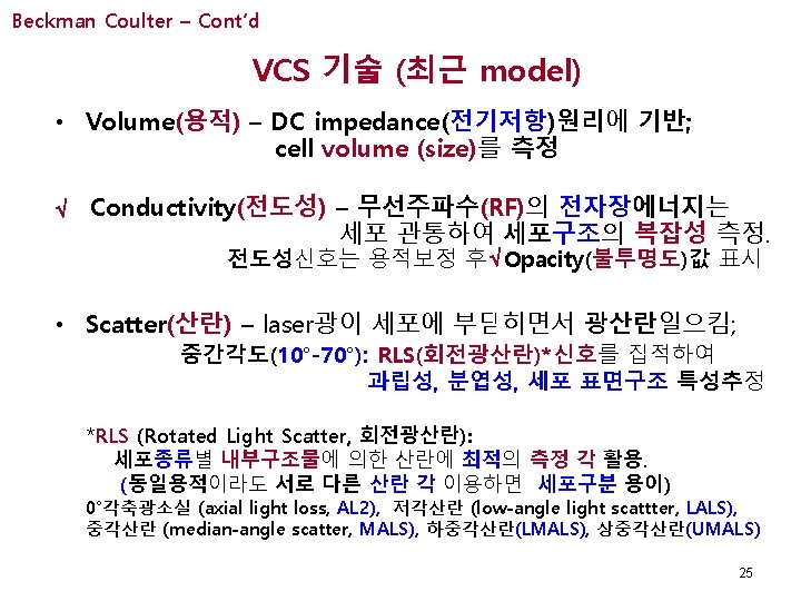
Beckman Coulter (VCS 기술) – Cont’d • 3차원 분석 - 3 D Scatterplot • 5 -part differential • V (volume): lymphs vs. monos 감별 • S (scatter)/RLS: neut vs. eos vs. baso 감별 • C (conductivity)/Opacity: lymph vs. baso 감별 Neutrophil Monocyte Eosinophil Lymphocyte Scatter (RLS) Co (O nd pa uc ci tiv ty it ) y Vol Basophil 26
Beckman Coulter – Cont’d LALS V V UMALS V OP LLSn AL 2 RLSn V AL 2 OP Figure 15 -6: Printout from Coulter STKR , Rodak Hematology Keohane EM, 2016, p. 215
Sysmex- Cont’d Sysmex 2100 WDF* SFL Mono Lymph Neut + Baso Eo Eo SSC 적혈구 용혈시키고, WBC막에 구멍을 낸 후, 형광염료로 DNA, RNA를 염색 *: WDF, WBC differential; SFL, side fluorescence 30
Sysmex- Cont’d WNR: WBC, Baso, n. RBC WBC perforated WBC Mono Lymph Neut + Baso Debris Baso n. RBC Eo After RBC lysing, DNA, RNA stained with fluorescent dye Polymethine stains the nuclei with n. RBC low / WBC high √ RBC IRF LFR MFR-HFR IPF Figure 15 -7: Printout from Sysmex XN-1000 Rodak Hematology Keohane EM, 2016, p. 217
Sysmex- Cont’d IRF in Automated Retics Counting (Sysmex XE-2100…) FSC IRF, immature reticulocyte fraction; MFR, middle-fluorescence ratio; HFR, high-fluorescence ratio BM recovery: IRF >5% SFL http: //www. sysmex. ru/files/articles/Xtra_online_reticulocytes. pdf 32
Cell-Dyn – Cont’d • 5 part Differential • 다각편광산란분리 (MAPSS) 기술 • Polarized Helium Neon Laser √ 4 Scatter Measurements Taken • • 00 900 7/100 900 D Size (용적) Lobularity (분엽성) Complexity (복잡성) Granularity–depolarized (과립성) • 3개 Scatterplot [산점도]를 이용한 세포 감별 34
Cell-Dyn – Cont’d Cell-Dyn MAPSS Technology 35 Figure 15 -8 A: Printout from Coulter STKR , Rodak Hematology Keohane EM, 2016, p. 218
Cell-Dyn – Cont’d 다형핵구 – 단핵구 감별 [산점도 1] 900 (분엽성) • 분엽성(900) vs. 복잡성(70)으로 • 다형핵구 vs. 단핵구 감별 70 (복잡성) 36 Figure 15 -8 B: Printout from Coulter STKR , Rodak Hematology Keohane EM, 2016, p. 218
Cell-Dyn – Cont’d 호산구 – 호중구 감별 [산점도 2] 900 D (과립성) • Plot 1 결과의 PMN cell 이용, 과립성(900 D) vs. 분엽성 (900)으로 • 호산구 vs. 호중구 감별 900 (분엽성) 37 Figure 15 -8 C: Printout from Coulter STKR , Rodak Hematology Keohane EM, 2016, p. 218
Cell-Dyn – Cont’d [산점도 3] 백혈구 감별 00 (용적) • 단핵구 분석 • 용적 (00) vs. 복잡성(70)으로 • Lymph, Mono, Baso를 구분 70 (복잡성) 38 Figure 15 -8 D: Printout from Coulter STKR , Rodak Hematology Keohane EM, 2016, p. 218
Cell-Dyn – Cont’d Eos Neut WBCs Mono Neut Reti-PLts PLTs Figure 15 -9: Rodak 혈액학, Keohane EM, 2016, p. 226 Retic RBCs
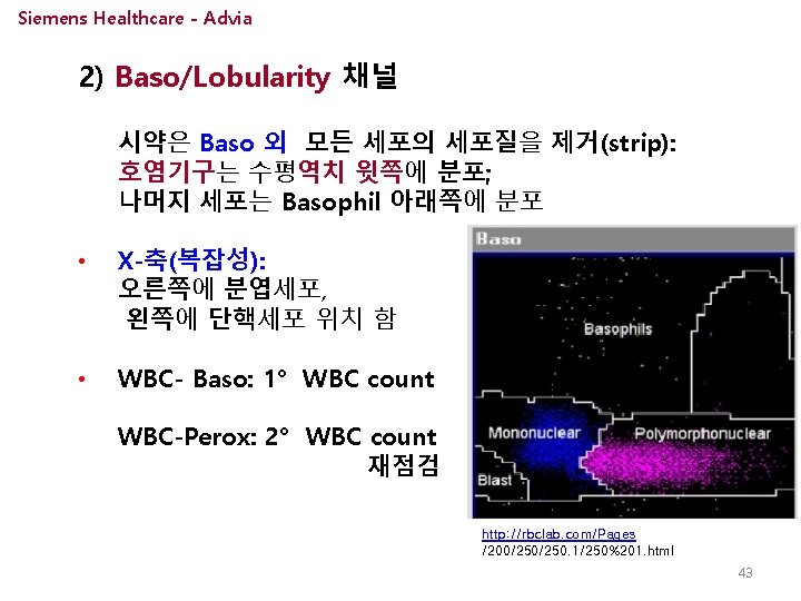
[Siemens Healthcare – Advia]- Cont’d • WBC/Differential - 광학(Tungsten Lamp), 세포화학 • 2 채널 (Peroxy채널 vs. Baso채널) 1) Peroxy 채널 (호중구, 단구, 호산구의 과립 내 존재) Step 1: 반응조 내에서 Sod dodecylsulfate에 의해 RBC 용해 Step 2: WBC는 Perox 1 sol (fixative)에 의해 고정 Step 3: WBC Perox 2 (4 -chloronaphthol), Perox 3 (H 2 O 2)에 의해 염색 peroxidase in granules • • • H 2 O 2/4 -chloro-1 -naphthol Y-축: 전방산란 (FS-세포크기); X-축: 흡광도 (Peroxidase활성도)를 플롯 과립구-우측 상단; 림프구-좌측 하단에 위치 ppt 41
Siemens Healthcare - Advia Peroxidase 채널 (FSC vs. Peroxidase OD) FSC Scatter Neut LUC Imm Gran LUC Atyp lymp Blast Granulo Monos Eos Lymphs Debris NRBC PLTs Eos n. RBC/Plts less Peroxidase Absorbance more Debris LUC = Large Unstained Cells (such as blasts, reactive lymphs) Peroxy activity 42
Siemens Healthcare - Advia 44 Figure 15 -12: Printout from Coulter STKR , Rodak Hematology Keohane EM, 2016, p. 222