Lecture 1 OVERVIEW CELLULAR COMPONENTS Hematopoiesis Chapters 1

 Date Lecture Topics")


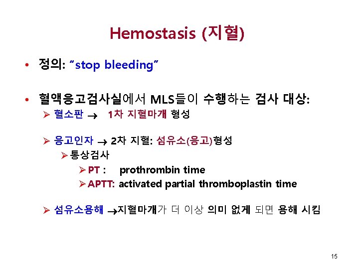
라고 하는데, 이 혈장은 출혈을 저지하는 응고인자 (coagulation factor)들을 포함")

 √ 혈액검사학의 기본인 CBC에 포함되는 항목 • • • WBC")

 • 골수생검 (Biopsies) • 세포화학 염색 • Myeloperoxidase")


 • 세포막의 특성 – 탄력성 (Elastic) – 변형가능성 (deformability) √ 이중지질막")
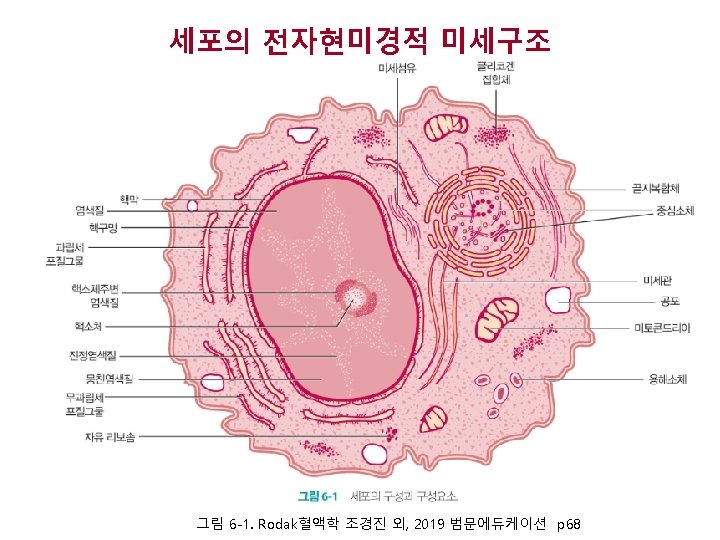
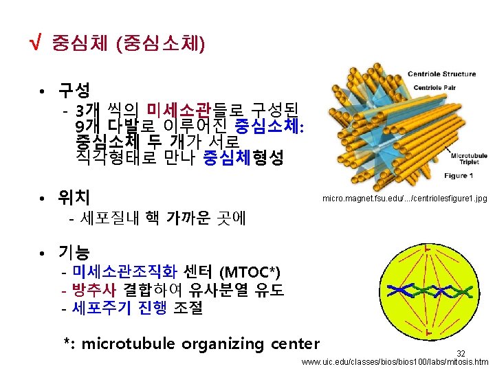
 √ Nucleus – 세포의 통제센터 - 주로 DNA로 구성 - DNA복제와 전사가")

 • Composition - 세포내막계에 해당되며, cisternae이라 불리는 여러 겹으로 겹쳐친")
 • 납작한 판, 주머니, 관의 형태로 나타나는 세포내막계(endomembrane")


- Slides: 39
Lecture 1 OVERVIEW; CELLULAR COMPONENTS; Hematopoiesis Chapters 1, 6 March 8, 2019 Elaine M. Keohane, Ph. D, MLS(ASCP)SH keohanem@shrp. rutgers. edu Rutgers-The State University of New Jersey School of Health Related Professions and Kyung Jin Cho, Ph. D, Professor Emeritus chokj@korea. ac. kr Dept. of Medical Laboratory Science College of Health Sciences, Korea University
Hematology – Spring 2018 (Rodak’s Hematology 5 th Ed, Elsevier, 2016) Date Lecture Topics Chapter Assignment 3/ 7 Overview of Hematology; Cellular Components and Function Ch 1, 6 3/14 Hematopoiesis; Erythrocyte Production & Destruction Ch 7, 8 3/21 RBC Metabolism; Membrane Physiology; Hemoglobin Metabolism; Ch 9, 10 3/27 Iron Kinetics; Leukopoiesis Ch 11, 12 4/ 4 Platelet Production, Structure, Activation; Ch 13 4/11 Laboratory Safety Ch 2 4/18 Specimen Collection Ch 3 4/25 Midterm Exam 2
Hematology - Spring 2017 – Cont’d Date Lecture Topics Chapter Assignment 5/ 2 Hemocytometry; White Blood Count (manual), Red Blood Count; Platelet Count Ch 14 5/ 9 Hb, Hct, RBC index Ch 14 5/16 ESR, Retics, Blood Film Ch 14 5/23 Wright Stain, Differential Count Ch 16 5/30 Automated Cell Counting Ch 15 6/ 6 현충일 6/13 Normal Hemostasis and Coagulation Ch 37 6/20 Final Exam 3
OVERVIEW OF CLINICAL LABORATORY HEMATOLOGY Chapter 1 Chapter Author: George A. Fritsma
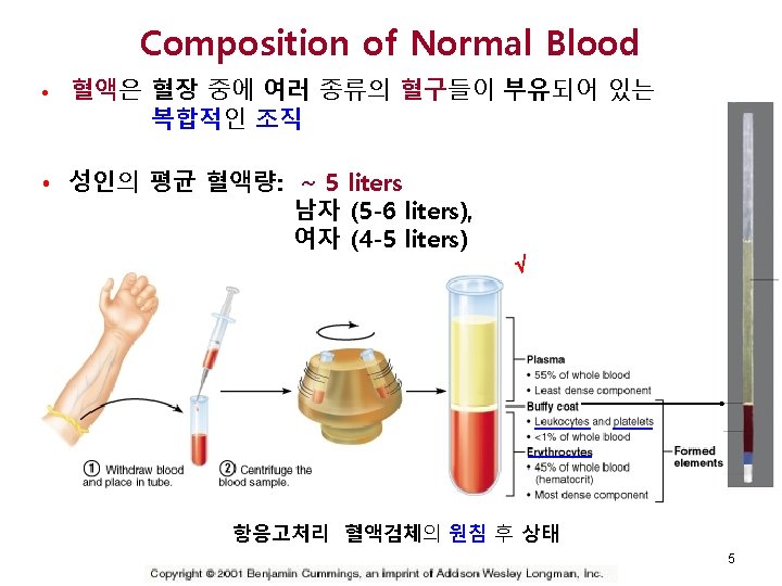
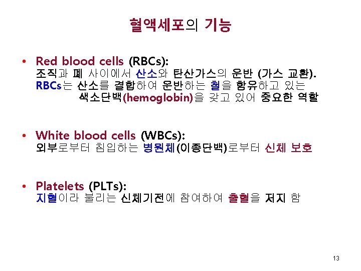
• 혈액의 액체성분을 혈장(plasma)라고 하는데, 이 혈장은 출혈을 저지하는 응고인자 (coagulation factor)들을 포함 • 혈장은 고형성분인 혈구를 운반하고 혈구에 영양을 공급 - Red Blood Cells - White Blood Cells: neutrophils, eosinophils, basophils, lymphocytes, monocytes - Platelets 7
History of Hematology – Cont’d • 1900년 이전부터 의사와 MLS*는 일정 량의 혈액 내 RBC를 계수하여 빈혈을 진단 • 1953, Wallace/Joseph Coulter형제는 미립자계수기 개발 • 1958, automated cell counters를 임상검사실 활용 • 21 stcentury: 자동혈구계산, Flow cytometry, Cytogenetics, Molecular analysis 분야 등이 크게 발전 *: medical laboratory scientists 10
Complete Blood Count (CBC) √ 혈액검사학의 기본인 CBC에 포함되는 항목 • • • WBC 계수 (WBC 종류별혈구수 포함) RBC 계수 Hemoglobin 농도 Hematocrit Platelet 계수 RBC 지수 – 적혈구의 크기 및 형태에 대한 정보 제공 • Mean Cell Volume (MCV) • Mean Cell Hemoglobin (MCH) • Mean Cell Hemoglobin Concentration (MCHC) • 혈구형태학적 검경 12
Cells Normally Found in Circulation A: RBC B: Segmented neutrophil C: Band D: Eosinophil E: Basophil F: Lymphocyte G: Monocyte H: Platelet Peripheral blood, Wright stain Fig 1 -1 Rodak BF, Fritsma GA, Keohane EM. Hematology-Clinical Principles and Applications, Elsevier, 2012, p 2. Copyright © 2012, 2007, 2002, 1995 by Saunders, an imprint of Elsevier Inc. 14
고급 혈액검사항목 • 골수검사 • 골수흡인(Aspirates) • 골수생검 (Biopsies) • 세포화학 염색 • Myeloperoxidase stain (MPO) • Sudan Black B stain (SBB) • Esterase tests • Acid and alkaline phosphatase (LAP) 16
고급검사항목 - 계속 • 유세포분석 • 양적 분석: cell count • 질적 분석: 표지자이용한 세포계열분류 • 분자검사 Fig 33 -9 A, B Rodak BF, Fritsma GA, Keohane EM. Hematology-Clinical Principles and Applications, Elsevier, 2012, p 498. Copyright © 2012, 2007, 2002, 1995 by Saunders, an imprint of Elsevier Inc. • Polymerase chain reaction(PCR) • Reverse Transcriptase(RT)-PCR • FISH(fluorescent in situ hybridization) Piazza RG, et al. Leukemia (2005) 19, 1986
CELLULAR STRUCTURE AND FUNCTION Chapter 6 Chapter Author: Elaine M. Keohane 18
Plasma Membrane (원형질막) • 세포막의 특성 – 탄력성 (Elastic) – 변형가능성 (deformability) √ 이중지질막 -인지질(극성)머리: Hydrophilic -지방산 꼬리: Hydrophobic Fig 13 -10 Keohane EM. Hematology- Clinical Principles and Applications. √ Functions: – 외부환경과의 물질교환 조절 – 많은 수용체분자를 갖고 있어 세포-세포 간의 인식 – 표면 표지자(당단백질)을 통해 세포정체성을 나타냄 https: //commons. wikimedia. org/wiki/User_talk: Superscience 71421
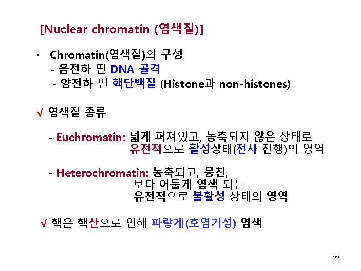
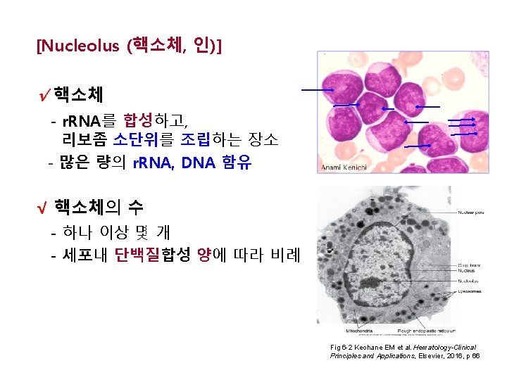
Nucleus (핵) √ Nucleus – 세포의 통제센터 - 주로 DNA로 구성 - DNA복제와 전사가 이루어지는 곳 • 세가지 기본 요소 - 염색질 - 핵막 - 핵소체 Fig 12 -7, B. Keohane EM. Hematology-Clinical Principles and Applications, Elsevier, 2016, p 154. 21
√ Golgi Complex (골지체) • Composition - 세포내막계에 해당되며, cisternae이라 불리는 여러 겹으로 겹쳐친 납작한 주머니 형태 Fig 8 -4. Keohane EM et al. Hematology. Clinical Principles and Applications, • Function Elsevier, 2016, p 100 - 다른 소기관에 대분자량 물질의 분비/전달을 위해 가공/포장 함 - Directs traffic in cell • 핵 근처에 위치 • 무색으로 환하게 염색 http: //biology. tutorpace. com/ golgi-complex
√ Endoplasmic Reticulum (내형질세망, ER) • 납작한 판, 주머니, 관의 형태로 나타나는 세포내막계(endomembrane system) Fig 6 -3. Endoplasmic retitulum. Keohane EM et al. Hematology-Clinical Principles and Applications, Elsevier, 2016, p 69 • 내형질세망(ER)의 종류 - Rough ER: 단백질합성, ribosomes보유 - Smooth ER: 새로생성된 단백질의 저장 장소 26
√ Ribosomes • Composition - 단백질 과 r. RNA • Location - 세포질 내 유리형 - 세포질그물(ER)표면에 결합형 Fig 6 -3. Endoplasmic retitulum. Keohane EM et al. Hematology-Clinical Principles and Applications, . Elsevier, 2016, p 69 • Function – 단백질 합성장소: 유전정보 보유한 m. RNA의 코돈에 상보적인 anticodon을 가진 t. RNA 가 특정amino acid를 ribosome 으로 옮겨와 peptide사슬 연장 (유전정보에 따른 AA서열로 단백질합성) http: //www. dna-sequencing-service. com/wpcontent/uploads/2010/07/translation-dna. gif 27
√ Mitochondria • Function 미토콘드리아 효소는 산화적 인산화를 통해 ATP생성 함 Fig 6 -4. Mitochondria. Keohane EM et al. Hematology-Clinical Principles and Applications, Elsevier, 2016, p 69 • 자기복제 능력 있으며, 별도의 mitochondrial DNA 존재 • Wright stain으로 염색되지 않음 • 이중 막을 가짐 (inner and outer membrane) 28
√ Lysosomes • Composition – 세포내막계로 가수분해 효소 간직 • Function: phagocytosis • 과립으로 염색 됨 https: //www. genome. gov/dmd/img. cfm? node=Photos/Graphics&id=85195 29
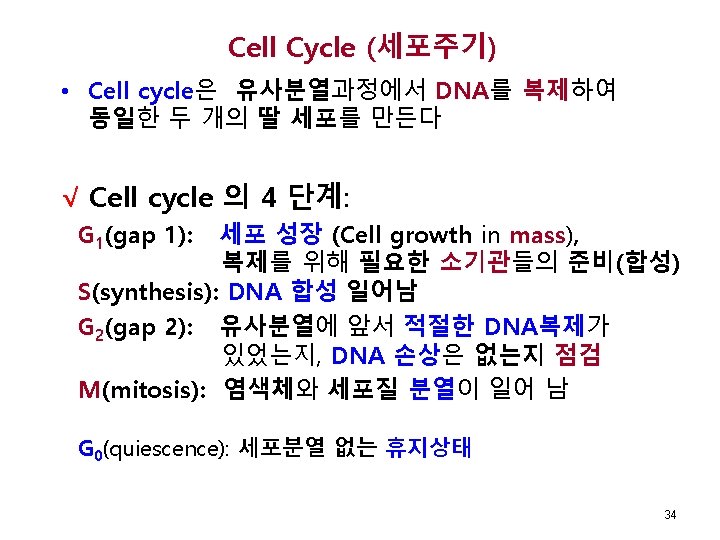
Cell cycle and Check points √ Fig 6 -5. Stages of cell cycle. Keohane EM et al. Hematology-Clinical Principles and Applications, Elsevier, 2016, p 71 35
CDK 2 CDK 1 CDK 4/6 https: //www. boundless. com/biology/textbooks/boundless-biology-textboo 37