Lec 23 URINAlysis A urinalysis is a test


: n n n A urinary tract infection occurs where bacteria")


 The most common sites of urinary tract infection (UTI)")
 Physical examination")
 Physical examination: n n n 1 -Urine volume: This")

: n Clear – freshly voided urine is clear. Cloudy –")
: determined by refractometer and indicator paper stripes. Normal value")

 concentration,")


 (ketones): The ketone bodies include acetone, acetoacetic acid (diacetic acid) and")


- Slides: 26
Lec. 2&3 URINAlysis
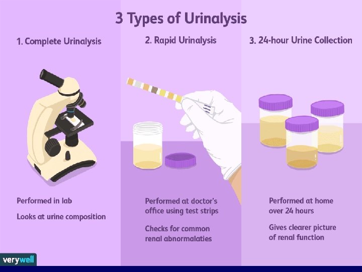
A urinalysis is a test of urine , used to detect and manage a wide range of disorders, such as urinary tract infections, kidney disease and diabetes. A urinalysis involves checking the appearance, concentration and content of urine. Abnormal urinalysis results may point to a disease or illness.
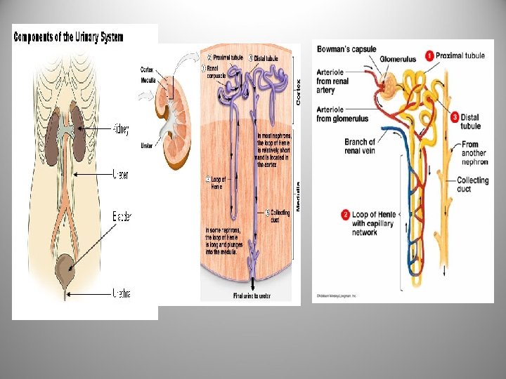
Urinary system n n n Urinary system consists of pair of kidneys and urinary tracts which includes (two ureters, urinary bladder and urethra). Each kidney contains 1. 3 million urinary units called nephrons. Each nephron consist of glomerulus and urinary ducts (Bowman's capsule, proximal convoluted tubule, Henley loop, distal convoluted tubule and collecting duct). Nephrons responsible of urine formation through three sequence process: Glomerular filtration, the Glomerular filtration rate(GFR) reach 180 liter /day. Reabsorption Secretion
Urination: is a voluntary process depends on person's choice to the suitable time and place to empty the urinary bladder from storage urine, without pain. Disturbances in urination include: 1 - Painful urination, frequency, Urgency. Because of microbial UTI. 2 - Impairment of urine flow, hesitancy, drubbingurine, incomplete emptying. Because of urinary bladder obstruction. 3 - Urinary retention, a sign of benign prostate enlargement (hypertrophy), urinary incontinence (enuresis) due to disfunction of bladder muscles or sphincter muscles.
Urinary tract infection (UTI): n n n A urinary tract infection occurs where bacteria and white blood cells are present in the urine of a patient with symptoms of infection of the urethra, urinary bladder, or the kidney. Bacteriuria occurs when bacteria are present in the urine; pyuria occurs when white blood cells are present in the urine. The lower urinary tract includes the urethra (urethritis) and urinary bladder (cystitis). The upper urinary tract includes the ureters and kidneys, infection in the upper urinary tract called (pyelonephritis). The bacteria that cause urinary tract infections are usually of fecal origin (e. g. , Escherichia coli). n Host factors that are important in protection from urinary tract infections: n These include: the normal daily flow of urine, the constant sloughing of the uroepithelial cells lining the urinary tract, and the presence of large numbers of Lactobacillus in the vaginal mucosa.
Etiology: n The most common cause of urinary tract infections (i. e. , urethritis, cystitis, and pyelonephritis) is E coli. Staphylococcus saprophyticus is the second most common cause of these infections in females between the ages of 13 and 40. In complicated cases of urinary tract infections such as those resulting from anatomic obstructions or from catheterization, the most common causes are E coli, Klebsiella pneumoniae, Proteus mirabilis, Enterococcus, and Pseudomonas aeruginosa. n n n Manifestations: Urethritis: is an infection of the urethra that causes pain and discomfort during voiding (dysuria). Cystitis: is an infection of the urinary bladder. Signs and symptoms of cystitis include urgency and frequency of urination, voiding small volumes of urine, and supra pubic tenderness just before or immediately after voiding. Pyelonephritis: is an infection of the lower urinary tract ascends the ureters to enter the kidneys, the signs and symptoms include fever, flank pain and tenderness, cost vertebral angle tenderness, and nausea and vomiting. Kidney stones: can serve as a location in which bacteria can escape antibiotics and cause recurrent urinary tract infections.
Epidemiology: n n n ■ Urinary tract infections rank second only to respiratory infections in their incidence. ■ Most cases of urinary tract infection occur in women (female to male ratio is 30: 1). The incidence of urinary tract infections increases with age. ■ Postmenopausal women have higher rates of infection because of bladder or uterine prolapse; loss of estrogen, which causes a change in the vaginal flora; loss of lactobacilli in the vaginal flora, which results in periurethral colonization with gram-negative aerobes (e. g. , E coli); and higher of concomitant medical illness (e. g. , diabetes mellitus). ■ Males experience a rapid increase in the incidence of urinary tract infections sometime in the fifth decade of life due to obstruction of the urethra following development of benign prostatic hypertrophy. ■ Urinary tract infections are usually endogenous; infection follows contamination of the distal end of the urethra with bacteria from the patient’s own fecal organisms. ■ Urinary tract infections occasionally occur following bacteremia or due to hematogenous spread of a distant infection to the urinary tract.
Kidney Function Test (URINE ANALYSIS) The most common sites of urinary tract infection (UTI) are the urinary bladder (cystitis) and the urethra (urithritis). n From these sites the infection may ascend into the ureters (ureteritis) and subsequently involve the kidney(pyelonephritis). n
Kidney Function Test (URINALYSIS) Physical examination
Kidney Function Test (URINE ANALYSIS) Physical examination: n n n 1 -Urine volume: This is dependent normally up on fluid intake, environmental condition, diet and activity of the human. Value above or below the normal value (1. 5 L/Day) can be considered as pathological disorder but it should be combined with clinical and laboratory examination. - above normal (polyuria) urine volume (> 2. 5 -3 L/Day) due to large quantities intake of liquids, diuretics, alcohol, in sufficient of urinary ductsin reabsorption of water and urine concentrated as in diabetes mellitus or diabetes insipidus. - under normal (Oligourea) urine volume (< 400 ml/Day) - Anuria, urine volume ( < 50 ml/Day), due to: hot weather, sweating, low water intake, or due to disease in kidney or urinary ducts.
n n n n 2 -Color: Can be observed in a test tube or in a urinometer tube. The following designations are used to observe the sample and correlatedto the following terms. *Colorless, Greenish yellow, Blue, Pale yellow, Green, Milky, Yellow, Red, Dark yellow, Reddish brown, Yellow brown, Brown. Interpretation Yellow to amber (Normal); the color comes primarily from the presence of urobilin. Urobilin is a final waste product resulting from the breakdown of heme from hemoglobin during the destruction of aging blood cells. Colorless to pale yellow; dilute urine with low specific gravity and polyuria. Dark yellow or yellow brown; concentrated urine with a high specific gravity and small quantity. Yellow brown or greenish yellow; yellow green foam when urine is shaken. Urobilinoids – chromagon derived from heme , yellow-brown-billirubin-and urobilin. Cloudy; hematuria (clearer after centrifugation). Translucent; hemoglobinuria. Brown to brownish black; hemoglobin up on standing bile large amounts. Green; bile biliverdin Red to pink; phenothiazine (beet root) Blue; medication contain methylene blue or food with blue dyes.
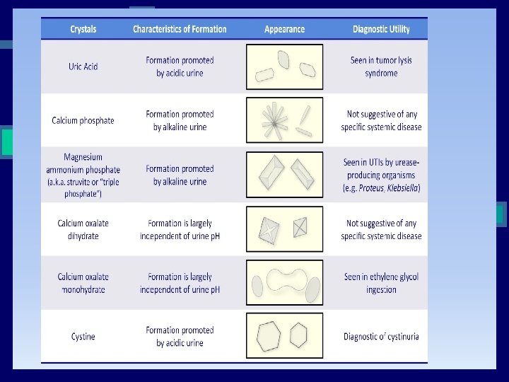
n 3 -Transparency (clarity): n Clear – freshly voided urine is clear. Cloudy – not necessarily pathological as many samples may become cloudy. Epithelial cells – present in large numbers. Blood – red to brown color and smoky. Leukocytes – may produce milky, ropy appearance if large number. Bacteria – produce a uniform turbidity if in large number; the turbidity doesn’t settle out and cannot be removed by filtration. n n n n n 4 -Mucus: 5 -Crystals: a salt compounds organized in geometrical shapes and looks like crystals, important in stones formation (cysteine crystals and oxalate crystals). Amorphous urate – white or pink cloud in acid urine Amorphous phosphate – white cloud in alkaline urine
n 6 -Specific gravity (SG): determined by refractometer and indicator paper stripes. Normal value in Man 1. 010 – 1. 030(Average normal = 1. 025). depending on SG the urine will be diluted (SG˂ 1. 002) or concentrated (SG˃1. 065) depend on the solvents in urine. SG used to determine the kidney efficiency keeping water balance in urine. n 7 -Odour: Normal odor – aromatic or acetone Abnormal odor – aromatic odor of ketone bodies (sweet fruit odor) as in starvation and diabetes. In UTI the odor of urine is bad smell. n n n 8 -Foam: Shake the sample and observe: • If the amount of foam produced is in excess and slow to disappear – proteinuria • If the color of the foam great – yellow or brown – bile pigments • If the color of the foam red to brown – hemoglobinuria
Microscopic Examination of Urine Sample n n n It is of great clinical importance and should never been omitted. Important structure to be include casts, erythrocytes, leukocytes, epithelial cells, budding yeasts, protozoa and bacteria. Casts: Cylindrical bodies performed in distal collecting tubules from RBCs or WBCs or fatty compounds or waxes. Dignosis of cast type aid in diagnosis of the disease. Types of casts: Hyaline casts, density granulated casts, finely granulated casts, red cell casts (hematouria), and white cell casts (inflammation), wax casts, fat casts, and epithelial casts. Erythrocytes: and leukocytes present in urine in case of UTI, diseases, inflammation. n n Epithelial cells: presence normally due to sloughing the lining layer of urinary tubules, bladder and urethra or because of some renal diseases. Budding yeasts: Candidaalbicans, found in diabetes patients urine because of the low PH and the presence of sugar necessary for the growth. Protozoa: like. Trichomonasvaginalis that infect the vagina in women and urethra in men and cause trichomoniasis.
Chemical Examination of Urine: -PH of urine The normal hydrogen ion (p. H) concentration, in the urine (5 -8) depends on the type of diet. Vegetable diet, citrus fruits (also bacterial infections) produce alkaline urine, while high protein diet (also blood acidosis where PH˂7. 35, some microbial infections, ketones elevation due to diabetes or aspirin intake) produce acidic urine. PH measured by paper strip or p. H meter.
-Proteins of urine: a little quantity, of protein are found normally in urine (150 mg/day)any access in protein called proteinuria which is an indication for many diseases like kidney diseases , fever and pregnancy. Types of protein in urine: 1 -Albumine: is the first protein appearing in urine due to its low molecular weight and size (albuminuria), this protein appears in Diabetes and hypertenation. 2 - Immunoglobulins: appear in urine due to inflammations and microbial infections 3 -Hemoglobine: found in urine due to blood hemolysis. Test: (Robert’s test) Principle: Precipitation of protein by strong acid A positive test is indicated by a white ring at the zone of concentration, which should be read against a dark back ground and reported as: Negative - No ring at the zone of concentration Note: In many clinical laboratories, Robert’s test is routine method as it is simple, quick and easy to read even when only a small amount of protein presents.
-Glucose in urine: No glucose is present in the urine normally which passes glomerular filter, because it is completely absorbed in the tubules. It present when the blood glucose level elevated to(180 mg/ml) which is called renal threshold, when blood glucose elevated the glucose present in urine as in diabetes. Glucose Test Method: Qualitative method (Benedicts test) Principle: Reducing sugars present in the urine react with the copper sulphate to reduce the copric ions to cuprous oxide giving a Color change from blue (negative) to green, yellow and red depending on the amount of reducing substances present
Acetone (Ketone body) (ketones): The ketone bodies include acetone, acetoacetic acid (diacetic acid) and betahydroxybutric acid. A state in which these substances are present in increased amount in the blood and urine is called ketosis. Acetoacetic acid and beta hydroxyl butric acid from which acetone is derived is normal intermediate product of fat metabolism. When greater amounts of fatty acids are utilized with the production of more acetoacetic acid and beta-hydroxybutric acid can be oxidized by thetissues. These bodies accumulate in the blood and are excreted in the urine (ketonuria). This bodies present in urine in starvation or low blood glucose levels.
# most of urine macroscopic examination can be done using special strips for rapid urinalysis
Urine Culture: n n n Urine is the specimen most frequently submitted for culture. The most common sites of urinary tract infection (UTI) are the urinary bladder (cystitis) and the urethra. From these sites the infection may ascend into the ureters (ureteritis) and subsequently involve the kidney (pyelonephritis). Females are more prone to infection of the urinary tract than are males In both males and females, UTI may be asymptomatic, acute, or chronic. -Asymptomatic infection can be diagnosed by culture. -Acute UTI is more frequently seen in females of all ages; these patients are usually treated on an outpatient basis and are rarely admitted to hospital. -Chronic UTI in both males and females of all ages is usually associated with an underlying disease (e. g. pyelonephritis, prostatic disease, or congenital anomaly of the genitourinary tract) and these patients are most often hospitalized. Since urine itself is a good culture medium, all specimens should be processed by the laboratory within 2 hours of collection, or be kept refrigerated at 4 C until delivery to the laboratory and processed no longer than 18 hours after collection
The examination procedure includes the following steps: n n 1. Examination of a Gram-stained smear. ( for the presence or absence of bacteria, polymorphonuclear leukocytes, and squamous epithelial cells). One or more bacterial cells per oil-immersion field usually implies that there are 105 or more bacteria per milliliter in the specimen. The presence of one or more leukocytes per oil-immersion field is a further indication of UTI. 2. A screening test for significant bacteriuria. The absence of leukocytes and bacteria in a Gram-stained smear of a clean catch urine sample prepared as described above is good evidence that the urine is not infected. A urine specimen that is “negative” on careful examination of the Gram-stained smear does not need to be cultured. 3. A definitive culture for urine specimens found to be positive in the screening test, and for all specimens obtained by cystoscopy, suprapubic bladder puncture (SBP), or catheterization. 4. Susceptibility tests on clinically significant bacterial isolates should only be performed on well-isolated colonies of similar appearance. Susceptibility tests are generally more importanton cultures obtained from patients who are hospitalized or have a history of recurring UTI.
Tha nk y ou