Learning Outcome Give a definition of homeostasis Homeostasis


 This is the hormone that controls how much water is reabsorbed")


- Slides: 73
Learning Outcome Give a definition of homeostasis.
Homeostasis is the maintenance of the body’s internal environment despite changes to the external environment. • We need to keep a constant internal environment for the body to work properly
What factors do you think must remain constant in our bodies?
water balance temperature blood sugar
Success Criteria I can give a definition of homeostasis.
Learning Outcome State that the kidneys are the main organs responsible for regulating water balance.
Water Balance The human body is made up of 70% water In order to maintain water balance the amount of water we gain must equal the amount of water we lose on a daily basis
Osmoregulation Kidneys regulate the water content of the body. This process is called osmoregulation.
In your groups you have 2 mins to write down all of the ways which you think we GAIN and LOSE water.
Water Gain Food
Water Loss SWEAT Urine Faeces Breath
Human Urinary System video
vena cava aorta RENAL ARTERY delivers unpurified blood to the kidney RENAL VEIN carries purified blood away from the kidneys KIDNEYS filter blood for useful and harmful substances + regulate the volume of water in the body URETER carries urine from the kidney to the bladder urethra URETHRA releases urine from the body BLADDER short-term storage of urine video
The kidneys
The kidney
The kidney As the renal artery enters the kidney it splits up into a large number of tiny blood vessels called capillaries.
The kidney
Two important jobs 1. Filtration 2. Reabsorption
Success Criteria I can state that the kidneys are the main organs responsible for regulating water balance.
Learning Outcome Label the different parts of a kidney nephron. Describe the functions of the different parts of the kidney nephron.
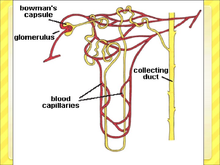
Nephron
Glomerulus The capillary forms a tight knot of blood vessels called the glomerulus. Blood flows through the glomerulus and is filtered.
The blood capillary entering the glomerulus has a wider diameter than the capillary leaving it.
Filtration
Bowman’s capsule The Bowman’s capsule surrounds the glomerulus. It collects the liquid filtered out of the blood. This liquid is called the glomerular filtrate
Filtered or not? Filtered Glucose Water Urea Salts Not filtered Protein Blood cells
Reabsorption Completely reabsorbed Variable amounts are reabsorbed None is reabsorbed Glucose Water Urea Salts
How does a kidney work?
http: //www. biologymad. co m/resources/kidney. swf
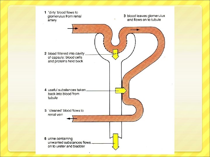
Blood contains useful + waste substances e. g. Glucose, urea, salts and water Kidney Tubule- Useful substances are reabsorbed back into the blood (glucose, some water and some salts) Blood capillary. Reabsorbs useful substances Blood is filtered and filtered substances enter the bowman’s capsule Filtrate enters Only waste substances remain- Urea, some salt, some water Blood leaving the kidney contains only useful substances (glucose, some water and some salt
From renal artery Glomerulus Bowman’s capsule • Water • Glucose • Urea all filtered into Bowmans capsule and down into tubule GLUCOSE Proteins and reabsorbed. blood cells too big and remain in blood Tubule WATER reabsorbed. Collecting duct to renal vein URINE: Urea + excess water To the URETER
Success Criteria I can label the different parts of a kidney nephron. I can describe the functions of the different parts of the kidney nephron.
Samples Experiment Measure 5 mls of each sample into 3 test tubes labelled A, B and C 2. Add a few drops of Benedicts solution and place in the water bath. IF SAMPLE TURNS ORANGE GLUCOSE IS PRESENT 3. Place 0. 5 ml of each sample AND 0. 5 ml of urease in a dimple tile 4. Test each with p. H paper. IF PAPER TURNS GREEN UREA IS PRESENT 1.
Renal Artery, Renal Vein or Urine? Glucose present Renal artery Renal vein Urine Urea present
Results and Conclusion Renal Artery = Sample _______ Renal Vein = Sample _______ Urine = Sample _______ Choose correct answer from the words underlined: • I know sample _____ is from the renal artery because it contained no/both urea and glucose. • • I know sample ____ is from the renal vein because it only contained urea/glucose. • • I know sample _______ is urine because it contained glucose/urea but did not contain glucose/urea
Learning Outcome Explain the role of ADH in water balance.
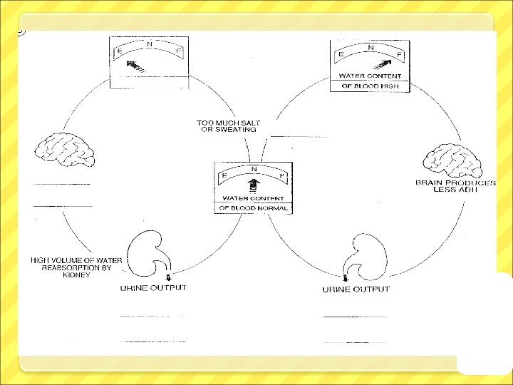
Anti-Diuretic Hormone (ADH) This is the hormone that controls how much water is reabsorbed by your kidneys. ADH
ADH increases the permeability of the kidney tubules which means more water is reabsorbed into the blood stream.
Blood capillary Tubule
Blood capillary Tubule
When the water concentration of the blood is too low, low a message is sent to the hypothalamus and ADH is released. This increases the volume of water reabsorbed. Think – ADH means Anti Drying out
Success Criteria I can explain the role of ADH in water balance.
Learning Outcome State that the hypothalamus is the area of the brain that controls body temperature.
Monitoring body temperature Body’s temperature monitoring centre is the hypothalamus.
Hypothalamus It receives nerve impulses from thermoreceptors in the skin and the body’s core (i. e. Lungs and heart) The hypothalamus central thermoreceptors are sensitive to change in blood temperature.
Hypothalamus The hypothalamus then responds by sending out signal via nerve impulses to effectors. These effectors are mainly in the skin and can bring about correction of body temperature to normal.
Success Criteria I can state that the hypothalamus is the area of the brain that controls body temperature.
Learning Outcome Explain the body’s response to overheating. Explain the body’s response to overcooling.
Skin and temperature change If the body temperature rises the hypothalamus sends a signal to the skin, this causes three responses: Vasodilation Increased sweating Relaxation of hair erector muscles
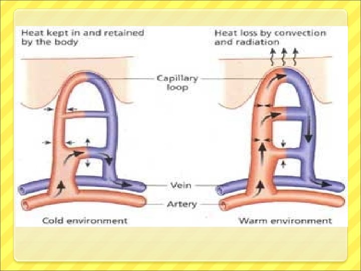
Vasodilation is the process where small arteries leading to the skin surface dilate. This increases the diameter of the vessel meaning more blood can flow through. Heat loss can be increased by conduction and radiation.
Increased sweating More sweat is released from the sweat glands. This coats the skin, and it evaporates by removing heat energy from the skin.
Investigating Sweat Aim: to investigate the effects of sweat on the rate at which a body loses heat Results: Time (mins) Temperature (°C) Dry Tube Method: Draw a diagram of your experiment. (25 ml of hot water in each boiling tube, one wrapped in dry paper towel, other in damp paper towel. Take temperature then add paper towels and start stop clock - take a temperature reading every 2 minutes) Wet Tube 0 2 4 6 8 10 Conclusion: The water in the wet/dry towelled tube cooled the fastest. This shows that sweat increases/decreases the rate at which the body loses heat
Hair erector muscles control the hairs on your body. When they relax, the hairs lie flat against your skin, this means there is no layer of air to insulate the skin.
Skin and temperature change If the body temperature starts to fall the hypothalamus sends a signal to the skin, this causes three responses: Vasoconstriction Decreased sweating Contraction of hair erector muscles
Vasoconstriction is the process where small arteries leading to the skin surface constrict. This decreases the diameter of the vessel meaning less blood can flow through. Heat loss by conduction and radiation is therefore decreased.
Decreased sweating Less sweat is released from the sweat glands. This keeps the skin dry and avoids heat loss by evaporation.
Hair erector muscles control the hairs on your body. When they contract, the hairs stand up against your skin, this means there is a layer of air to insulate the skin.
Success Criteria I can explain the body’s response to overheating. I can explain the body’s response to overcooling.
LEARNING INTENTIONS Describe how the body regulates blood glucose levels
Blood Glucose Levels Blood glucose levels in the body must stay at a constant level so that there is always enough glucose in the blood to be delivered to cells for respiration. • 2 hormones produced by the pancreas control blood glucose levels – INSULIN and GLUCAGON. •
Insulin This is produced by the pancreas. It is carried in the blood to the target organ - liver. • Insulin causes the liver to take up glucose from the blood and convert it to a storage carbohydrate called glycogen and therefore reduces the levels of glucose in the blood. • Glucose Insulin Glycogen
Glucagon This is also produced by the pancreas. It is carried in the blood to the target organ liver. Glucagon causes the liver to convert the storage carbohydrate glycogen to glucose and therefore increases the levels of glucose in the blood. Glucagon Glucose Glycogen
Glucose in the blood increases which pancreas detects Pancreas makes MORE INSULIN and Insulin tells the liver to convert more glucose into glycogen LESS GLUCAGON Eating Less glucose in blood Normal level of glucose in the blood More glucose in blood Exercise Glucose in the blood decreases which pancreas detects Pancreas makes LESS INSULIN and MORE GLUCAGON Glucagon tells the liver to convert more glycogen to glucose
LEARNING INTENTIONS Discuss diabetes and its causes
Diabetes is a condition caused by a communication pathway failure which results in; Ø a fault in release of insulin (type 1) or Ø a failure to respond to insulin (type 2)
Research Task