Learning Objectives The structure of proteins and their
in our cells")
Learning Objectives The structure of proteins and their possible jobs (including enzymes)in our cells n How and Where Transcription and Translation occur. n How DNA, m. RNA, t. RNA, r. RNA all work together to go from Gene Protein n

A Brief Review: Monomer Amino Acids Polymer Proteins
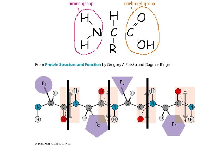
 is called a Amino acids protein (polymer) Peptide")
A chain of amino acids (monomers) is called a Amino acids protein (polymer) Peptide Bonds Protein -but proteins fold in on themselves… …. to make a specific 3 D shape that allows proteins to do their jobs
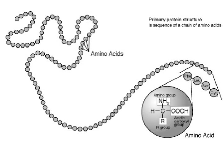

-but proteins fold in on themselves… …. to make a specific 3 D shape that allows proteins to do their jobs **REMEMBER** Amino Acid Protein’s order shape Function Or simply Order Shape Function
")
Proteins: the hardest working molecule Possible Protein Functions: Structural support (helps keep cell’s shape) Storage Transport across cell membrane Sensory reception (communication between cells) Contractions (movement) Defense (t-cells, red blood cells) Gene Regulation Building bone, brain, muscle, organ cells Transporting Oxygen in blood ENZYMES***

Enzymes Pg 52 Act as CATALYSTS that can speed up some reactions by more than a billion times! Enzymes work by a physical fit (Lock and Key) between the enzyme molecule and its SUBSTRATE, the reactant being catalyzed. Enzymes reduces the activation energy for the chemical reaction to occur. After the reaction, the enzyme is released and is unchanged, so it can be used many times Enzyme names end in –”ase”
 Pg 52 p.")
Enzyme & Substrate fit like a lock & key (Shape specific) Pg 52 p. H or temperature can change the active site shape on any enzyme Active site is where the reactants bind to the enzyme

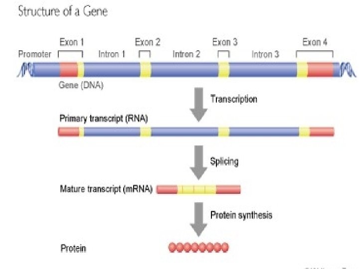
Gene: Gene a specific segment of DNA that codes for a protein Ex: Gene 1 Gene 2 Gene 3 3 genes on one DNA strand coding for 3 separate proteins

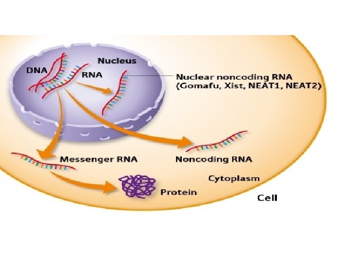
Gene 1 Gene 2 Transcription: Copying one side of DNA and creating a strand of m. RNA. This happens inside the nucleus all the time -when completed, the m. RNA that was transcribed from a specific gene will then travel outside the nucleus to be Translated

m. RNA can bind to single DNA strand DNA Transcribed Strand Un-Transcribed Side m. RNA DNA Codon A T G G A T A C A A T T A C C T A T G T T A U A C C U A U G U U A m. RNA Codon

Gene 1 Gene 2 Gene 3 Gene Transcription to m. RNA In Eukaryotes the m. RNA is “processed” before it leaves the nucleus -Introns (Junk RNA) are removed -Exons (working RNA) are spliced together to make a full m. RNA chain that then will leave the nucleus

Exon 1 Exon 2 Exon 1 Exon 3 Exon 2 Exon 3

Transcription Animations: http: //www. stolaf. edu/people/giannini/flashanimat/molge netics/transcription. swf http: //www-class. unl. edu/biochem/gp 2/m_biology/animation/gene_a 2 https: //www. youtube. com/watch? v=28 mgfg 8 n. RT 4 http: //www. youtube. com/watch? v=zt. Pkv 7 wc 3 y. U&feature=related Translation Animations: http: //www. stolaf. edu/people/giannini/flashanimat/m olgenetics/translation. swf http: //www. dnalc. org/view/15501 -Translation-RNA-to-protein-3 Danimation-with-basic-narration. html https: //www. youtube. com/watch? v=n. HM 4 UUVHPQM
 to create")
After m. RNA leaves the nucleus: Translation: using m. RNA (from transcription) to create a chain of Amino Acids (a. k. a. Proteins) -the process requires: m. RNA t. RNA Ribosomes COPY THE NEXT DIAGRAM Remember cells use proteins for all kinds of different jobs: -Transportation across membrane, digestion, building organelles & other cell structures, and of course. . ENZYMES

COPY DIAGRAM with labels

Pg 303 The genetic code reads for the m. RNA codon, NOT the t. RNA Anticodon

DNA: m. RNA Codon t. RNA Anti. Codon Amino Acid U C A A GU Serine A G T U C A Translation TRANSCRIBED SIDE Transcription

Ex Problem: DNA Codon DNA Un-transcribed Strand T A C C T A T G T T A A T G G A T A C A A T Transcribed Side m. RNA Codon t. RNA Anti. Codon Amino Acids m. RNA U A C C U A U G U U A A U G G A U A C A Tyrosine Leucine Cysteine

The genetic code reads for the m. RNA codon, NOT the t. RNA Anticodon

Transcription & Translation http: //highered. mcgrawhill. com/sites/0072437316/student_view 0/chapter 15/animat ions. html# http: //www. stolaf. edu/people/giannini/flashanimat/cellstructures/endome mbrane%20 protein%20 synthesis. swf https: //www. youtube. com/watch? v=28 mgfg 8 n. RT 4 https: //www. youtube. com/watch? v=n. HM 4 UUVHPQM
- Slides: 26