Learning Objective Know about liver structure Success Criteria

Learning Objective • Know about liver structure Success Criteria • Describe the gross structure and histology of the liver • Examine and draw stained sections to show the histology of liver tissue

Starter: Review your FLIP learning, discuss what you learnt with the person next to you

Terms never to confuse!! �Excretion versus egestion �Metabolic waste – substances produced from chemical reaction that may be toxic at high levels in the body Carbon dioxide Nitrogenous waste (urea)

Week 5 A diagram showing the positions of the main excretory organs

Liver Gallbladder Pancreas

Week 5 The liver and its connections to the blood system © Pearson Education Ltd 2009 This document may have been altered from the original

Liver

Liver basics Lies to the right side of body just under diaphragm Largest internal organ- holds 13% total blood at any one time - can store & release blood acting as a reservoir to compensate for smaller changes in blood volume Uses up to 20% total energy in body Made of left & right lobes enclosed by fibrous capsule (Glissons Capsule) Each lobe formed from hexagonal lobules (100, 000) Dual blood supply: receives blood from 2 blood vessels

Liver Lobules Inter-lobular vessel Liver lobule Intra-lobular vessel

Key Skill: Drawing Microscope Slides Looking at liver slides
![Low Power Drawing Mark Scheme [10] Annotations [3] Whilst a label might be the](http://slidetodoc.com/presentation_image_h2/bac9927c6b47a17642fb9c03b97a6f8c/image-12.jpg "Low Power Drawing Mark Scheme [10] Annotations [3] Whilst a label might be the")
Low Power Drawing Mark Scheme [10] Annotations [3] Whilst a label might be the name of a tissue, an annotation adds a descriptive quality such as shape, size or colour. Drawings from a microscope [7] Single, clear lines drawn with a sharp pencil. No shading or colour on the diagram. Informative title to be included. Scale included (e. g. high power, low power, x 80, x 10) to show approximate magnification. Low power tissue plans may not include cells. Tissues should be in correct proportions. Label lines drawn in pencil using a ruler. You will be PA on your drawings

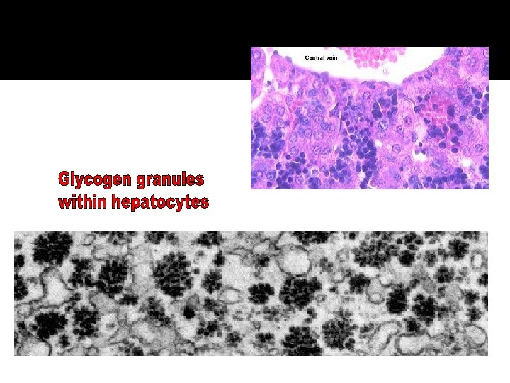
Hepatocytes Make up roughly 80% of the mass of the liver Nuclei are distinctly round, most single nucleus, some binucleate cells. Hepatocytes are exceptionally active in synthesis of protein and lipids for export therefore have large quantities of both rough and smooth endoplasmic reticulum and Golgi membranes. Glycogen granules and vesicles containing very low density lipoproteins are readily observed.

SINUSOID DELIVERS BLOOD IN TO LIVER LOBULE OUTSIDE OF LOBULE SPECIALISED MACROPHAGE CELL SPECIALISED LIVER CELLMICROVILLI CHANNEL SPACE BETWEEN LIVER CELL AND SINUSOID (BLOOD VESSEL PLASMA) INSIDE OF LOBULE

The arrangement of liver cells into cylindrical lobules Blood moves from outside of lobule from HPV and HA HV Blood from HPV and HA mix and enter channels called SINUSOIDS Hepatocytes lining the SINUSOIDS absorb products in blood and secrete products into the blood as it flows over them. The branches of HV have thin vessel walls and receive more blood from the sinusoids from each plate (layer) of lobule: link to from bigger branches Interlobular vessels (triad) © Pearson Education Ltd 2009 This document may have been altered from the original

The arrangement of liver cells in a lobule © Pearson Education Ltd 2009 This document may have been altered from the original
 Release:")
The sinusoids Hepatocytes have microvilli (increase surface area of liver cells: x 6) Release: plasma proteins (prothrombin, fibrinogen, albumin), lipoproteins (endocrine function of liver), cholesterol Absorb: insulin, glucose, minerals, vitamins, blood borne toxins for detox. Kupffer cells in SINUSOID: phagocytic cells (derived from monocytes). Ingest bacteria from blood. Breakdown bilirubin for absorption into hepatocytes
 Hepatocytes close to bile")
Bile Canaliculus Hepatocytes separated by second channel: bile cannuli (plural) Hepatocytes close to bile canuli rich in Golgi vessels reflecting transport of bile constituents into the channels Contents drained into bile duct giving bile to the duodenum

Plenary: PPQ

Markscheme
- Slides: 22