Learning Aim B Know about the musculoskeletal system

Learning Aim B: Know about the musculoskeletal system and cardiorespiratory system and the effects on the body during fitness training Lesson Objectives: To be able to Identify and name different muscles within the body and know where they are located. Extension: To start to identify bones on the skeleton and know where they are located. Numeracy Challenge: Literacy Key Words: Muscular Skeletal Pectoralis Major Biceps Cranium Femur Humerus Exit Note: At the end of each lesson write down something you are unsure about and I will set it as your starter task


Musculoskeletal System What does this mean? Muscular system Muscles- major muscles, muscle groups and also including tendons Skeletal system Skeleton- including bones, cartilage and ligaments

Muscular System • There are over 640 named muscles in the human body and these make up approximately 40% of your body mass. • The muscles that move your bones during activity are called skeletal muscles/voluntary muscles (Remember these muscles are under conscious control). • There are different types of muscles and they have very specific functions. • When a muscle contracts, it exerts a pulling force on a tendon, which then pulls on the bone causing the movement to occur. • Many muscles work in antagonistic pairs to create a movement. Don’t panic you only need to know the location of the major muscles in the body! Remember Muscles are connected to bones via tendons

Can you label any muscles on the body?

Pectoralis major Deltoid Biceps External obliques Triceps Latissimus dorsi Gluteus maximus Hamstrings Quadriceps Tibialis anterior These are the names of all the muscles you need to know Gastrocnemius

Major Voluntary muscles Some muscles work in antagonistic pairs Name: BICEPS Name: TRICEPS Location: Front of the upper arm Location: Back of the upper arm Role: Flexion at the end of the elbow Role: Extension of the arm at the elbow Example: Upwards phase of a biceps curl Example: Straightening the arms in a chest press FLEXION means bending a joint EXTENSION means straightening a joint

Major Voluntary muscles Name: QUADRICEPS Location: Front of the upper leg Role: Extension of the leg at the knee Example: The following through of a kicking motion in football Name: HAMSTINGS Location: Back of the upper leg Role: Flexion of the leg at the knee Example: The preparation phase of kicking a football

Major Voluntary muscles Name: LATISSIMUS DORSI Location: Side of the back Role: Adducts the upper arm at the shoulder/rotates the humerus Example: Bringing arms back to the sides during a straight jump in trampolining Name: PECTORALIS MAJOR Location: Front of the upper chest Role: Adducts the arm at the shoulder Example: Follow- through from a forehand drive in tennis ADDUCTS means to move towards the midline of the body

Major Voluntary muscles Name: GLUTEUS MAXIMUS Location: The largest muscle in the buttocks Role: Extension of the leg at the hip Example: Lifting the leg back at the hip when running Name: GASTROCNEMIUS Location: Back of the lower leg Role: Pointing toes towards the floor (plantarflexion) Example: Pointing toes when performing a straddle jump in trampolining Remember always use the correct name for the gastrocnemius, not the calf. Remember it has a ‘C’ sound in it Gast…. . ro. C nem……ius.

Major Voluntary muscles Name: DELTOID Location: The top of the shoulder Role: Abducts the arm at the shoulder Example: Lifting your arms above your head to block the ball in volleyball Name: EXTERBAL OBLIQUES Location: On the sides and front of the abdomen Role: Supports the rotation of the spine Example: Twisting crunches or standing twists using a resistance band

Major Voluntary muscles Name: TIBIALIS ANTERIOR Location: Front of the lower leg Role: Responsible for dorsiflexing and inverting the foot Example: Toe raises pointing toes towards the sky. DORSIFLEXION means pointing the toes upwards

Can you label any bones? The human skeleton An adult human skeleton is made up of 206 bones

Clavicle Sternum Humerus Cranium Scapula Ribs Radius Pelvis Ulna Femur Patella Tibia Fibula These are all the names of the bones you need to know!

Cranium= Skull Pelvic= Pelvis Leg= Femur, Patella, Tibula and Fibula Arm= Humerus, Radius and Ulna Shoulder girdle= Clavicle + Scapula Chest= Sternum + Ribs

Functions of the skeletal system Protection Muscle attachment and movement Storage of minerals Function s Blood production Shape Support

Protection The skeleton provides PROTECTION to VITAL ORGANS. For example, a rugby player’s: • Rib cage protects their heart and lungs when being tackled • Cranium protects their brain if they fall or if they are tackled illegally • Vertebral column protects their spinal cord

Muscle attachment and movement Muscles are attached to bones via tendons. Bones have joints that permit MOVEMENT. The skeleton is joined to allow us to move when the muscles attached to them contract The vertebrae allow us to bend, stretch and rotate our body The bones and joints work with muscles to enable us to walk, run and sprint. When a muscle contracts, the tendon it is connected to pulls on the bone and produce movement.

Support The skeleton provides a FRAMEWORK for your body and therefore SUPPORTS you. The skeleton provides the support that enables a gymnast to balance when performing a handstand. Without the skeleton, the body would be flabby and shapeless.

Shape Your skeleton gives you SHAPE. When your skeleton grows as you change from a child to an adult, your shape changes with it. Body shape is particularly important in certain sports- such as basketball, where players need to be tall.

Blood production Red blood cells are produced in the BONE MARROW of long bone e. g. the ends of the femur and humerus. Having more red blood cells means you have the ability to carry more oxygen, which can help sport performance. Femur: located in the upper part of the leg Humerus: located in the upper part of the arm

Storage of minerals Bones store four minerals: • CALCIUM: important for bones and teeth formation, clotting blood and muscle contraction. • PHOSPHOROUS: important for bones and teeth formation, and energy metabolism. • SODIUM: important for muscle contraction and nerve impulses • POTASSIUM: important for muscle contraction and the functioning of the nervous system.
 allow us the free movement to")
Synovial Joints • Synovial joints (freely movable joints) allow us the free movement to perform skills and techniques during physical activity. • This is a joint which has the greatest range of movement. • Synovial joints have synovial fluid in the joint cavity that lubricates or 'oils' the joint so it moves smoothly. Synovial fluid is made by the synovial membrane. • In synovial joints, the ends of the bones are covered with cartilage which cushions the joint and prevents friction and wear and tear between the bone ends. Cartilage is a soft, spongy connective tissue it prevents wear and tear on the bones. A joint is a place where two or more bones meet and is also called an articulation. The role of joints and connective tissue Connective tissues consist of ligaments, cartilage and tendons. A joint is held together by ligaments which give the joints their stability. Cartilage is found at the ends of bones and where joints meet. Tendons attach muscles to the skeleton.
 Hinge joint Ball and socket Found at")
Types of synovial joints (freely moveable joints) Hinge joint Ball and socket Found at the elbow and knee. The range of movement is limited to one plane. Found in the shoulder and hip. This joint allows for the greatest range of movement.

Synovial fluid Knee joint Synovial membrane Bursa Ligament Joint synovial capsule Tendon Articular cartilage

Parts of Synovial Joint Functions Synovial Capsule Keeps synovial fluid in place. Synovial Membrane Acts as a lining for the joint capsule and produces synovial fluid which lubricates the joint. Synovial Fluid Acts as a lubricant between bones and nourishes the joint. Bursa A fluid filled sac between the tendon and bone that helps to reduce friction. Cartilage Soft, slippery, slightly elastic tissue covering heads of bones allows friction free movement, acts as a shock absorber, reduces friction and prevents wearing of the bones at the joint. Ligament (elastic) Tough connective tissue that joins bone to bone and holds bones together forming the joint. Made up of tough tightly packed elastic fibres that can withstand sudden stresses and provide strength to prevent injuries such as dislocations. Tendon (non-elastic) Connects muscle to bone: transmit force created by the muscles to the bones to allow movement. They are tough and strong.

Elbow joint

Clavicle Tendon Shoulder joint Ligament Bursa Cartilage Scapula Synovial Membrane Humerus

Easily bent, flexible What is one of the reasons that we do a warm up before exercise? As the muscles get warmer the become more pliable. This increases the range of movement available at a joint because the muscles are able to stretch more once they are warm. There is an increased range of movement because our muscles become warmer the reason they get warmer is because there is blood getting pumped to them more quickly. Planning for progressive overload When we exercise there are tiny tears in the muscle fibres. These tears are necessary breaks in the muscle to encourage your body to rebuild and repair the muscle to make it become bigger and stronger and over a long period of time the muscle will go through a process of hypertrophy. After exercise your body needs rest time in order to recover. This recovery period is when micro tears are repaired.

Cardiorespiratory System What does this mean? This word is broken into two to make up the two systems below The cardio-respiratory system works together to get oxygen to the working muscles and remove carbon dioxide from the body. During exercise the muscles need more oxygen in order to contract and they produce more carbon dioxide as a waste product. Think HEART Think BREATHING Cardiovascular system Respiratory system This is made up of your heart, blood and blood vessels This is made up of your lungs and airways

• The heart is the central part of the cardiovascular system, which transports oxygen around the body. • It is important to understand the structure of the heart in order to understand the effects of shortterm and long-term exercise on it. • The cardiovascular system is sometimes referred to as the circulatory system and consists of the heart, blood vessels and blood. • The cardiovascular system is the major transport system in your body, carrying food, oxygen and all other essential products to cells, and taking away waste products of respiration and other cellular processes, such as carbon dioxide. • Oxygen is transported from the lungs to body tissues, while carbon dioxide is carried from the body tissues to the lungs for excretion. The heart has four chambers. The two atria collect blood and the two ventricles pump the blood out of the heart. These are three essential components the body needs to survive

Right Atrium Can you label the heart Arteries take blood Away. Left Atrium Ventricles are the lower chambers of the heart. Right Ventricle Atriums are the upper chambers of the heart. Left Ventricle Aorta largest artery in the body Vena Cava Pulmonary Vein Pulmonary Artery Veins take blood INto the heart Vena cava largest vein in the body The right side of the body pumps deoxygenated blood (blood with no oxygen in it) to the lungs. The left side pumps oxygenated blood (blood with more oxygen in it) from the lungs to the rest of the body.

Aorta Pulmonary Artery Right Atrium Pulmonary Veins Left Atrium Left Ventricle Right Ventricle Vena Cava

• Vena cava: brings deoxygenated blood from the body • The right atrium receives it • Passes through to the right ventricle • Deoxygenated blood leaves pulmonary artery • Then reaches the lungs • Pulmonary vein brings oxygenated blood from lungs • Left atrium receives it • Passes through to the left ventricle • Oxygenated blood leaves the aorta • Travels to the rest of the body The left ventricle has a much thicker wall than the right ventricle, because it needs more blood to pump blood around the whole body. Blood in the left ventricle is under higher pressure.

Name Function Atria Collect blood Ventricles Pump blood from the heart Aorta Carries oxygenated blood away from the heart (left ventricle) towards the rest of the body Vena Cava Carries deoxygenated blood back into the heart (right atrium) from the body Pulmonary Artery Carries oxygenated blood to the lungs to allow it to be re-oxygenated Pulmonary Vein Carries oxygenated blood back to the heart from the lungs

The lungs Deoxygenated blood Right atrium Left atrium Right ventricle Left ventricle The body Blood flow through the heart, body and lungs The cardiovascular system and respiratory system work together to help us take part in sport. Oxygenated blood

Deoxygenated blood Pulmonary Artery Vena Cava Right Atrium Right Ventricle Oxygenated blood Pulmonary Vein Aorta Left Atrium Left Ventricle

The function of the respiratory system is to get oxygen into the body and carbon dioxide and waste products from out of the body. This happens through the act of breathing. Breathing in (inhalation) gets the oxygen in, so it can be used by the body to release energy. Breathing out (exhalation) removes the carbon dioxide so it does not build up and poison the body. Muscles need oxygen to function, which is provided through RESPIRATION.

You have 5 key areas you need to locate and label within the respiratory system Lungs Diaphragm Bronchioles Alveoli Does anyone know what these might be?

Bronchi Lungs Alveoli Bronchioles Diaphragm

Structure Description Function Lungs A pair of spongey air filled sacs located on either side of the chest Is the process of gas exchange called respiration (breathing) Diaphragm A muscle that separates the chest from the abdomen Contracts continually as you breathe in and out Bronchi Two smaller tubes leading to each lung and the bronchioles Carries air into the lungs Bronchioles Smaller branches leading from the bronchi Ensure air is supplied to each alveoli Alveoli Small sacs at the end of the bronchioles Where gaseous exchange takes place

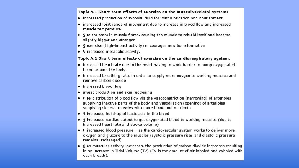
Increased heart rate and breathing rate during fitness training activities to supply oxygen to working muscles. the breathing rate and volume of each breath increases to bring more oxygen into the body and remove the carbon dioxide produced the heart rate increases, to supply the muscles with extra oxygen and remove the carbon dioxide produced Increased build-up of lactic acid as a result of increased intensity in the main component. If insufficient oxygen is available to the muscles, for instance the exercise is vigorous and/or prolonged, the heart and lungs are unable to supply sufficient oxygen. Muscles begin to respire anaerobically. Lactic acid is produced from glucose, instead of carbon dioxide and water. Muscles continue to contract, but less efficiently.

Scenario: When doing any form of physical activity what is the first thing you notice happen in relation to your cardiorespiratory system? What happens when performing exercise of a vigorous intensity Increased heart rate and breathing rate during fitness training activities to supply oxygen to working muscles. the breathing rate and volume of each breath increases to bring more oxygen into the body and remove the carbon dioxide produced the heart rate increases, to supply the muscles with extra oxygen and remove the carbon dioxide produced Increased build-up of lactic acid as a result of increased intensity in the main component. If insufficient oxygen is available to the muscles, for instance the exercise is vigorous and/or prolonged, the heart and lungs are unable to supply sufficient oxygen. Muscles begin to respire anaerobically. Lactic acid is produced from glucose, instead of carbon dioxide and water. Muscles continue to contract, but less efficiently.

Lactic acid is a waste product we make when we exercise. The harder we work the more lactic acid we produce. This can stop our muscles working because they burn and we feel like we cannot carry on. If in the warm up we did sprints we might find that we will have sore or heavy legs from the lactic acid we might not be able to do the main part of the session very well. This is because the warm up was too intense. During the warm up your body will increase in temperature as your muscles start to move. The synovial fluid in your joints gets thinner which gives you a bigger range of movement in the joint. Doing stretches will increase the range of movement in certain muscles and joint. You should stretch the main muscles you will use in the exercises you are going to do.

e v i s s e r g Pro d a o l r e v O Doing the same training in every session will not make you fitter. You need to mix up the training and make it harder each time to make sure you improve. When you overload your muscles (make the session harder each time) you should get micro-tears in your muscles. These are not big tears by small ones which are the first step in building bigger or stronger muscles. Increasing the amount of repetitions, sets or amount of time you train for could be an example of progressive overload if you do this bit by bit. Heart a nd brea thing rate Both of these will go up when you start exercising. This is to make sure your muscles have enough oxygen and to take away carbon dioxide. The blood delivers oxygen to the muscles and takes deoxygenated blood to the lungs to get fresh oxygen. The amount they will go up will depend on how hard you exercise. A warmup should be a steady increase in heart rate to slowly prepare your body rather than a fast increase.

Engaging learners in physical activities to highlight the obvious short-term effects of exercise on the body would be beneficial. For example, jogging around a sports field or completing timed sprints in a sports hall and then noting the physical effects: such as, becoming out of breath, getting hot, and having a flushed complexion. Physiological data could also be collected analysed, for example heart rate and breathing rate pre- and post-activity.


Can you label the heart Right Atrium Left Atrium Right Ventricle Left Ventricle The right side of the body pumps deoxygenated blood (blood with no oxygen in it) to the lungs. Veins take blood INto the heart Arteries take blood Away. Ventricles are the lower chambers of the heart. Atriums are the upper chambers of the heart. Aorta Vena Cava Pulmonary Vein Pulmonary Artery The left side pumps oxygenated blood (blood with more oxygen in it) from the lungs to the rest of the body.

Aorta Vena Cava Pulmonary Artery Left Atrium Right Atrium Pulmonary Vein Left Ventricle Right Ventricle
- Slides: 50