LEAF ANATOMY The two parts of leaf the


- Slides: 30
LEAF ANATOMY
The two parts of leaf, the petiole and the lamina, are considered for studying the internal structure of a complete leaf.
Epidermis Petiole It is single layered composed of compactly arranged barrelshaped cells. However, cells may be radially elongated in certain aquatic plants such as Eichhornia and Nymphaea. Epidermis is as usual covered with cuticle in terrestrial plants. In Banksia and Cucurbita, several multicellular hairs arise from epidermis. Hypodermis It lies next to epidermis and is usually collenchymatous in nature. In Mirabilis jalapa, it consists of patches of collenchyma and sclerenchyma.
Petiole Ground tissue It consists of loosely arranged parenchymatous cells with well developed intercellular spaces. In Mangifera, sclerenchymatous cells occur scattered in this region. In certain cases, laticifers and resin canals may be found. In aquatic plants, large air chambers are present.
Vascular bundles Petiole The arrangement of vascular bundles in petiole shows variation. In Ricinus, Platanus and Quercus, these arranged in a single ring while in Prunus and Galphimia, they are in the form of a girdle with adaxial xylem and abaxial pholem. The vascular bundles found in petiole of Cucurbita are bicollateral and arranged in two rings, the smaller below the grooves and larger opposite the ridges.
Vascular bundles Petiole In members of leguminosae, a swollen basal portion called pulvinus is present. This region is largely made up of thin walled parenchymatous cells with intercellular spaces. The vascular tissue is represented by only one concentric strand, although several bundles are present above and below the pulvinus. It is involved in drooping movements of the leaf, brought about by changes in the turgidity of the cells.
Lamina The internal organization of leaf lamina shows differentiation into vascular tissues. epidermis, mesophyll and
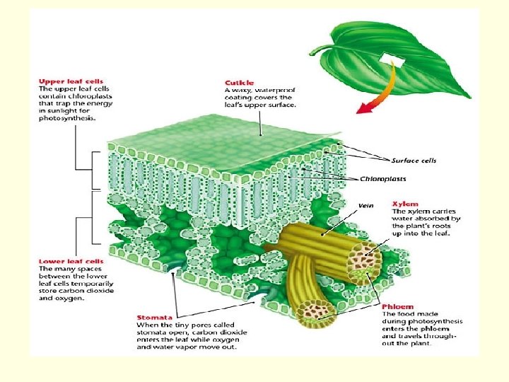
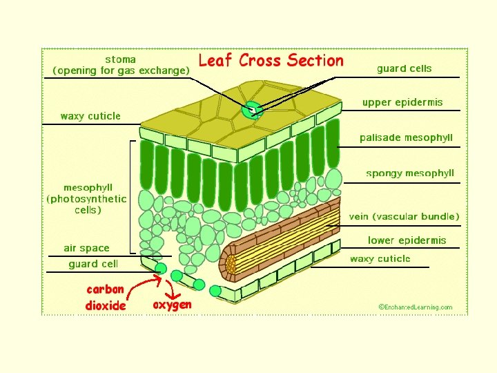
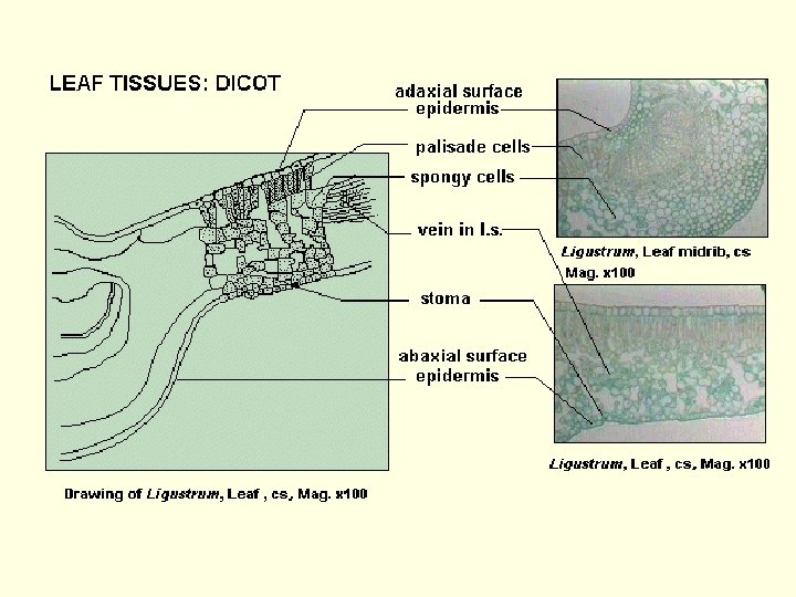
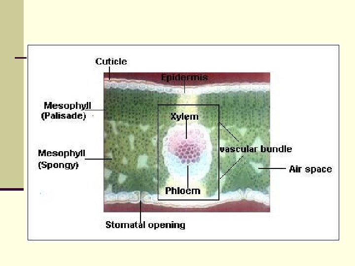
Epidermis Lamina Two layer epidermis forms the dermal tissue system of the leaf. Both upper and lower epidermis are usually uniseriate composed of compactly arranged cells. Usually, all cells of epidermis are of similar type but in certain monocots, groups of large cells but with thin flexible walls are found in longitudinal furrows which are called bulliform cells or motor cells. These help in rolling of leaves during dry weather, thus preventing the loss of water via transpiration.
Lamina The outer walls of epidermal cells have deposition of cutin, which is very thick and distinct among xerophytic leaves and relatively less distinct in mesophytic leaves. Cuticle is absent in case of submerged, aquatic leaves. Stomata is an important feature of the epidermis. If these are present only on upper surface, the leaf is called epistomatic as in case of Nymphaea, if on only lower surface it is hypostomatic as in mango, oleander and banyan and if on both the surfaces, it is amphistomatic as in isobilateral leaves.
Lamina Stomata remain surrounded by two highly specialized cells called guard cells which may be reniform or kidney-shaped as in most dicots or monocot leaves. The stomata may be present at the same level to epidermis as in mesophytic leaves, or may be sunken as in Cycas, Pinus or Allium or in stomatal cavities as in Nerium. In certain cases, glandular and non-glandular hairs arise from epidermis.
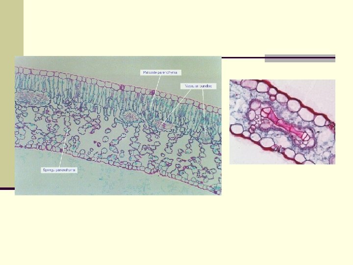
Mesophyll It is the most important part of leaf and forms the bulk of internal tissue of leaf extending from upper epidermis to lower epidermis. In most dicotyledonous leaves, it shows remarkable differentiation in to palisade and spongy cells. Palisade cells are columnar and more or less compactly arranged with their long axis at right angles to the leaf epidermis. The amount of palisade varies in different plants which also correlate with external environment. In centric leaves as in Allium and Juncus, the palisade occurs all around the periphery of leaf.
The arrangement of palisade cells at the right angles to the leaf surface protects chloroplasts from excessive heat of sun rays. Spongy cells are of variable shapes and are loosely arranged with abundant intercellular spaces. The intercellular spaces which maintain continuity with stomatal chamber facilitate the gaseous exchange.
In monocotyledonous leaves, mesophyll tissues are not differentiated into palisade and spongy cells and there occur only one type of cells. These cells contain abundant chloroplasts and intercellular spaces. In aquatic plants, mesophyll region accommodates large air chambers. The cells surrounding them have chloroplasts. Sometimes, various types of idioblasts are found in mesophyll region.
Vascular tissues The vascular tissues in leaf show no much variation. The mid rib in most dicot leaves have large bundles with adaxial xylem and abaxial phoem. This bundle is conjoint, collateral and open. A thin strip of cambium is present between xylem and phloem Vessels in xylem and sieve tubes in phloem are found in larger bundles but in smaller bundles, xylem is represented by tracheids and phloem by some sieve elements.
It is interesting to note that larger veins in case of some dicotyledons such as Ficus religiosa show a limited amount of secondary growth as a result of activity of cambium. In monocotyledonous leaves also, the bundle is conjoint, collateral and closed.
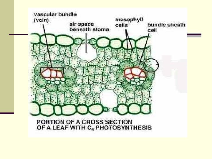
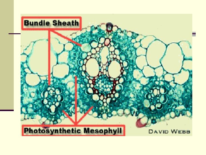
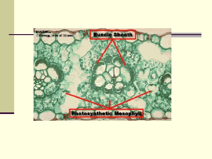
The vascular bundles in leaf have characteristic bundle sheath. The bundle sheath is usually composed of parenchymatous cells which are colorless and morphologically distinct. In dicotyledons, the parenchmatous cells extend in the direction parallel to veins. Mostly the bundles have uniseriate sheath of parenchyma cells.
The sheath in dicots usually extends upto epidermis on one or both sides of leaf. In monocotyledons, the bundles may remain partly or completely surrounded by one or two bundle sheath, each consisting of a single layer of cells. The cells of bundle sheath are achlorophyllous and have thick walls. smaller and
Dicot leaf 1. The stomata on epidermis are Monocot leaf 1. Stomata are found on both the generally present only on lower surfaces of leaves, thus leaves are surface, thrs leaves are hypostomatic amphistomatic types. 2. Guard cells are dumble shaped. 2. Guard cells are kidney shaped. 3. Bulliform cells are generally present. 3. In epidermis, bulliform cells are not 4. Mesophyll tissues are not differen- found. tiated into palisade and spongy cells. 4. Mesophyll tissues are generally well differentiated into palisade and spongy cells. 5. Intercellular spaces are large and numerous. 5. Intercellular spaces are fewer and smaller.
Xylem
Bulliform Tissue Mesophyll Tissue Bundle sheath Xylem Phloem Bundle sheath Phloem xylem
Bulliform Tissue Mesophyll Tissue Bundle sheath Phloem Xylem Epidermis