Late pregnancy bleeding Placental Abruption Presented by Aya

Late pregnancy bleeding Placental Abruption Presented by : Aya Deeb Al. Ma’aitah Supervised by : Dr. Seham Abu fraijeh

Placental abruption Clinical Presentation • A 25 -year-old primigravida at 36 weeks gestation is brought to the emergency department with constant, excruciating abdominal and back pain and sudden vaginal bleeding for the past 3 hours. Her blood group is B+ • Blood pressure is 160100 mm. Hg and Pulse is 118min. Physical examination shows a firm, distended, and tender uterus. Speculum shows approximately 75 m. L of red blood in the vagina. FHT shows a baseline 108min and no variability.
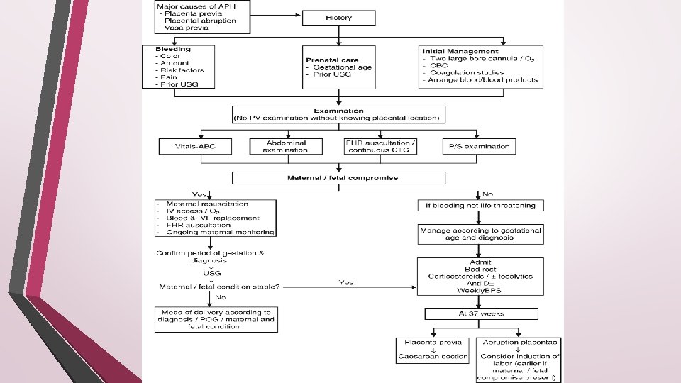

Initial evaluation : History… What is the nature and duration of the bleeding Age? Gestation History of trauma ? Is there pain or contraction ? Fetal movement? Examination. . Hypovolemia? ? What the patient vital sign? Are fetal heart tones present ? And fetal status? Acute placental abruption : Abrupt onset of : 1) Vaginal bleeding. (visible or concealed) 2) Abdominal pain (sudden constant localized to uterus) 3) Uterine contractions : usually high frequency and low amplitude ( by tocodynamometry) 4) The uterus is often firm, and tender. The amount of vaginal bleeding correlates poorly with the degree of placental separation and does not serve as a useful marker of impending fetal or maternal risk. Maternal hypotension and fetal heart rate (FHR) abnormalities, however, suggest clinically significant separation that could result in fetal death and severe maternal morbidity.
")
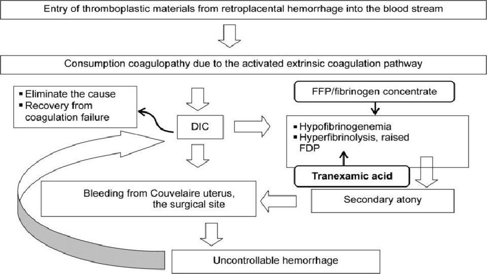
Initial investigation : • Complete blood count • Disseminated intravasculer coagulation(platletes, ptt, fibrinogen, D-dimer) • Blood type and cross match • Sonogram for placental location • Apt test ( Alkali denaturation test) • Never perform a digital or speculum examination until you rule out placenta previa by ultrasound The degree of maternal hemorrhage correlates with the degree of hematological abnormality; fibrinogen levels have the best correlation with severity of bleeding. Initial fibrinogen values of ≤ 200 mg/d. L are reported to have 100 percent positive predictive value for severe postpartum hemorrhage, while levels of ≥ 400 mg/d. L have a negative predictive value of 79 percent. - Mild separation/hemorrhage may not be associated with any abnormalities of commonly used tests of hemostasis. - Severe abruption can lead to DIC, which occurs in 10 -20 % of severe abruptions with death of the fetus.

Placental abruption ● Bleeding at the decidual-placental interface that causes partial or total placental detachment , after 24 weeks of gestation and prior to delivery of the fetus. - Placental abruption is the premature separation ( partial or complete ) of a normally situated placenta from the uterine wall, resulting in haemorrhage before the delivery of the fetus. ● 40 -60 % of abruptions occurred before 37 weeks of gestation and 14% occurred before 32 weeks. The perinatal death rate is approximately 12 % (versus 0. 6 % in nonabruption births). The majority of perinatal deaths (up to 77 %) occur in utero; deaths in the postnatal period are primarily related to preterm delivery.

Thrombin plays a key role in the clinical consequences of placental abruption, and may be important in its pathogenesis, as well. ● The production of thrombin can lead to the following clinical sequelae: 1) Uterine hypertonus and contractions, as thrombin is a potent, direct uterotonic agent. 2) Triggering of coagulation e. g DIC.
 Previous abruption : the most")
The major risk factors of placental abruption : 1) Previous abruption : the most important risk factor. The risk of recurrence has been reported to be 5 -15 %, . After two consecutive abruptions, the risk of a third rises to 20 -25 %. 2) Hypertension : 5 fold increased risk of severe abruption 3) Premature rupture of membranes 4) Chorioamnionitis 5) Abdominal trauma/accidents 6) Cocaine abuse 7) Polyhydramnios 8) Smoking during pregnancy : it is associated with a 2. 5 -fold increased risk of abruption 9) Maternal age 10) Parity 11) Multi-fetal gestation. 12) Thrombophilias.

Imaging : ● Identification of a retro-placental hematoma is the classic ultrasound finding of placental abruption. Retro-placental hematomas have a variable appearance; they can appear solid, complex, and hypo-, hyper-, or iso-echoic compared to the placenta. ● The sensitivity of ultrasound findings for diagnosis of abruption is only 25 -50 %, but the positive predictive value is high (88 %) when ultrasound findings suggestive of abruption are present. ● The absence of retro-placental hematoma does not exclude the possibility of severe abruption because blood may not collect behind the placenta. ● MRI can detect abruptions missed by ultrasound examination.

complications The risks to the fetus are related to both the severity of the separation and the gestational age at which delivery occurs. Mild abruption : less than 25% of placental surface is seperated , no fetal abnormality Moderate abruption ; from 25% to 50 % of placental surface is seperated , fetal monitor may show tachycardia , decreased variability or late deceleration. Sever abruption: greater than 50% of placental surface is seperated , fetal monitor show severe late deceleration, bradycardia, or even fetal death, and severe DIC may occur. Fetal morbidity and mortality related to hypoxemia, asphyxia, low birth weight, and/or preterm delivery and Fetal growth restriction FGR (with chronic abruption). Maternal: For the mother, the potential consequences of abruption are primarily related to the severity of the placental separation…. . ● Excessive blood loss and DIC generally necessitate blood transfusion and can lead to hypovolemic shock, renal failure, adult respiratory distress syndrome, multi-organ failure, peripartum hysterectomy and, rarely, death. ● Emergency cesarean delivery for fetal or maternal indications. ).

Management: Initial interventions : Any woman complaining of antepartum hemorrhage should be admitted. If women presents with potentially severe acute abruption: she is admitted to the labor room and stabilized
 Stabilization of the mother: 1) Call for Help. 2) I. V fluid :")
A) Stabilization of the mother: 1) Call for Help. 2) I. V fluid : two, wide-bore intravenous lines. 3) Closely monitor the mother's hemodynamic status (heart rate, blood pressure, urine output). Urine output should be maintained at above 30 cc/hour and monitored with a Foley catheter. 4) Keep maternal oxygen saturation >95 percent and keep the patient warm. 5) Draw blood for a complete blood count, blood type and Rh ( preparation of 4 units PRBCs), and coagulation studies. Repeat coagulation tests in patients with clinical signs of severe abruption as coagulopathy may develop or worsen over time. 6) Notify the anesthesia team. Anesthesia-related issues in these patients include management of hemodynamic instability, technical issues related to bleeding diathesis, and the potential need for emergency cesarean delivery. 7) Notify the blood bank so blood replacement products (red blood cells, FFP, cryoprecipitate, platelets) will be readily available, if needed. B) Immediately initiate continuous fetal monitoring. :
 Hospital stay: There are no compelling data to guide the")
Expectant management : 1) Hospital stay: There are no compelling data to guide the length of a hospital stay in these pregnancies. A reasonable approach is to monitor the patient in the hospital until the bleeding has subsided for at least 48 hours, fetal heart rate tracings and ultrasound examinations are reassuring, and the patient is asymptomatic. At that point, discharge may be considered. Importantly, the patient should be counseled to return immediately should she experience further bleeding, contractions, reduced fetal movement, or abdominal pain. 2) Single course of antenatal corticosteroid ( 26 -35 weeks of gestation). 3) Serial assessment of fetal well being tests : NST / biophysical profie / doppler studies for fetal blood vessels / sonographic estimation of fetal weight to assess growth. 4) Anti-D immune globulin for Rh(D)-negative women. 5) Schedule delivery at 37 -38 weeks because of the increased risk of stillbirth. - Delivery before 36 -37 weeks is indicated if additional complications arise ( FGR, preeclampsia, PROM, nonreassuring fetal assessment, recurrent abruption with maternal instability).

→ In women with DIC, transfuse blood and blood products to achieve the following minimum levels: ● Platelet count ≥ 50, 000/micro. L ● Fibrinogen ≥ 100 mg/d. L ● Prothrombin (PT) and partial thromboplastin time (PTT) less than 1. 5 times control ● Hematocrit 25 -30 %

PLACENTA PREVIA DONE BY : ASEEL KHRIESAT SUPERVISED BY: DR-SEHAM ABU FREIJEH

What is placenta previa? • the placenta that covers the internal os either partially or completely and associated with high risk of hemorrhage (rupture of placental vessels) and birth complications.
 new classification: Nowadays, the diagnosis and classification")
Classification of placenta previa (old vs new) new classification: Nowadays, the diagnosis and classification of Placenta Previa depends on the actual distance from the placental edge to the internal cervical OS at TVUS: using standard terminology of millimeters away from the OS or millimeters of overlap. A placental edge exactly reaching the internal os is described as 0 mm. If the placental edge is more than 20 mm away from the internal os this excludes Placenta Previa.

• Associated conditions : placenta previa has been associated with an increased risk of several other pregnancy complications: • Placenta accrete : placenta accrete complicate 1 -5% of pregnancies with placenta previa and an unscarred uterus • Malpresentation: the large volume of placenta in the lower portion of the uterine cavity predisposes the fetus to assume a non –cephalic presentation • Preterm labor and rupture of the membranes : antepartum bleeding from any cause is a risk factor for preterm labor and premature rupture of membranes • Vasa previa and velamentous umbilical cord : they are uncommon , but when present they are often associated with placenta previa • Congenital anomaly: population – based cohort studies have reported an increase in the overall rate of neonatal congenital anomalies in pregnancies complicated by placenta previa, but no single anomaly or syndrome was associated with the disorder

MAJOR RISK FACTOR FOR PLACENTA PREVIA? • Previous placenta previa : recurrence rate is 4 -8% • Previous cesarean delivery • Multiple gestation • Multiparity • Advanced maternal age • Infertility treatment • Previous abortion • Previous intrauterine surgical procedure • Maternal smoking • Maternal cocaine use

CASE 1 • A 30 year old Gravida 5 Para 4 women at 31 weeks gestation come to the emergency department because of an episode of vaginal bleeding that morning. The bleeding has subside. she had no prenatal care. her previous child was delivered with a caesarean section because of a breech presentation. • Her temperature is 37. 1, pulse is 88/min, respirations are 14/min , and blood pressure is 125/85 mm hg. the abdomen is non tender. no contractions are felt. the fetal heart rate is 145/min. her hemoglobin concentration is 12 g/dl, leukocyte count is 13000/mm^3 , and platelet count is 350. 000/mm^3.

What is the approach for this p. t? • History • Physical examination • Investigations • Management

History • Age? gravida? Para? • Vaginal bleeding+duration? Painful vs painless? • Colour? bright red, associated with mucous ? • Onset: spontaneous vs triggerd (following abdominal trauma, or intercourse) • Amount: spotting or excessive (presence of clots) number of pads/day (soaked or not)? • Course: single attack/recurrent, continuous or intermittent? • Severity: dizziness, loss of consciousness, shortness of breath, palpitation? • Fetal movement? • History of placenta previa with a previous pregnancies ? • risk factors : 1. Scar in uterus (previous c/s, D&N, myomectomy), ? 2. Multiple pregnancy? (large placenta) 3. Smoking? 4. History of infertility treatment?

• Examination • General examination including consciousness , signs of hypovolemia, signs of anemia. • Vital signs • abdominal examination? Tis it tender or not ? presence of uterine contractions (regular or not /frequency in 10 minutes/ duration)? • Obstetric examination • Pv is contraindicated in any antepartum hgg till placenta previa is excluded by us. • Assessment of fetal heart rate ?

Investigations Initial investigation : 2 large pore IV cannulae and draw blood samples for : Complete blood count Coagulation profile (pt, ptt, fibrinogen, platletes, d-dimer) Blood type and RH , cross match Preparing 4 unites of blood (and notify the blood bank for the possible need of FFP, platelets and cryoprecipitate) • KFT • • Immediately initiate continuous fetal monitoring. • What is your next step?

• Next step is : transvaginal ultrasound to assess the position of the placenta

1 -What is the most likely diagnosis? Placenta previa 2 -What will be the long term management of this patient?

Long-term management in this case : • expectant management ( the patient is hemodynamically stable , no active heavy bleeding or fetal distress) • Symptomatic women often remain hospitalized from their initial bleeding episode until delivery üCorrection of anemia ü 4 units of packed PRBCs should be available üAnti –D immune globulin for RH(D)-negative women üIf gestational age is<34 weeks: fetal lung maturity induction with corticosteroids(betamethasone) Route of delivery : cesarean delivery (bcz ideally scheduled at 36 -37 weeks gestation
")
CASE 2 • A 29 -year-old women, gravida 3 para 2 (previous 1 cs) , comes to the emergency department due to sudden-onset , heavy vaginal bleeding that has soaked clothes(spontaneous), (not stop). The patient has had inconsistent prenatal care and says she is about 8 months pregnant. she has had some non painful contractions. • four years ago, she had a low transverse cesarean delivery. • The patient smokes a pack of cigarettes a day and takes no medications. • On examination : • Temperature is 37, blood pressure is 96/70 mm hg , pulse is 118/min, and respiration are 16/min. • fetal heart monitoring shows a baseline of 150/min, moderate variability, acceleration , and no deceleration.
 Stabilization")
• • • Management: stabilization and emergency cesarean delivery (sever active bleeding) Stabilization of the mother: Call for help I. V fluid –secure intravenous access with two wide –bore intravenous lines Closely monitor the mother hemodynamic status (heart rate, blood pressure, urine output ). urine output should be maintained at above 30 ml/hour and monitored with a foley catheter Draw blood for a complete blood count, blood type and RH (preparation of 4 units PRBCs), and coagulation studies Notify the anesthesia team. anesthesia –related issues in these patients include management of hemodynamically instability and the potential need for emergency delivery Notify the blood bank so blood replacement products(red blood cells, fresh frozen plasma, cryoprecipitate, platelets ) will be readily available, if needed Immediately initiate continuous fetal monitoring

CASE 3 • A 39 -year –old women , gravida 3 para 2 (all by vaginal delivery) , comes to emergency department at 37 weeks gestation due to leakage of fluid for the last 2 hours she has also had some vaginal bleeding and contractions for the last hour. • Fetal movement is normal the patient immigrated to the united states 6 months ago and has had no prenatal care this pregnancy. • Her previous pregnancies were uncomplicated and delivered vaginally at term. • On examination : • blood pressure 100/60 mm hg. pulse is 102/min, and respirations are 15/min. on speculum examination , rupture of membrane is confirmed and the patient has small blood clots at the cervical os. • An ultrasound reveals active fetus in cephalic presentation whose growth is consistent with gestational age and placental tissue covering the cervix. • fetal heart rate baseline is 150/min with acceleratoions and no deceleration.

• The best plan for placenta previa in this patient ? • stabilization and immediate cesarean delivery • Indications for immediate cesarean delivery in placenta previa • 1 -mature fetus (term) • 2 -non reassuring fetal heart tracing • 3 -persistent bleeding (unstable hemodynamic status) • 4 -complete previa • 5 -labor

CASE 4 • A 39 year old women , gravida 3 para 2 (all by vaginal delivery), comes to the office at 24 weeks gestation for prenatal care and follow up of a prenatal ultrasound. • the patient has had no pelvic pain , leakage of fluid or vaginal bleeding. • fetal movement has been normal. • prior pregnancies were uncomplicated and delivered vaginally at term. • this pregnancy was conceived via in vitro fertilization with normal preimplantation genetic testing. • On examination : • blood pressure is 110/60 mm hg and pulse is 78/min. • ultrasound performed today showed a single fetus in the breech presentation. a placenta covering the internal cervical os.

Management: Initial management is with routine obstetric care, pelvic rest and abstinence from intercourse (due to potential cervical contact) are recommended, and clinicans should refrain from digital cervical examination. The majority (90%) of cases resolve spontaneously due to lower uterine segment lengthening and or placental growth toward the fundus. Therefore, initial management is with obstetric care. Repeated ultrasound is performed in the third trimester (28 weeks gestation and more ) and patients with previa resolution can continue routine care without pelvic restriction. Asymptomatic patients (no vaginal bleeding) with persistent previa undergo scheduled cesarean delivery at 36 -37 weeks gestation

CASE 5 • A 36 year old women , gravida 4, para 3, at 35 weeks gestation is brought to the emergency department for the evaluation of a sudden , painless, bright red vaginal bleeding for the last hour. she has had no prenatal care. Her third child was delivered by lower segment transverse cesarean section because of a preterm breech presentation: her first two children were delivered vaginally. The patient pulse is 100/min , and blood pressure is 105/70 mm hg. Examination show soft non tender abdomen: no contractions are felt. there is blood on the vulva, the introitus, and on the medial aspect both thighs bilaterally. the etus is in a cephalic presentation. the fetal heart rate is 140/min. one hour later, the bleeding stop. what is the most likely diagnosis ?

Placenta accrete spectrum and vasa previa Presented by: Joman Awwad supervised by: Dr-Siham abu Freijeh

Case scenario: • A 37 -year old woman gravida 4 para 2 +1 at 38 wks of gestation, comes to the hospital with labor pains. The patient received care at another clinic, but her medical records are unavailable. She delivered her first child vaginally at age 16 after an uncomplicated, full-term pregnancy. The patient had previous dilatation and curettage at the age of 25. She had a lower transverse cesarean delivery at term for breech presentation at the age of 30. On physical examination, her cervix is 6 cm dilated and 90% effaced with the fetal head at +1 station. She is hospitalized, dilates quickly to 10 cm, and vaginally delivers a healthy 4540 -g (10 b) boy. The umbilical cord avulses from the placenta, necessitating a manual extraction. The placenta is extracted in pieces. During placental removal, the patient develops profuse vaginal bleeding. Her uterus is firm, and the bleeding is unresponsive to uterine massage and uterotonic medications. • what is the most likely diagnosis? • What is the next step?
 is a general")
what is the most likely diagnosis? • Placenta accreta spectrum (PAS) is a general term used to describe abnormal trophoblast invasion into the myometrium of the uterine wall. Ø Considerations: • This patient has had two previous uterine surgery , which increases the risk of • placenta accreta. ( dilatation and curettage and previous cesarean section), other risk factor in this patient is maternal age above 35 years. • There are 2 clinical presentation: • Clinically, patients present with delayed placental detachment and massive, lifethreatening postpartum hemorrhage at the time of attempted manual separation of the placenta, as seen in this patient. • During the prenatal period, patients are often asymptomatic and diagnosis is generally made via routine prenatal ultrasound.

RISK FACTORS The most important risk factor for placenta accreta is placenta previa after a prior cesarean delivery. In women with placenta previa, the frequency of placenta accreta increases with an increasing number of cesarean deliveries as follows: ● No previous cesarean birth, 1 -5 %. ● One previous cesarean birth, 11 -25 %. ● Two previous cesarean births, 35 -47 %. ● Three previous cesarean births, 40 %. ● Four or more previous cesarean births, 50 -67 %. In the absence of placenta previa, the frequency of placenta accreta still increases with an increasing number of cesarean deliveries, but the incidence is much lower. In women without placenta previa, the frequency of placenta accreta is : ● One previous cesarean birth, 0. 3 %. ● Two previous cesarean births, 0. 6 %. ● Three previous cesarean births, 2. 4 %.

v according to the depth of invasion : ●Placenta accreta 63% – Anchoring placental villi attach to the myometrium (rather than decidua). ●Placenta increta 15% – Anchoring placental villi penetrate into the myometrium. ●Placenta percreta 22% – Anchoring placental villi penetrate through the myometrium to the uterine serosa or adjacent organs, if invades the bladder it can cause hematuria.

What is the next step? • In the rare event that a placenta accreta is first diagnosed after vaginal delivery, as in this patient, emergent laparotomy for hysterectomy is indicated to control the bleeding.

The 2 nd clinical presentation is prenatal diagnosis in asymptomatic women: ● Prenatal diagnosis of placenta accreta is typically based upon the presence of characteristic findings on ultrasound examination. - MRI can be more useful than ultrasound in two clinical scenarios: (1) Evaluation of a possible posterior placenta accreta because the bladder cannot be used to help clarify the placentalmyometrial interface. (2) Assessment of the depth of myometrial and parametrial involvement, and, if the placenta is anterior, bladder involvement. ● Postnatal diagnosis is based on histological examination of the placenta or the placenta and uterus. → Women with a placenta previa or a low anterior placenta and prior uterine surgery should have thorough sonographic evaluation of the interface between the placenta and myometrium between about 18 and 24 weeks of gestation. At this gestational age, the diagnosis is suspected or excluded in virtually all cases. MRI can be useful when the ultrasound findings are uncertain.

• Findings that were considered suggestive of placenta accreta included loss of the retroplacental hypoechoic clear zone, loss of the bladder wall-uterine interface, presence of placental lacunae (vascular spaces), and presence of hypervascularity of the interface between the uterine serosa and the bladder wall on color Doppler imaging.

The management: There are two options: 1. Cesarean hysterectomy 2. uterine conservation with the placenta left in situ. has a high risk of bleeding and sepsis, nevertheless in the rare case of a younger patient who strongly desires more children, this option may be entertained. - All patients with placenta accreta should be counseled about the suspected diagnosis and potential sequelae (Hemorrhage, blood transfusion, cesarean hysterectomy, maternal ICU admission). Consultation with a maternal-fetal medicine specialist is desirable. - Management by a multidisciplinary team and delivery in a tertiary care facility improve outcomes and lower complication rates. The team includes maternal-fetal medicine specialists, anesthesiologists, neonatologists, blood bank and nursing personnel. It is desirable to have a surgeon present who has extensive experience with wide dissection of the lower uterine segment and parametrial areas in the event such dissection or bladder resection is required.

- Delivery should be scheduled electively for optimal availability of necessary personnel and facilities. Planned delivery is associated with less intraoperative blood loss than emergency delivery. - The optimum gestational age for scheduled delivery is controversial. Some experts have recommended delivery of previaaccreta at 34 to 36 weeks of gestation. - Adequate blood and clotting factors should be available at the time of delivery. The magnitude of blood loss is difficult to predict antepartum. v. Preoperative placement of balloon catheters into the internal iliac arteries. The catheters may be inflated intermittently during hysterectomy, thus potentially decreasing blood loss and providing optimum exposure of the operative field. They may also be used for embolization of persistent bleeders.

Case scenario: • A 29 -year-old woman, gravida 1 para 0, at 33 weeks gestation is an inpatient on the antepartum unit for management of known vasa previa few minutes ago, the patient had rupture of membranes with leakage of blood-tinged fluid She has no contractions or heavy vaginal bleeding. The pregnancy was the result of in vitro fertilization and has otherwise been uncomplicated. A fetal growth ultrasound performed yesterday revealed a cephalic fetus with an estimated fetal weight at the 50 th percentile and a normal amniotic fluid index. Temperature is 36. 7 C (98. 1 F), blood pressure is 110/70 mm. Hg and pulse is 70/min. Fetal heart rate is 100/min. The uterus is nontender and has no palpable contractions. • What is the best next step in management of this patient?
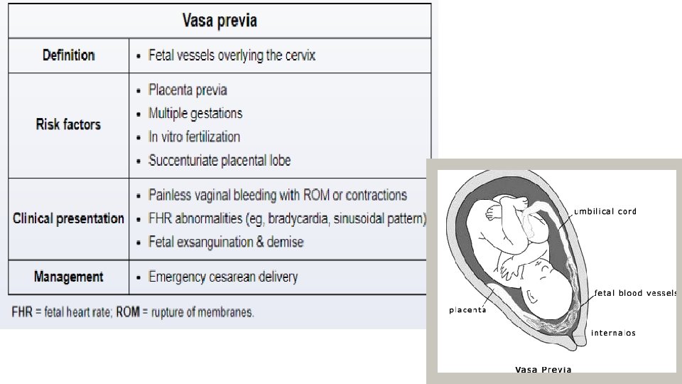

• Normal fetal vessels travel in the umbilical cord surrounded by thick, gelatinous tissue (ie, Wharton jelly) that protects them. In contrast, vasa previa is an aberrant condition in which the fetal vessels overlie the cervix, surrounded only by thin fetal membranes, making them prone to tear with rupture of membranes or contractions. Risk factors include in vitro fertilization as in this patient, and placenta previa.

Ø Cause of this condition: The exact pathophysiologic mechanism of vasa previa is not known, but it is associated with a velamentous insertion of the umbilical cord or joining an accessory (succenturiate) placental lobe or it may be associated with placenta previa and multiple pregnancy. If these fetal vessels rupture the bleeding is from the fetoplacental circulation, and fetal exsanguination will rapidly occur, leading to fetal death.

Prenatal diagnosis : • Antenatal diagnosis is based primarily on identification of membranous fetal vessels passing across the internal cervical os by real-time and color Doppler ultrasound, and it can be detected as early as 16 weeks GA. • ***In the absence of prenatal diagnosis, a clinical diagnosis of vasa previa should be suspected in the setting of vaginal bleeding that occurs upon rupture of the membranes and is accompanied by fetal heart rate abnormalities, particularly a sinusoidal pattern or bradycardia. v Confirmation that the blood is fetal via Apt, Kleihauer-Betke tests, or other tests (Ogita, Londersloot) supports the diagnosis; however, there is usually no time to wait for test results before performing an emergency cesarean delivery for fetal distress.

Vilamentous cord insertion

The best next step in management is emergency cesarean delivery. • Because total fetal blood volume is low (eg -250 ml or 1 cup), even minimal fetal bleeding can lead to rapid exsanguination and fetal demise Therefore, these patients require third-trimester inpatient management to monitor for acute changes that require immediate delivery. This patient with vasa previa and rupture of membranes likely has a ruptured fetal vessel, as evidenced by the leakage of blood-tinged vaginal fluid and fetal bradycardia. This obstetric emergency requires immediate delivery. Because vaginal delivery can cause further fetal vessel tearing and fetal compromise, induction of labor is contraindicated.
 Admission in the third trimester till delivery. 2) Single course")
Management in general: 1) Admission in the third trimester till delivery. 2) Single course of corticosteroids. 3) Serial fetal assessment : NST two to three times daily. 4) Scheduled delivery early, between 35 -36 weeks. 5) Emergency cesarean section should be done if any of the following occur : • labor/premature rupture of membranes/fetal distress by NST.

Case scenario: • A 36 year old woman, gravida 3 para 2, at 35 weeks gestation comes to the emergency department due to leakage of fluid and painless vaginal bleeding. The patient had rupture of membranes 30 minutes ago the fluid was initially clear but became bloody a few minutes later. She had normal fetal movement earlier today. The patient has not received prenatal care this pregnancy, her 2 previous pregnancies resulted in term cesarean deliveries. She has hypertension but stopped taking her medication when she became pregnant. Temperature is 36. 1 C (07 F), blood pressure is 140/96 mm Hg, and pulse is 92 mmhg. The uterus is nontender. Speculum examination confirm rupture of membranes, the cervical os is 1 m dilated with minimal vaginal bleeding. Doppler ultrasound is unable to detect fetal heart tones. • What is the most likely cause of this patients presentation? • What is the next step?

• What is the most likely cause of this patients presentation? • Ruptured vasa previa complicated by fetal demise. • What is the next step? • cesarean section.
- Slides: 55