Large intestine INTRODUCTION Length 1 5 m Subdivision

Large intestine

INTRODUCTION � Length- 1. 5 m � Subdivision- caecum, ascending colon, transverse colon, descending colon, sigmoid colon, rectum and anal canal. � Becomes continuous with terminal part of ilium at ilioceacal junction and joined by narrow blind tube the vermiform appendix. � Peritoneal relationsascending & descending colon- retroperitoneal Transverse & sigmoidcolon – suspended by mesentry called transverse mesocolon & sigmoid mesocolon. Caecum- surounded all round by peritonium , posteriorly separated by a recess called retrocaecal recess. Rectum- partially covered by peritoneum. Anal canal- doesn’t come in contact with peritoneum

APPEARNCE External- characterized by haustrations made by 3 taenia coli but absent in caecum & more pronounced from middle of transverse colon. �Taenia coli. Ant. -Taenia libra Posteromedial- taenia mesocolia Posterolateral- taenia omentalis In t. c. these r rotated through 90° thus ant. Become inf. , p. m. become post. And p. l. become superior.
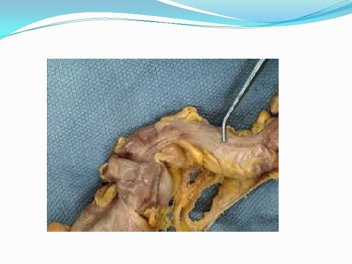

CEACUM � A large blind pouch lying in rt. ilac fossa below iliocaecal valve. � Continue proximally with distal ilium and distal with ascending colon. � Blind ending vermiform appendix arises on its medial side. � Length- 6 cm � Breadth- 7. 5 cm � Relations- posteriorly- rt. iliacus, psoas major Ant. - ant. abdominal wall, peritoneum Ilium opens into posteromedial aspect of large intestine.

Vermiform appendix � A narrow- worm like tube arises from posteromedial caecal wall 2 cm below end of ilium. � Length- 2 -20 cm � It may occupy one of several positions. 1. Retroceacal 2. Pelvic 3. Descending 4. Subceacal 5. Preileal 6. Postileal 7. Promonteric The three taenia converge on base of appendix into its longitudinal muscle. * Lumen of appendix is small & opens into caecum by an orifice lying posteroinferior to iliocaecal valve.

�Blood supplyant. & post. Branches of inf. Iliocolic artery of sup. mesentric artery & corresponding veins �Nerve supplysym. -sup. mesenteric plexus T 10 -L 1. Parasympathetic-vagus nerve Lymphaticsthrough ileocolic nodes to sup. mesenteric nodes.

Applied aspect �Appendicitis – inflammation of appendix Pain felt around umbilicus then shifts to rt. Iliac fossa. Rupture of appendix is serious complication that may spread to peritonitis. It is most common site for carcinoid tumour.

COLON �Part of large intestine from caecum to rectum. �Divisions: ascending, transverse, descending and sigmoid. �Ascending : 15 cm long �Lie vertically in rt. lateral region of abdomen. �Lower end continues with caecum at level of intertubercular plane. �Upper end meets t. c. at rt. Colic flexure.

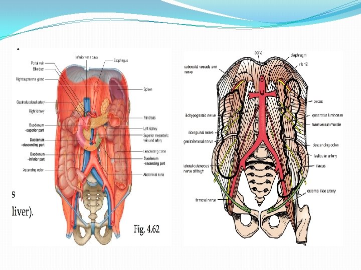
Relations �Posteriorly: iliacus, quadratus lumborum and transversus abdominis. �Lateral cutaneous nerve of thigh, iliac branch of iliolumbar artery. �Anteriorly: anterior abdominal wall. �Upper end: deep to liver, rt. of duodenum& g. b.
 begins at rt. colic flexure and ends at")
Transverse colon �Longest division (50 cm) begins at rt. colic flexure and ends at left colic flexure. �Forms loop of varying size and descends to level below umbilicus even may descend into pelvis. �Relations: ant. Greater omentum & ant. abd. wall �Post. : descending part of duodenum & pancreas, coils of jejunum & ileum. �Upper margin: liver, g. b. , stomach, spleen & tail of pancreas lt. kidney in lt. side.

Descending colon � 25 cm long, Begins at lt. colic flexure. �Lies in lt. hypochondrium descends through lt. lateral region and lt. inguinal region to reach lt. side of true pelvis. �It is covered by peritoneum on front and sides and anchored to diaphragm by fold of peritoneum called phrenicocolic ligament. �Relations : lt. kidney, transversus abdominis, quadratus lumborum, iliacus, psoas major. �Lt. iliohypogastric , ilioinguinal nerve , lateral cutaneous nerve of thigh, lt. iliac branch of iliolumbar artery. �Just above inguinal ligament it lies over ext. iliac artery, femoral nerve, genitofemoral nerve and testicular vessels.
 �Continuous with descending colon above and rectum")
sigmoid colon �Variable in length (40 cm) �Continuous with descending colon above and rectum below at level of S 3. �It forms convoluted loop enclosed all around by peritoneum and attached to p. a. w. by v shaped sigmoid mesocolon. �Relations : structures of pelvic wall- internal & external iliac vessels, obturator nerve, ductus deferens/ ovary �A part of s. c. lies btw. Rectum & urinary bladder in males and rectum and uterus in females.

Vascular supply

Thank you……
- Slides: 16