LARGE INTESTINE Extends from the distal end of


- Slides: 18
LARGE INTESTINE
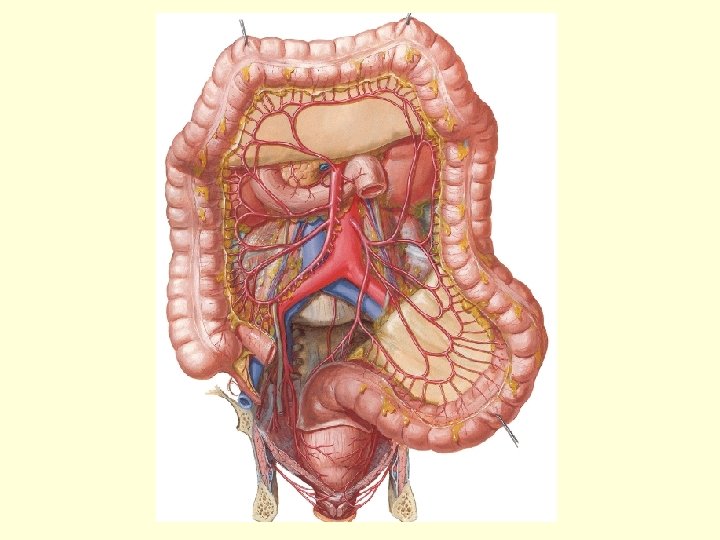
Extends from the distal end of the ileum to the anus. Consists of caecum, ascending colon, transverse colon, descending colon, sigmoid colon, rectum and anal canal. Absorbs fluids and salts from the gut contents.
HISTOLOG Y
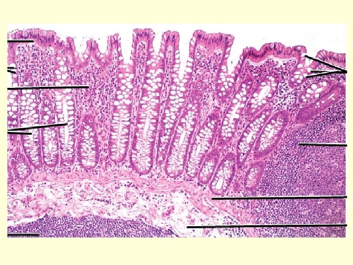
U can see numerous cresentric mucosal folds. Absence of villi
Large number of Goblet cells Enteroendocrine Columnarcells with brush Longitudinal – aggregatesborder. as longitudinal bands taenia coli. Three thick longitudinal bands of Stem cells. taenia coli. Tenia mesocolica, tenia libera & tenia omentalis Tubular intestinal glands / Crypts of Lieberkuhn
As the taenia are shorter in length the intestine here is sacculated called haustrations. appendices epiploicae pouch like processes filled with
Columnar cells with striated border absorb excess water and electrolytes. It also secrets Ig A antibodies. Goblet cell number increases caudally down the intestine. They secrete mucus that lubricates and facilitates the passage of the semisolid intestinal contents.
• Enteroendocrine cells/ enterochromaffin cells/argentaffin cells: – They produce amines like 5 -HT. – These cells are positive to chromaffin reaction (with potassium dichromate) and hence called enterochromaffin cells.
Microscopic view
HISTOLOGY OF APPENDIX
The longitudinal muscle coat is uniformly thick and has no taenia coli.
The lamina propria and submucosa is full of lymphoid nodules. The lymphoid tissue is absent at birth and reaches a maximum at ten years and then progressively decreases with age. Mucous membrane similar to large intestine: simple columnar with large number of Muscularis mucosa is not well developed and missing in goblet cells. some areas.
Lamina propria consists of much more lymphatic tissue extending onto submucosa and has few intestinal glands or Crypts of Lieberkuhn.
Summary of GIT: § Proximal to distal 1. Increase in lymphocytes. 2. Increase in Goblet cells.
IDENTIFICATION POINTS • Large intestine 1. Presence of numerous goblet cells in the epithelium 2. Outer longitidinal layer of smooth muscle if thickened to form taenia coli
IDENTIFICATION POINTS • Vermiform Appendix 1. lymphoid follicles in the lamina propria extending in to submucosa. 2. Few intestinal glands.