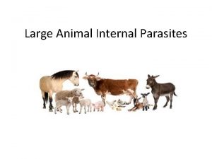
Large Animal Internal Parasites Routine fecal examination of


- Slides: 40

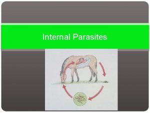
Large Animal Internal Parasites

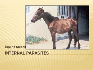
Routine fecal examination of horses will reveal parasitism by four types of parasites. Strongyloides westeri, Parascaris equorwn, Strongyles (large and small, sp. are not distinguishable by eggs), and Anoplocephala sp. (eggs of the two important sp. in the U. S. are indistinguishable). However, other sp. of parasites occur and will be reviewed. If a more thorough explanation of the “Salient Points” or “Features of the Life Cycle” are necessary please refer to your corresponding lecture notes from the second year or an appropriate text.

Strongyloides westeri eggs (40 -52 u x 32 -40 u) Thin walled, larvated egg, typical of Strongyloides

Strongyloides sp. Life Cycle: Parthenogenic parasitic females are found in the small intestine of foals, animals become immune and the adults are not generally found in the intestine after six months of age, infection occurs, via milk, skin penetration and ingestion, these parasites have a short prepatent period (5 -6 days) and may be found in very young horses.

Parascaris eguorum eggs (90 -100 u) thick-shelled , globular shape, eggs are not larvated in fresh feces, some eggs here contain the infective larvae.

Parascaris equorum emerging from a perforated portion of the small intestine, this may occur in heavy infections. Pathology due to migrating ascarid larvae is considered to occur.

Gastrophilus Almost all horses are infected with bots Gastrophilus intestinalis sometimes with G. nasalis and rarely in the U. S. , G. hemorrhoidalis. Bots cannot be diagnosed by fecal examination. Finding eggs on the horse's hair is an indication of internal infection. Unless treated most horses are infected.

Cutaneous habronemiasis: “Summer Sore” on the prepuce of horse, develop from larvae being deposited or escaping from vector flies feeding around a wound or abrasion. May also cause pulmonary granulomas if larvae invade the lungs. In the stomach Habronema sp. produce mucus exudate, and D. megastoma produce tumor- like lesions along the margoplecates

Oxyuris equi egg(42 -90 u) Eggs are elongated and slightly flattened, with a operculum like plug at one end, these are rarely seen in feces.

Oxyuris equi adult females: More commonly found in foals, females rupture during their migrations to the rectum and anus, eggs are released and attach to walls, fixtures, etc. , development to infective larvae within the eggs is fast, (3 -5 days), prepatent period -5 months

Loss of Hair: Due to pruritus caused by migrating female 0. equi, this irritation and resulting hair loss is the principle problem associated with the infection, and the common means of diagnosis

Anoplocephala sp. Eggs -(50 -60 u) Eggs possess pyriform apparatus, spp. identification is not possible on egg morphology.

A. magna in horse intestine: Largest (up to 12 in) of two common spp. in the U. S. found in posterior portion of small intestines may produce catarrhal or hemorrhagic enteritis

Anoplocephala perfoliata adults: Smaller than A. magna (1 -2 in. ) occur in larger numbers more , pathogenic of the two spp. , produces ulcers, and may induce occlusion of ileo-cecal valve, or intussusception in this area.

. Strongyle egg -{70 -85 u x 40 -47 u) Spp. cannot be identified by egg morphology , egg here can be compared to Parascaris equorwn and Strongyloides westeri.

The large strongyles are pathogenic because of their larval migrations, adults attach to the mucosa of the large intestine and cecum and suck blood. Some Triodontophorus spp. produce mucosal ulcers

Strongylus vulgaris caused aneurism in mesenteric artery due to larval migrations

Strongylus vulgaris related intestinal infarction Due to emboli produced by S. vulgaris larval migrations in mesenteric, arteries. Other large strongyle larvae (S. edentatus & S. equinus) produce lesions during their migrations in liver, diaphram and other viscera. .

Comparison of large and small strongyles

Symptoms of Parasitic Infestation in Cattle • Parasites to the stomach and intestines cause: • Anemia • Scouring • Depression • Death

Description of Parasitic Infestation • Roundworms: • Found in the digestive system • Most important parasites from an economic standpoint • Mostly in stomach and intestines

Stomach Worm • Several species of stomach worm • Twisted stomach worms and brown stomach worms are the most important. • Found in all classes of livestock • Most common in cattle, sheep and horses. • Penetrates the stomach lining • Causes severe damage

Calf Scours • • Diarrhea Two main forms Hypersecretion Malabsorption Result of Diarrhea Dehydration Dryed out mouth

Strongyles • • Several species Attack all species Greater affect on young Blood sucking parasites that attach to the lining of the intestines

Ascarids • Parasites of cattle, sheep, horses and hogs • Affects young mostly • The larvae burrow into the wall of the intestines and migrate through the liver, heart, and finally the lungs

Internal Parasites of Goats and Sheep

Strongyloides • Strongyloides stercoralis is an unusual "parasite" in that it has both free-living and parasitic life cycles. In the parasitic life cycle, female worms are found in the superficial tissues of the human small intestine; there apparently no parasitic males. The female worms produce larvae parthenogenically (without fertilization), and the larvae are passed in the host's feces. The presence of nematode larvae in a fecal sample is characteristic of strongylodiasis. Once passed in the feces, some of the larvae develop into "freeliving" larvae, while others develop into "parasitic" larvae. The "free-living" larvae will complete their development in the soil and mature into free-living males and females. The free-living males and females mate, produce more larvae, and (as above) some of these larvae will develop into "free-living" larvae, while other will develop into "parasitic larvae. "

Tapeworms • • • Broad tapeworm - Moniezia expansa Fringed tapeworm - Thysanosoma actinioides Hydatid cysts - Echinococcus granulosus Cysticercosis - Taenia ovis Taenia hydatigena Gid - Taenia multiceps

Broad tapeworm - Moniezia expansa The life cycle of Moniezia expansa involves sheep as the definitive host and soil mites as the intermediate host. The tapeworm's eggs are passed in the sheep's feces, and mites are infected when they eat the eggs; the metacestode stage in the mite is called a cysticercoid. Sheep are infected when then ingest infected mites. This species of tapeworm is unusual in that each proglottid contains two sets of female reproductive organs

Fringed tapeworm - Thysanosoma actinioides • Definitive hosts: ruminants • Site of infection: small intestine Typical size: up to 30 cm long • Distribution: cosmopolitan • Intermediate hosts: cysticercoids develop in psocid lice; these lice are ingested along with vegetation

Hydatid cysts - Echinococcus granulosus • The life cycle of Echinococcus granulosus includes dogs (and other canines) as the definitive host, and a variety of species of warm blooded vertebrates (sheep, cattle, goats, and humans) as the intermediate host. The adult worms are very small, usually consisting of only three proglottids (total length = 3 -6 mm), and they live in the dog's small intestine. Eggs are liberated in the host's feces, and when these eggs are ingested by the intermediate host they hatch in the host's small intestine. The larvae in the eggs penetrate the gut wall and enter the circulatory system. The larvae can be distributed throughout the intermediate host's body (although most end up in the liver) and grow into a stage called a hydatid cyst.

Taenia hydatigena • The life cycle of the Taenia tapeworm starts in the host’s intestine, the host being a dog or cat. The worm can be unbelievably long (up to 5 yards for Taenia hydatigena) and is made of segments. Each segment contains an independent set of organs with new segments being created at the neck and older segments dropping off the tail. As segments mature, the reproductive tract of the segment becomes more and more prominent until it consists of a bag of tapeworm eggs. These segments, called proglottids, are passed with the feces into the world where an unsuspecting intermediate host (mouse, rabbit, deer, goat etc. ) swallows one while feeding.

Gid - Taenia multiceps • The life cycle of this parasite involves warm blood vertebrates as both the intermediate and definitive hosts. • Infections with cenuri can cause pathology in the intermediate host. Human infections (acquired by accidental ingestion of eggs) have been reported. The cenuri of T. multiceps (sometimes called Cenurus cerebralis) can infect the brains of sheep/goats, causing a disease referred to as "gid" or "staggers. " These terms refer to the behavior of sheep/goats infected with this parasite.

Flukes - Trematodes • Common liver fluke - Fasciola hepatica

Flukes - Trematodes • The common name of this parasite, the "sheep liver fluke, " is somewhat misleading since this parasite is found in animals other than sheep (including cattle and humans), and the parasite resides in the bile ducts inside the liver rather than the liver itself. This species is a common parasite of sheep and cattle and, therefore, relatively easy to obtain. Thus, in introductory biology or zoology courses, it is often used as "THE" example of a digenetic trematode. This species has been studied extensively by parasitologists, and probably more is known about this species of digenetic trematode than any other. The adult parasites reside in the intrahepatic bile ducts, produce eggs, and the eggs are passed in the host's feces. After passing through the first intermediate host (a snail), cercariae encyst on vegetation. The definitive host is infected when it eats the contaminated vegetation. The metacercaria excysts in the definitive host's small intestine, and the immature worm penetrates the small intestine and migrates through the abdominal cavity to the host's liver. The juvenile worm penetrates and migrates through the host's liver and finally ends up in the bile ducts (view a diagram of the life-cycle). The migration of the worms through the host's liver, and the presence of the worms in the bile ducts, are responsible for the pathology associated with fascioliasis.

Flukes - Trematodes

Protozoa - Coccidia • Coccidia (coccidiosis) - Eimeria

Protozoa - Coccidia • The diseases caused by these parasites are referred to collectively as coccidiosis, and they vary tremendously in virulence. Some species cause diseases that result in mild symptoms that might go unnoticed (i. e. , mild diarrhea) and eventually disappear, while other species cause highly virulent infections that are rapidly fatal.

Protozoa - Coccidia A severe coccidiosis infection in a small animal

QUESTIONS? 40
 Are platyhelminthes acoelomates
 Multicellular animal parasites
 Stool microscopic examination
 Gse test
 Normal bilirubin
 Apa itu fecal oral
 Is used for
 Holliday segar
 Fecal loop
 Gasto fecal
 Marasmo y kwashiorkor
 Fecal fat qualitative
 Fecal
Fecal Fecal matter
 Fecal matter
 Fecal coliform
 Fecal impaction
 Hemosure ifob test directions
 Examples of symbiosis
 Kato katz technique
 Parasites classification
 Parasite
 Bacteria virus fungi and parasites
 Hookworm vs threadworm
 Arthropod
 Parasites alimentaires
 Parasites of medical importance
 What do parasites eat
 Herbivores carnivores omnivores scavengers decomposers
 Parasites
 Parasitic ova
 Trichinella
 Autoinfection parasites examples
 Protozoa are polyphyletic
 Sarcomastigophora are unicellular immotile parasites
 Parasites of livestock - vocabulary
 Bilateral symmetry worm
 What is ookinite
 Entamoeba histolytica infective stage
 Mastigophora movement
 Enslaver parasites
