LACRIMAL DRAINAGE SYSTEM ANATOMY The nasolacrimal drainage system


 1. 2. Deficiency of lacrimal secretion")


- Slides: 23
LACRIMAL DRAINAGE SYSTEM
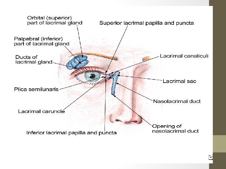
ANATOMY • The nasolacrimal drainage system serves as a conduit for tear flow from the external eye to the nasal cavity. • Tears drain into the upper and lower puncta upper and lower canaliculi common canaliculus lacrimal sac lower canaliculus nose • Tear drainage is active process • Each blink will pumps tears through the system
ABNORMALITIES OF LACRIMAL SYSTEM Abnormalities are found is • Tear composition • Drainage of tear
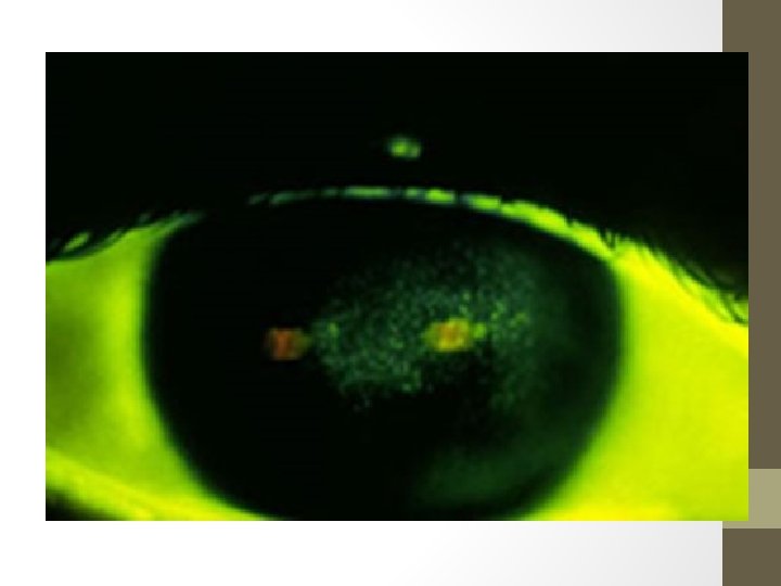
1. ABNORMALITIES IN COMPOSITION AQUEOUS INSUFFICIENCY (DRY EYES) 1. 2. Deficiency of lacrimal secretion resulting in Keratoconjunctivitis sicca (KCS). If associated with dry mouth or mucous membrane = Sjogren’s Syndrome Symptoms • Grittiness, burning, and photophobia • Lids heaviness and ocular fatigue. May worse in evening • Visual acuity may be reduced Signs • Small dots of fluorescence over exposed corneal & conjunctival surface. • Tags of abnormal mucus may attach to cornea causing pain. (filamentary keratitis) Treatment • Supplementation of tears (artificial tear) • Humid environment around the eyes using shielded spectacles • Occlude the puncta with plug or surgery to conserve the tears
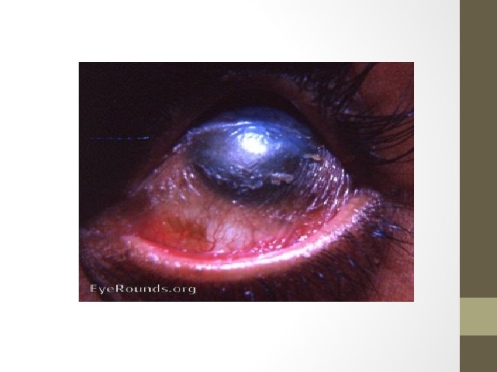
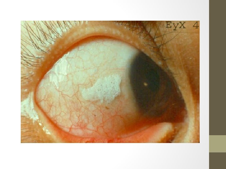
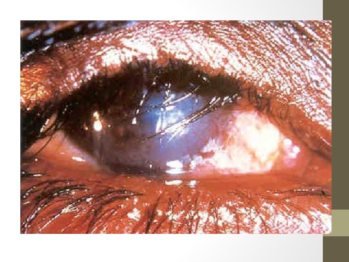
INADEQUATE MUCUS PRODUCTION STEVENS-JOHNSON’S SYNDROME • Acute episodes inflammation causing macular target lesion on skin and discharging lesion on the eye, mouth and vulva. • Causes conjunctival shrinkage with adhesion forming between the globe, aqueous and mucin deficiency. Similar symptoms to those seen in aqueous deficiency. XEROPHTHALMIA • • • Vit A Deficiency, causing childhood blindness on a worldwide scale. Goblets cells are lost from the conjunctiva and ocular surface become keratinized. Aqueous deficiency causing dry eyes can lead to corneal ulceration and lastly blindness Bitot’s spot : clumps of keratin debris build up inside conjunctiva preceedes ulceration Prevented by early treatment with vitamin A OTHER CAUSES • Chemical burns of the eyes by alkalis. • Trachoma causes by Chlamydial infection roughening the inner surface of the eyelid.
MALPOSITION OF EYELID MARGIN • Ectropion or insufficient closure of eyes (7 th nerve palsy or proptosis in dysthyroid eyes) preocular tear film cannot form adequeately dry eyes Treatment • Correction of lid deformity by LATERAL TARSHORRHAPHY • If temporary, use artificial tears and lubricant • Or induced temporary ptosis by local injection of botulinum toxin
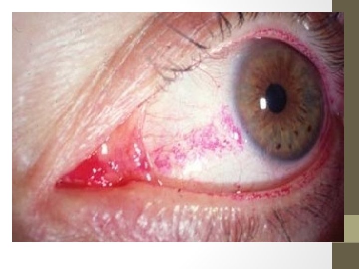
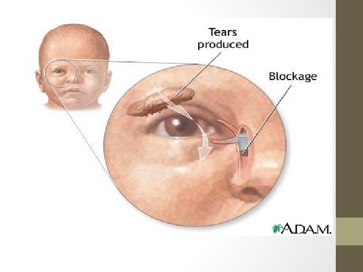
2. DISORDERS OF TEAR DRAINAGE • Tear production exceed the capacity of drainage system. It may caused by : 1. 2. • Irritation of ocular surface, e. g. by foreign body Occlusion of any part of drainage system. OBSTRUCTION OF TEAR DRAINAGE – Nasolacrimal duct is common site for tear drainage system to get blocked. – Usually block by infection or direct trauma. – In peadiatric age, congenital obstruction of the duct is common at the distal end watering eye • The sac may become infected accumulate as mucocele or causing dacrocystitis. • Conjunctiva is not inflamed. • Most obstruction resolved spontaneously in first year of life. • If epiphora persist, patency is achieved by passing probe via the punctum to open the obstruction.
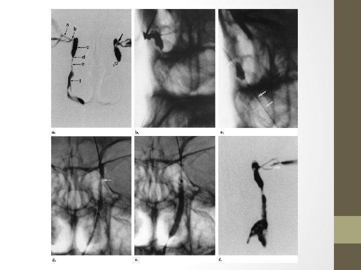
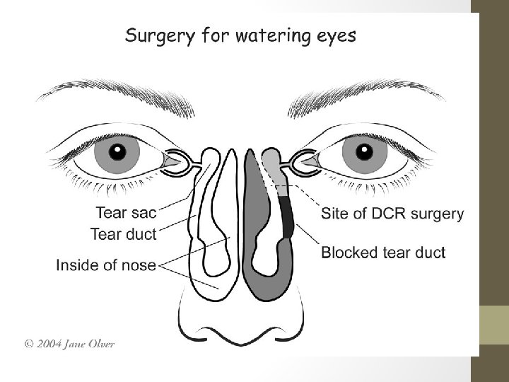
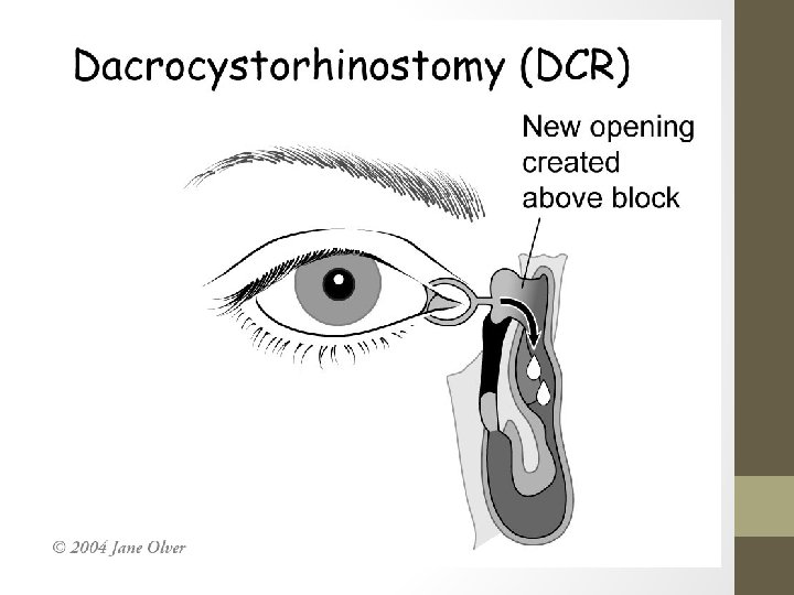
SYMPTOMS • Watering eyes associated with stickiness • Eye is white. • Symptoms may get worse during windy or cold weather SIGNS • Stenosed punctum may apparent on slit lamp examination • Obstruction may diagnosed by syringing the nasolacrimal system with saline the system is patent if the patient taste the saline as it reached the pharynx. • Injecting radio-opaque dye to confirmed the exact location into the nasolacrimal system. Then, X-rays is used to follow the passage of the dye until we find the blockage. TREATMENT • Treat the underlying cause such Blepharitis etc. • SURGERY : Dacryocystorrhinostomy (DCR), connecting the mucosal surface of lacrimal sac to the nasal mucosa by removing the intervening bone.
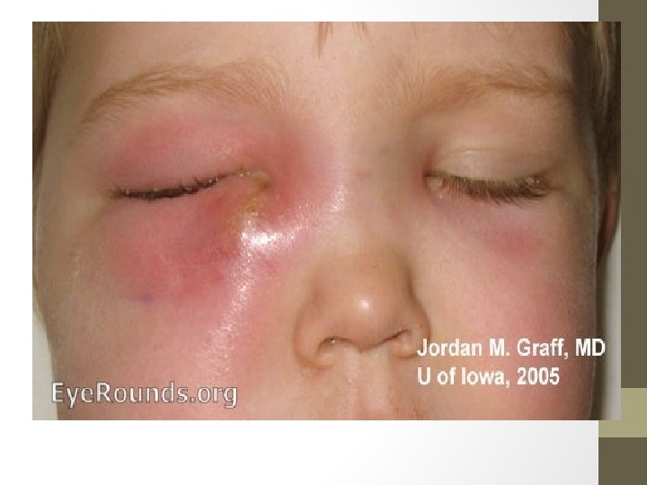
3. INFECTION OF THE NASOLACRIMAL SYSTEM • DARCOCYSTITIS • Infection of the sac cause by obstruction of the drainage system. • Organism involved usually Staphylococcus. Symptoms • Painful swelling on medial side. • Enlarged and infected sac. • Could resulting in formation of mucocele Treatment • Systemic antibiotic • DCR may be necessary to prevent recurrence.