LABORATORY EXERCISES FOR CARTILAGE Hyaline Cartilage DEMO SLIDE

LABORATORY EXERCISES FOR CARTILAGE Hyaline Cartilage
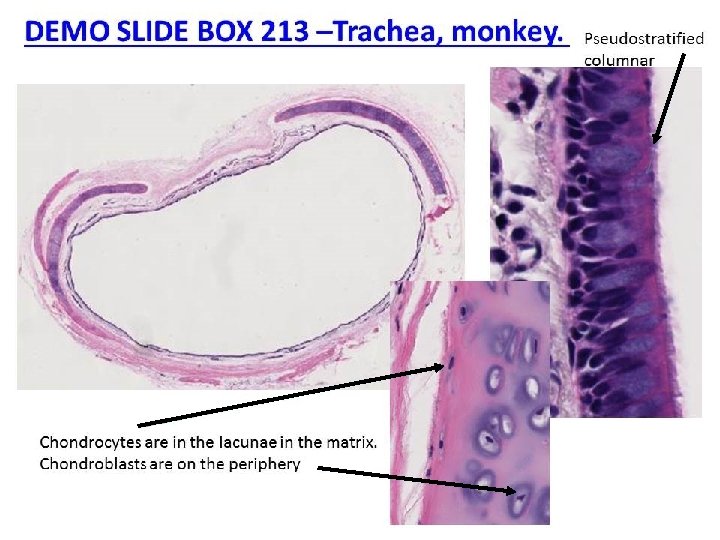
. Chondroblasts are located just")
DEMO SLIDE BOX 181– Comb and nasal cavity, chicken (954). Chondroblasts are located just below the perichondrium and appear as flattened cells. chondrocytes within lacunae. You may be able to see only the nuclei of the cells, as the cytoplasm may have undergone considerable shrinkage during preparation basophilia of the matrix, especially the matrix immediately surrounding the chondrocytes (this is called territorial matrix
. Fibers and Ground substance")
DEMO SLIDE BOX 181– Comb and nasal cavity, chicken (954). Fibers and Ground substance both have the same refractive index. This means you can’t visually distinguish fibers from ground substance.

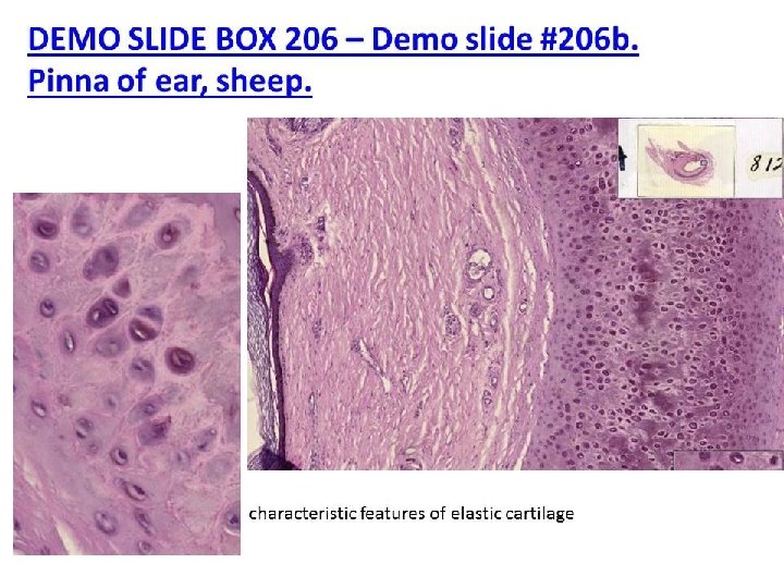
Slide #99 -Pinna of ear, Cow Looks like hyaline See elastic fibers

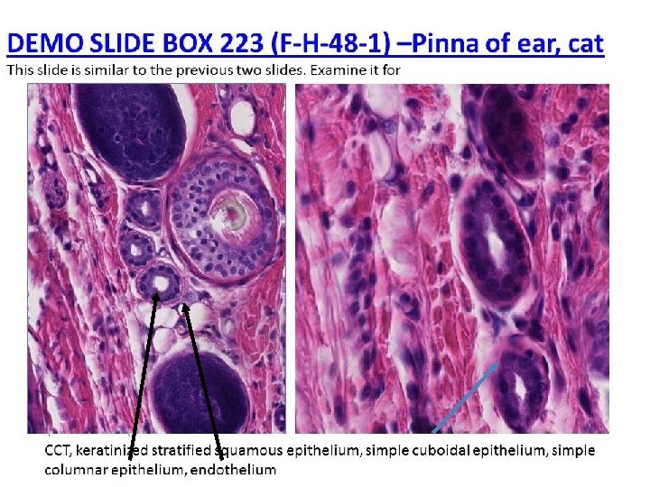
Slide #99 -Pinna of ear, Cow Keratinized stratified squamous epithelium CCT Simple cuboidal epithlium Simple columnar epithelium

Elastic cartilage in sweat gland
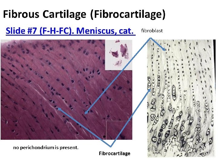
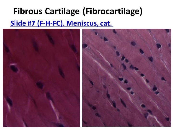
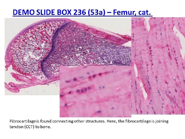
 – Femur, cat. Bone Chondrocytes in fibrocartilage Fibroblasts")
DEMO SLIDE BOX 236 (53 a) – Femur, cat. Bone Chondrocytes in fibrocartilage Fibroblasts in tendon Fibrocartilage is found connecting other structures. Here, the fibrocartilage is joining tendon to bone.

CELLS OF CT FIBROBLASTS MESENCHYMAL CELLS and RBC ADIPOSE CELLS MACROPHAGE PLASMA CELLS MAST CELLS and WBC CHONDROBLASTS CHONDROCYTES OSTEOBLASTS OSTEOCYTES OSTEOCLASTS

TYPES OF CARTILAGE CHONDROCYTES HYALINE CARTILAGE ELASTIC CARTILAGE Chondrocytes Lacunae = “space” FIBROCARTILAGE
- Slides: 18