Laboratory Equipment Microscope Indispensable instrument Used to view

Laboratory Equipment

Microscope � Indispensable instrument � Used to view objects too small to be seen with the naked eye. � Used: ◦ ◦ ◦ Evaluated stained blood smears Urine sediment Vaginal secretions Smears made from body fluids Microbiological cultures

Microscope � Have three components ◦ Magnification system ◦ Illumination system ◦ Framework: includes all components responsible for positioning the slide and focusing. � Magnification ◦ Ocular lens ◦ Objective lens System

Microscope
 Monocular one eyepiece Binocular two eyepieces")
Microscope � May be monocular or binocular (eyepiece) Monocular one eyepiece Binocular two eyepieces Located at top of the microscope Usual magnification of oculars is 10 x Compound microscopes have objective lenses that increase the magnification of the specimen ◦ Objective lenses are attached to the revolving nosepiece ◦ Most microscopes have 4 different objective each with a different magnifying power. ◦ ◦ ◦
")
Microscope � Objective ◦ ◦ lenses Shortest objective has the lowest power (4 x) Called a scanning lens. Next sized lens is 10 x High dry 40 -45 x Longest objective ( oil immersion) (100 x) �Uses special oil Immersion Oil The oil prevents refraction of the light and improves the resolution (clarity) of the magnified image. �Oil immersion is used to view cells and extremely small materials such as bacteria and platelets and to examine stained specimens. ◦ To determine magnification of the specimen multiply the magnification of the objective lens by 10 (magnification of the ocular).

Microscope � Arm of the microscope connects the objectives and the oculars to the base which supports the microscope and contains its light souce. � Stage of the microscope holds the slide to be viewed. � The light source, condenser, and the iris diaphragm make up the illumination system. � Condenser directs light up through the stage � The iris diaphragm regulates the amount of light passing through the specimen.

Microscope � Above the base are the focusing knobs ◦ Coarse adjustment is used only with scanning and low-power lenses ◦ Fine adjustment is used with high power and oil immersion lenses. � Maintenance of microscope ◦ Dependent on the amount of daily use. ◦ Dirt is the primary enemy of the microscope ◦ Oil, makeup, dust and eye secretions all can obstruct vision through the lens and may also transmit infective organisms. ◦ Always store in a plastic dust cover when not in use. ◦ Lenses should be cleaned before and after each use with lens paper and cleaner. Solvent cleaner such as xylene is not recommended.

Microscope � Should be placed in a permanent location � Moving a microscope: ◦ Carry securely with one hand supporting the base and the other holding the arm. ◦ When stored it should be left covered and with the low-power objective in the lowest position. Stage should be centered. � Using a microscope involves focusing and illumination ◦ Image is focused by moving the objective closer to the specimen ◦ illumination is accomplished by raising or lowering the condenser and by moving the specimen closer to or farther from the objective.

Microscope � Focused through movement of the objective or stage controlled by knobs on both sides of the microscope. � Proper focusing begins with the objective at lowest power. � Course adjustment moves the objective very quickly. � Course knob is used first to bring the specimen into approximate focus. � Fine adjustment brings the specimen into precise focus. Fine focus moves the objective more slowly to allow the viewer to zero in on the specimen with greater accuracy.

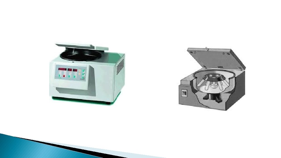
Centrifuge � Used when solids must be separated from liquids � Used increased gravitational force achieved by rapid spinning. � Separates: ◦ Blood cells from serum ◦ Cells and crystals from urine ◦ Used in many areas of clinical laboratory

Centrifuge � Types ◦ ◦ Bench-top Floor models Some may be refrigerated. Some may have rotors or heads that are interchangeable. � Centrifuges ◦ Fixed angle rotors: specimen cups are held in a rigid position at a fixed angle ◦ Horizontal head with swinging buckets that swing out horizontally during centrifugation. ◦ A third type that is used for centrifuging capillary tubes for microhematocrit determination. ◦ Centrifuges also may be equipped with timers to automatically stop at set time.

Centrifuge � Using a centrifuge usually are given in terms of revolutions per minute RPM � Spinning generates centrifugal force � General laboratory centrifuges operate at up to 6, 000 rpms generating a relative centrifugal force of up to 7, 300 times the force of gravity. � Conventional horizontal centrifuges attain speeds of up to 3, 000 rpm angle-head centrifuges can attain higher speeds (up to 7, 000 rpm)

Centrifuge � Mechanical hazard ◦ Centrifuges can be dangerous if not used correctly. ◦ Ensure that the centrifuge is balanced so that tubes of equal size and containing equal volume are directly across from on another in the rotor holders. ◦ Always an even number of tubes in the centrifuge. If a second specimen of the same volume in the same-size tube is not available for balance, a tube of water may be used to balance the load. ◦ Tube should be capped to prevent emission of aerosols. ◦ Rubber cups should be placed in the bottom of the carrier cups to prevent breakage of glass tubes.

Centrifuge � Mechanical hazards ◦ Never be opened while in operation ◦ Never slow a centrifuge with hands ◦ Use brake if equipped only in an emergency. Most common emergency is broken glass tube. Wait until centrifuges comes to a complete stop and follow the manufacturer’s instructions for disinfecting the unit. ◦ Follow Standard Precautions to prevent injury and disease transmission. � Maintenance ◦ Check, clean and lubricate regularly to ensure properation. ◦ Certified technician must use a photoelectric device or a strobe tachometer to ensure the centrifuges speed to comply with QA guidelines.

Incubator � Cabinets that maintain constant temperatures � Generally used in microbiology and sometimes in chemistry � Hold constant temperature of 95°F to 98. 6°F ( 35°-37°C). Other temperatures may also be used. � Some are enriched with Carbon Dioxide gas (CO 2) to enhance the growth of pathogenic bacteria. � A pressurized tank of CO 2 is attached to the cabinet and the concentration is maintained at 10%. � Incubators may have warning alarms that sound if the temperature exceeds or falls below a specified range

Incubator

Incubator � Maintenance ◦ Temperature should be checked daily ◦ Cabinets should be cleaned regularly with disinfectant approved by the manufacturer.

Autoclave

Autoclave � Strict QA methods must be followed when an autoclave is used. � A certified technician should regularly examine the autoclave and biologic and chemical indicators should be checked daily. � Biologic indicators include spore preparations that are wrapped in the autoclave load. � At the end of the sterilization period, they are incubated and checked for germination. � If the spores fail to germinate the autoclave reached the appropriate temperature.

The End
- Slides: 22