LAB 6 DIALYSIS KAUFaculty of Science Biochemistry department
: DIALYSIS KAU-Faculty of Science- Biochemistry department Analytical biochemistry lab (Bioc 343) 2012")
LAB (6): DIALYSIS KAU-Faculty of Science- Biochemistry department Analytical biochemistry lab (Bioc 343) 2012 T. A Nouf Alshareef nf. shareef@hotmail. com

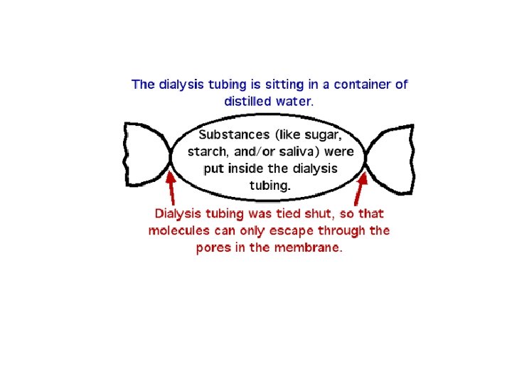
Introduction: • Dialysis is non chromatographic separation method used to separates molecules according to size through the use of semipermeable membranes. • dialysis process occurs at the membrane surface, depend on conc. differences between two solutions on both side of the membrane. • the porous membrane selectively allows smaller solutes to pass while retaining larger species

It is a simple process in which: High conc. solution “small solutes “ diffuse semi-permeable membranes until equilibrium Low conc. solution

Factors affect on dialysis: • Temperature, viscosity, and pressure gradient across the membrane. • Solvent, pore size, and the nature of the membrane. (rate of dialysis is greatest in distilled water)

Advantages of lab dialysis: • • • Very gentle conditions Easy operation Wide range of samples volume Many membrane types Inexpensive materials Disposable membranes & devices

Dialysis membrane materials: • Synthetic or natural membranes are used for diffusion or osmosis. • It allows the passage of small molecules but not larger ones (Filtration applications). • Membrane materials: regenerated cellulose, cellulose acetate, polysulfone, polycarbonate, polyethylene, polyolefin, polypropylene, and polyvinylidene fluoride. • Membrane size: wide range of pore size (MWCO: M. wt. cut-off). The main feature of this membrane is that it is porous.
: • is derived from cotton: • has a symmetric pore structure")
Regenerated cellulose (Cellu·Sep®): • is derived from cotton: • has a symmetric pore structure allows small molecules to migrate in either direction. Cellophane: • is most commonly use dialysis material. • is thin, transparent sheet made of regenerated cellulose. • contains traces of sulpher compounds, metal ions and some enzymes.

• Dialysis is best carried out with freshly prepared tubing (wet is very susceptible to attack by micro-organisms). • For storing: the tubing is best kept with a trace of benzoic acid in the solution

Dialysis membrane preparation: • Boil the bag for 30 min in alkaline EDTA (activation of membrane and to make the dialysis sac soft) • After boiling, the tubing is washed with distilled water and soaked in water.

Lab practice: Aim: • Separation of maltose from mixture solution using dialysis process. • Our sample: (solution of starch, salivary amylase and maltose) • dialysis bag: cellophane sealed with string by knotting. • Solvent: distilled water.
 • Starch consists of: Starch Amylopectin: (M.")
Principle: Amylose: (M. wt 50, 000 g) • Starch consists of: Starch Amylopectin: (M. wt 1000, 000 g) Salivary amylase neither of them will pass through dialysis membrane Maltose (M. wt 360 g) (Able to diffuses out into the dialyzate) - Maltose (smaller than membrane pores) - Starch (Larger molecule) moves freely across out of the membrane into the solvent is retained inside the dialysis bag

Dialysis process: • the volume of the solvent outside the bag is greater than inside, over time, most of maltose will leave the bag (Osmosis). • The passage of maltose into or out of the bag is in the direction of decreasing concentration, therefore displaying diffusion. • Equilibrium is an achieved when the concentration of maltose equal inside and outside of the bag.

Chemicals & other material: • Salivary amylase (1 ml saliva diluted to 5 ml with distilled water) • Iodine solution (5 mmol/L in 30 g/L Potassium iodide) • Soluble starch (20 g/L) • Sodium chloride solution (1 g/L) buffered with 0. 02 mol/L Sodium phosphate p. H 6. 8). • Fehling’s solution.

Procedure:

1 - Label 14 small test tubes as the following: add 10 drops of iodine solution 1 S 2 S 3 S 4 S 5 S 6 S (Test for starch). Starch + iodine (Blue color). 2 - Seal one end of the dialysis bag (the bottom) with a string. 7 S 1 ml of Fehling’s 1 M 2 M 3 M 4 M 5 M 6 M 7 M (Test for maltose). Maltose + Fehling’s Heating 5 min Red ppt 3 - Fill the sac with the reaction mixture: ( 2. 5 ml starch, 0. 5 ml buffer, 0. 5 ml amylase) 4 - Quickly (at 0 min) pipette 1. 5 ml of reaction mixture (0. 5 ml to tube 1 S) and (1 ml to tube 1 M)

5 - Immerse the dialysis bag in beaker contain 100 ml dis. H 2 O. 6 - Place the beaker on a stir plate (stir 90 min) (Stirring help in entry of water inside the sac and small molecules to getting out of the sac) 100 ml dis. H 2 O 7 - every 15 min, pipette 1. 5 ml from the solvent (outside) and examine each fraction for the presence of starch or maltose. Stirrer 8 - At the end of experiment (90 min): cut the dialysis sac carefully pipette 1. 5 ml from solution inside, examine them for starch and maltose.
")
Test for starch: Change of the color of iodine during separation 75 min (inside) yellow color 7 S 15 to 75 min (outside) yellow color 2 S- 6 S 0 min (inside) blue color 1 S
 red")
Test for maltose: Formation of red precipitate during the separation 75 min (inside) red ppt 7 M 15 to 75 min (outside) Red ppt 2 M to 6 M 0 min (inside) red ppt 1 M
, and observe result for")
Result sheet • Make a table of time intervals (min), and observe result for each fraction with iodine and Fehling’s tests. • Note observation in table for Fehling’s test as follows: (+) for a few ppt, (++) for more ppt, (+++) for much more ppt and so on.
 Inside tubing Fehling’s I 2 test Comment Outside tubing")
Tabel : 1 Time (min) Inside tubing Fehling’s I 2 test Comment Outside tubing I 2 test Fehling’s test Comment 0 Tabel : 2 Time (min) 15 30 45 60 75 90 Tabel : 3 Time (min) 90 Inside tubing I 2 test Fehling’s Comment test Outside tubing I 2 test Fehling’s test Comment
- Slides: 21