Lab 5 The Respiratory System 1 Functions of

Lab # 5 The Respiratory System 1

Functions of Respiratory System 1 - O 2 and CO 2 exchange between blood and air 2 - Speech and other vocalizations (laughing, crying) 3 - It provides the sense of smell 4 - It helps to control the p. H of body fluids by eliminating CO 2 5 - It helps to regulate blood pressure by synthesis of a vasoconstrictor called angiotensin II 6 - Breathing creates pressure gradients between thorax and abdomen that promote the flow of lymph and venous blood 7 - Breath-holding helps expel abdominal contents during urination, defecation, and childbirth

Principal Organs of the Respiratory System Nose Pharynx Larynx Trachea Lungs Bronchi

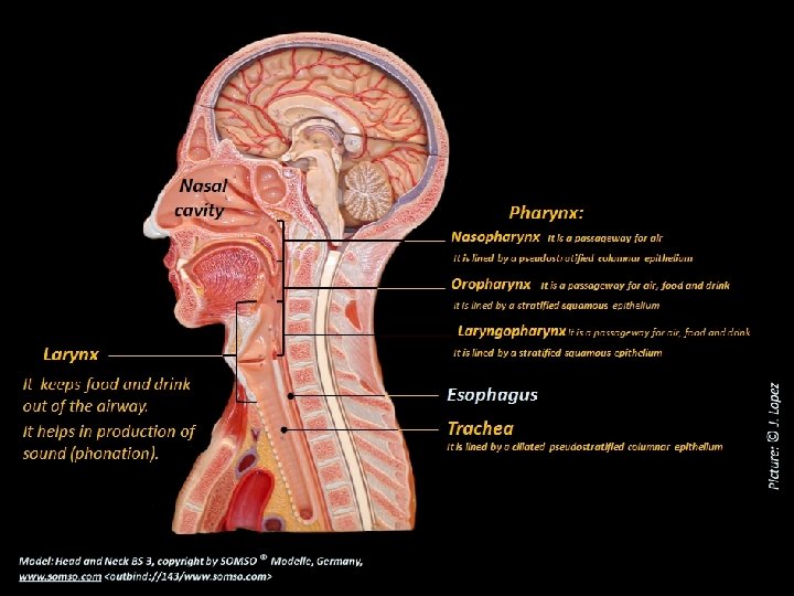
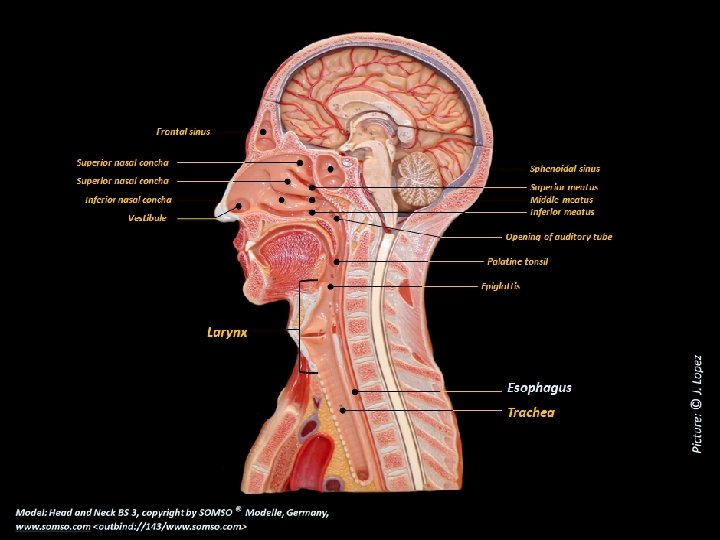
The Nasal Cavity Meatuses: Superior Nasal conchae: Superior Middle Inferior Posterior nasal aperture Functions of the nose 1 - It warms, cleanses, and humidifies inhaled air 2 - It detects odors in the airstream 3 - It serves as a resonating chamber that amplifies the voice Vestibule The respiratory epithelium lines the rest of nasal cavity except vestibule. It is a ciliated pseudostratified columnar epithelium with goblet cells
 Auditory")
The Pharynx Nasopharynx (posterior to nasal apertures Pharyngeal tonsil and above soft palate) Auditory tube Oropharynx (space between soft palate Palatine tonsil and epiglottis) Laryngopharynx (from the epiglottis to the cricoid cartilage)

The Pharynx Posterior nasal aperture Pharynx: Nasopharynx It is a passageway for air It is lined by a pseudostratified columnar epithelium Oropharynx Larynx Trachea It is lined by a stratified squamous epithelium It is a passageway for air, food and drink Laryngopharynx It is a passageway for air, food and drink It is lined by a stratified squamous epithelium Esophagus
")
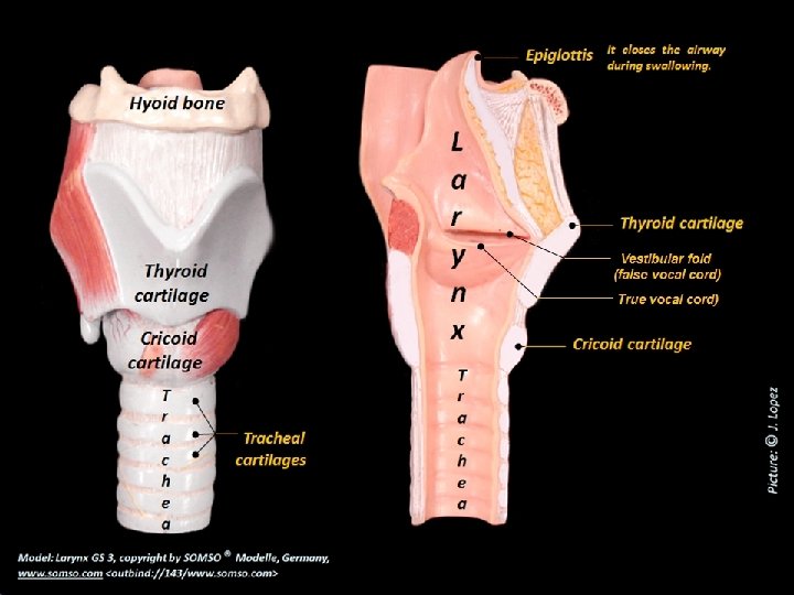
The Larynx It is a cartilaginous chamber about 4 cm (1. 5 in. ) 1 - To keep food and drink out of the airway Functions: 2 - Production of sound (phonation) Epiglottis It closes the airway during swallowing Hyoid bone Epiglottic cartilage Hyoid bone Thyroid cartilage Arytenoid cartilage Corniculate cartilage Vestibular fold Vocal cord Cricoid cartilage Arytenoid cartilage Cricoid cartilage Trachea (a) Anterior Tracheal cartilage (b) Posterior (c) Median

The Larynx Vestibular fold They play no role in speech but close the larynx during swallowing Vocal cord (from the thyroid cartilage to the arytenoid cartilage) They produce sound when air passes between them Median
 is a rigid tube about 12 cm (4. 5")
The Trachea The trachea (windpipe) is a rigid tube about 12 cm (4. 5 in. ) long and 2. 5 cm (1 in. ) in diameter. It is found anterior to the esophagus and it is supported by 16 to 20 Cshaped rings of hyaline cartilage, which reinforces the trachea and keeps it from collapsing when you inhale Ciliated pseudostratified columnar epithelium with goblets cells Trachea

Mucociliary escalator It is a mechanism that moves debris-laden mucus to the pharynx to be swallowed Ciliated pseudostratified columnar epithelium with goblets cells Goblet cell Ciliated cell Mucus Mucous gland

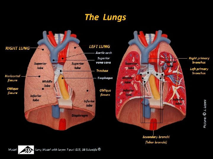
The Lungs They are conical organs with a broad, concave base, resting on the diaphragm, and a blunt peak called the apex projecting slightly above the clavicle Apex of lung Superior lobe Costal surface Superior lobe Horizontal fissure Middle lobe Mediastinal surface Oblique fissure Inferior lobe Base of lung Diaphragmatic surface

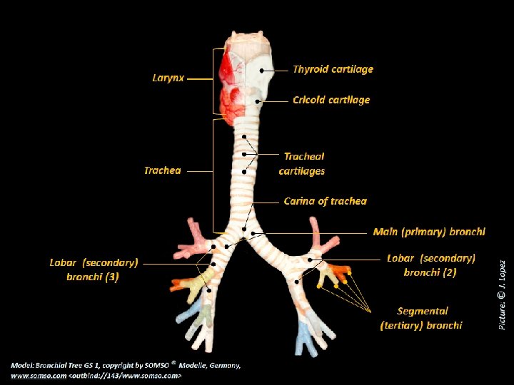
Thyroid cartilage Larynx Cricoid cartilage Bronchial Tree All bronchi are lined with ciliated pseudostratified columnar epithelium The lamina propria has an abundance of mucous glands and lymphocyte nodules (bronchus-associated lymphoid tissue, BALT) positioned to intercept inhaled pathogens Trachea Carina Main bronchi (primary) Superior lobar bronchus (secondary) Middle lobar bronchus Segmental bronchi (10 on right) Inferior lobar bronchus (secondary) Inferior lobar bronchus Segmental bronchi (tertiary) (8 on left) Bronchopulmonary segment: It is a functionally independent unit of the lung tissue
 Conducting Division of Respiratory System It consists of those passages that")
Main bronchus (lung) Conducting Division of Respiratory System It consists of those passages that serve only for airflow: 1 - Nostrils 2 - Nasal cavity 3 - Pharynx 4 - Larynx 5 - Trachea 6 - Main (primary) bronchi (lungs) 7 - Lobar (secondary) bronchi (lobes) 8 - Segmental (tertiary) bronchi (segments) 9 - Bronchioles (lobules) 10 - Terminal bronchioles (the final branches) Lobar bronchus (lobe) Bronchioles and terminal bronchioles lack of supportive cartilages) Segmental bronchus (segment) Bronchiole (pulmonary lobule) Respiratory Division of Respiratory System It consists of those structures that participate in gas exchange Terminal 1 - Respiratory bronchioles 2 - Alveolar duct (final branches of conducting division) 3 - Atrium 4 - Alveoli

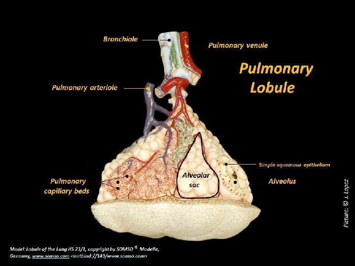
Pulmonary arteriole Pulmonary venule Openings of alveolar ducts Bronchiole Alveolar sac Terminal bronchioles Respiratory bronchioles Every respiratory bronchiole divides into 2 to 10 alveolar ducts, which end in the alveolar sac Alveoli

The Respiratory Membrane Squamous alveolar cell O 2 O 2 Respiratory membrane O 2 CO 2 CO 2 Shared basement membrane Capillary endothelial cell

Epithelium Type Changes in the Respiratory System Nasal cavity Ciliated pseudostratified columnar epithelium Nasopharynx Ciliated pseudostratified columnar epithelium Oropharynx Stratified squamous epithelium Laringopharynx Stratified squamous epithelium Larynx (superior part) Stratified squamous epithelium Larynx (inferior part) Ciliated pseudostratified columnar epithelium Trachea Ciliated pseudostratified columnar epithelium Bronchioles Ciliated simple columnar epithelium Terminal bronchioles Simple cuboidal epithelium Alveoli Simple squamous epithelium (with 5% of round or cuboidal cells (type II alveolar cells)

The Respiratory Muscles
- Slides: 23