LAB 5 GEL FILTRATION CHROMATOGRAPHY T A Nouf
: GEL FILTRATION CHROMATOGRAPHY T. A Nouf Alshareef KAU-Faculty of Science- Biochemistry department")
LAB (5): GEL FILTRATION CHROMATOGRAPHY T. A Nouf Alshareef KAU-Faculty of Science- Biochemistry department Analytical biochemistry lab (Bioc 343) 2012 nf. shareef@hotmil. com False colour scanning electron micrograph of Sephadex beads.

Background • Gel-filtration is liquid chromatography which separates molecules according to their size. • Also known as: Size-Exclusion Chromatography (SEC) Gel - Permeation Chromatography Molecular sieve chromatography • Used in separation of macromolecules such as proteins, peptides, nucleic acids and carbohydrates.

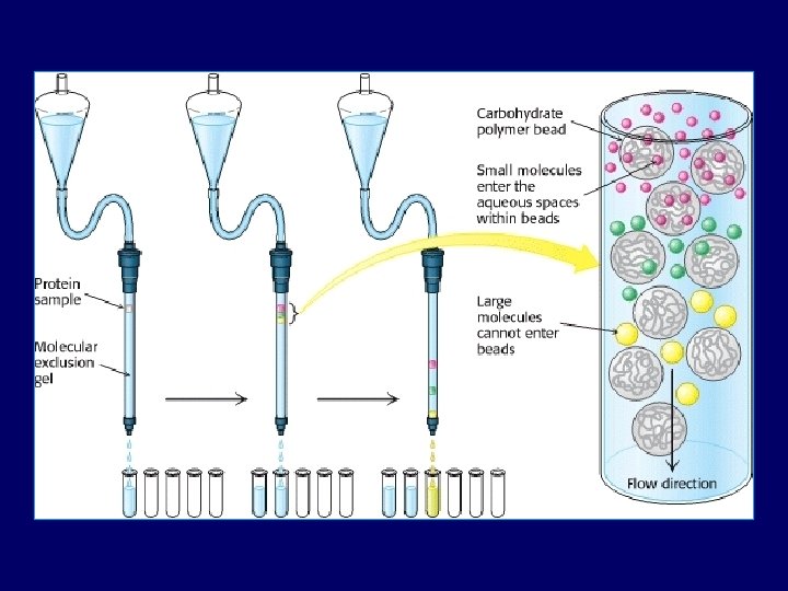
Stationary phase • Is gel beads which contain pores of specific size. • Usually gel is polysaccharides (dextran) or other polar polymers formulated into small beads • Theses beads varying in degrees of cross-linking of the polysaccharide within the bead. • Beads allowing smaller molecules to pass through their pores, while larger molecules are excluded.

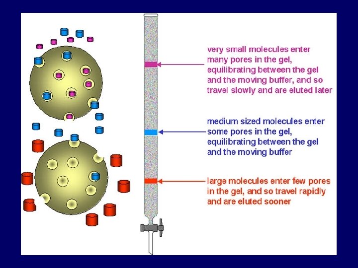
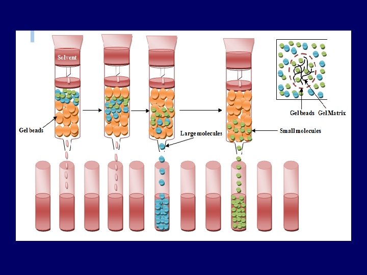
Principle: • Sample pass through a column packed with a swollen gel. • Separating of molecules occurs according to molecular weight: - Large molecules (that are larger than the largest pore): can’t penetrate gel pores move around the beads excluded from gel pores pass through the column quickly elute first - Smaller molecules (the are retained because) Smaller molecules penetrate the pores to different degrees depends on their size molecules are eluted in order of decreasing size. So, Gel-filtration works opposite to sieve.
: volume between gel beads (Vo")
Important parameters: • Exclusion volume or void volume (Vo): volume between gel beads (Vo = elution volume of large molecules they do not enter the pores) • Internal pore volume Vi: volume inside the beads. (Vi = elution volume of small molecules) • Elution volume (Ve): volume required to elute a particular molecule • Total volume (Vt ): is total volume of mobile phase in the column Vt = Vi+Vo

 • Kd: is the partition coefficient for solute, (the extent to")
Partition coefficient (Kd) • Kd: is the partition coefficient for solute, (the extent to which the molecules can penetrate the pores in stationary phase) • its value range between 0 and 1 Kd = Ve – Vo Vi • it is difficult to measure Vi precisely, the equation modified to determine available part of the resin (Kav). Kav = Ve – Vo Vt – Vo (Vt = π r². h) • Sample components (solutes) are easily separated if their (Kav) value different from each other.

Types of stationary phase • The media used for gel exclusion chromatography are: – – dextran (Sephadex™), polyacrylamide (Bio-Gel P™) dextran-polyacrylamide (Sephacryl™) agarose (Sepharose™ and Bio. Gel A™) • Sephadex G-25 is most common gel used in gel-filtration chro. • Each is available with a different ranges of pore size in the beads, permitting separation of macromolecules of different size.

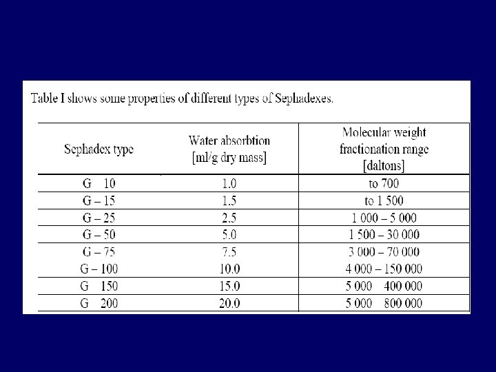
Different types of matrix forming stationary phase: Cross-linked dextran polymer It is a strongly hydrophilic polymer, and swells in water dextran (Sephadex G-10 to G-200) before a column is prepared, the gel must be full hydrated. Cross-linked polyacrylamide (Biogel P-2 to G-300) They are hydrophilic but are chemically more stable than dextran gels. Agarose-the largest pore size Agarose They are also hydrophilic but are sold in the swollen form. Mixed gels of polyacrylamide The polyacrylamide provide a three-dimensional and agarose (Ultragel) structure which supports the interstitial agarose gel. agarose - Controlled-pore glass beads can use as porous gel
. • Sephadex")
Sephadex-25 • It is an inert, bead-formed, cross-linked dexran (polymer of glucose). • Sephadex beads are porous, Molecules larger than largest pores cannot enter the gel and are eluted first, Smaller molecules enter the beads and are retard. • Sephadex G-25 exclude all molecules with a molecular weight greater than 5000, thereby eluting them first.

Advantages of gel filtration • Reliable and simple • Little equipment is required • The procedures are straight forward • Good separation and yields
 •")
Application of gel-filtration: Used in: • Separate molecules of different sizes (biological molecules) • Determination of the relative molecular mass: using a calibration curve prepared from the elution volumes of several reference substances of known relative molecular mass. • Desalting or buffer exchange: The removal of solutes of low relative molecular mass from preparations of macromolecules.

Lab Practice • In this lab, you will separate a mixture of blue dextran and cobalt chloride molecules through a gel-filtration column • collecting elutes (Run-off), and calibrating a curve to show the column separates molecules by molecular weight. • Then, you will use the calibration curve to identify the molecular weights of the proteins.

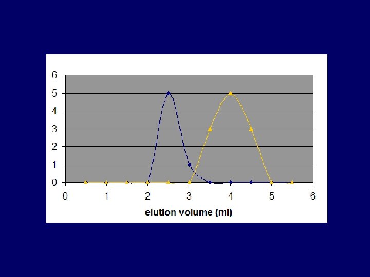
Blue dextran: • is a glucose polymer with high M. wt (M. wt =2000000 daltons) • It is too large and can’t get into the beads and therefore excluded from the gel (Kd=0), and pass the column in the void volume (space between gel beads). • Blue dextran is often used as a marker to measured void volume. Cobalt chloride: • low molecular weight (Small molecule) is freely accessible to the gel particles (Kd=1), and elute at a volume equal to Vt
")
• Both molecules (Blue dextran & Co. Cl 2. 6 H 2 O) are colored so the progress of the filtration can be followed by observing the separation of the colored bands. Fraction analysis: • The completed fraction is then analyzed by measuring the extinction of each fraction at 625 nm and 510 nm Blue dextran ʎmax = 625 nm Co. Cl 2. 6 H 2 O ʎmax = 510 nm

Chemicals & other material: • • Sephadex G 25 Blue dextran in saline Cobalt chloride in saline Sodium chloride (0. 9 %), (Saline)

Procedure: • Number 15 test tubes and arrange them in order on a rack. Prepare the gel bead column: • Column (12 cm) is filled with semisolid (Swollen) gel beads of Sephadex G-25 [Sephadex gel soaked in the elution buffer 3 -4 hrs]. Be gentle; do not allow gaps or bubbles to form. • Allow small amount of saline to flow through the column between additions of beads (help the beads to settle) • Equilibrate the column with saline by passing about 10 ml of saline through the column beads after it has completely settled.

• It is important that the gel should be homogenous, free from bubbles, free from crack, and free from spaces between the walls. • And it should be covered by the liquid "mobile phase" all the time. • Avoid stirring up the top of the column bead when adding saline or samples, as this will give poor resolutions of the samples.
")
• Drain saline solution down (keep 1 mm of buffer above the gel) before adding the sample mixture • Carefully add the sample mixture to the top of the column using a pasture pipette. (do not stir up the top of the gel) • Turn off the stopcock to allow the mixture to enter the gel beads. • Then add saline solution to the top, filling the space at the top of the column.
 beginning with tube # 1 (Early fractions")
• Collect the fractions (3 ml/tube) beginning with tube # 1 (Early fractions contain large molecules while later fractions contain smaller ones). • Measure the absorbance spectrum of each fravtion in order to identify each molecule (Blue dextran at 625 nm, Co. Cl 2. 6 H 2 O at 510 nm).
, (Saline)")
Cobalt chloride Blue dextran Sephadex G 25 Sodium chloride (0. 9%), (Saline)

Small molecule Large molecule
 Volume Tube No. fraction (ml) 1 2")
Results: Absorbance at 510 nm (Blue dextran) Volume Tube No. fraction (ml) 1 2 3 4 5 6 7 8 9 10 11 12 13 14 15 16 17 18 3 6 9 12 15 18 21 24 27 30 33 36 39 42 45 48 51 54 Absorbance at 625 nm (Cobalt chloride )
- Slides: 28