Lab 4 Hook worms A Old world hook

Lab. 4: Hook worms A. Old world hook worm: Ancylostoma duodenale B. New world hook worms: Necator americanus

Life cycle of Hookworms

Ancylostoma duodenale: Female & Male

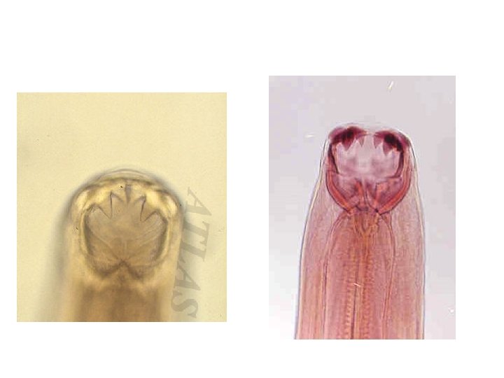
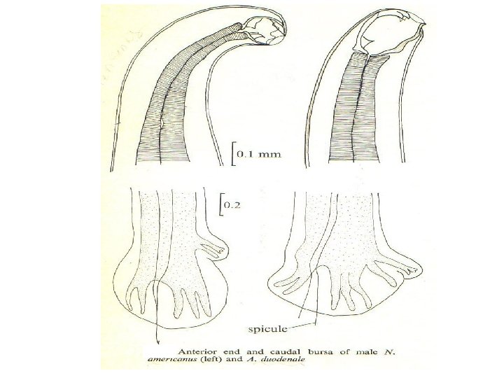
Ancylostoma duodenale: 1 -Buccal capsule: in male & female ü anterior end, enlarged, chitinized with: • 2 pairs of subequal ventral teeth-like thickenings, � 1 pair of dorsal small tooth-like thickenings.

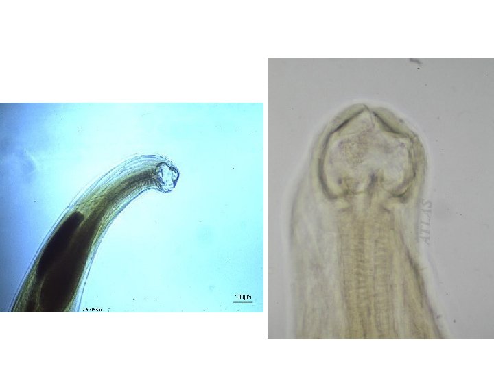
Ancylostoma duodenale: buccal capsule

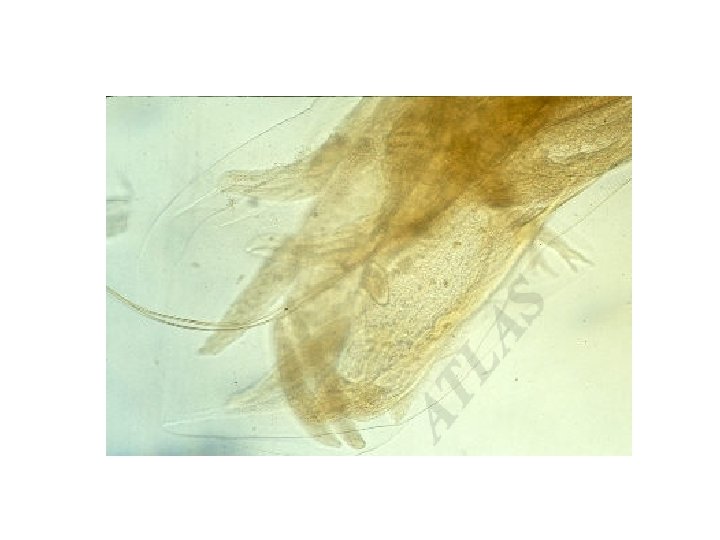
2 -Capulatory bursa in male: q Fan-like expansion of posterior part of cuticula, q With rib-like thickenings & 2 thin separated copulatory bursa. 2 separated spicules
 of A. duodenale")
posterior end in male (Bursa) of A. duodenale

Necator americanus: 1 -Buccal capsule: in male & female • anterior end, enlarged, • chitinized with 2 cutting plates on the ventral side.

Capulatory bursa in male: § Fan-like expansion of posterior part of cuticula, § with rib-like thickenings & 2 thin fused copulatory barbs.
 of N. americanus")
posterior end in male (Bursa) of N. americanus
 of")
Egg: ►Thin shelled transparent, ovoid & measures 6476 µm in early stages(4 -celled) of cleavage when laid in several hours may reach the early larval stage, then hatching in 24 -48 hrs. to L 1. 1 - celled 4 - celled

Eggs of hookworms in unstained wet mount

C- Rhabditoid larva: Mouth open Esophagus short with bulb.

Open mouth Bulb Rhabditoid larvae of hookworms

D- Filariform larva: more delicate closed mouth long esophagous

Bulb Closed mouth Filariform larvae of hookworms

Ancylostoma duodenale Filariform Larva in sputum
 Trichinella spiralis The blood- and tissue dwelling nematodes: Filariae")
Lab. 5 Strongyloides stercoralis (Threadworm) Trichinella spiralis The blood- and tissue dwelling nematodes: Filariae Wuchereria bancrofti

Life cycle Strongyloides stercoralis :

5 - Strongyloides stercoralis: A-Adult male: free living curved posterior end with 2 spicules.
. Free")
Free-living adult male S. stercoralis. Notice the presence of the spicule (red arrow). Free living adult male S. stercoralis, showing a spicule (red arrow). A smaller, rhabditiform larva lies adjacent to the adult male.

B-Adult female: tapering posterior end parthenogentic uterus with eggs.

Adult free-living female S. stercoralis alongside a smaller rhabditoid larva. Notice the developing eggs in the adult female. Adult free-living female S. stercoralis. Notice the row of eggs within the female’s body.

Male Female Free living adult of strongyloides stercoralis

Hematoxylim - eosin s. Strongyloides stercoralis Adult Parasitic Female Small Intestine Section

In Iodine s. Strongyloides stercoralis Rhabditiform Larva in fresh stool smear 200 – 250 X 16 µm note : 1 - this larvae with short mouth and double – bulb oesophagus 2 - in hook worm the rhabdiform larva detected in old stool ( 24 – 48 hr. ) or when we used sugar with concentration method

filarial – form larvae of strongyloides stercoralis, 500µm Note : more slender than rhabditiform larva have shourt mouth and cylinderical oesophagus about ½ of the body

Trichinella spiralis Disease: Trichinosis

Life cycle

6 -Trichinella spiralis: a- female: small size stichocytes club-shaped posterior end.
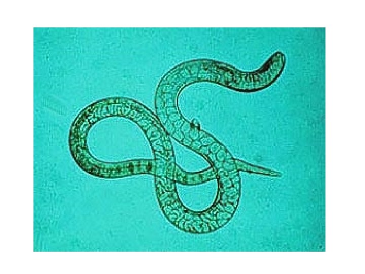

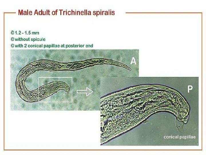
6 -Trichinella spiralis: b- Male: Small size Stichocytes With 2 conical papillae at posterior end.

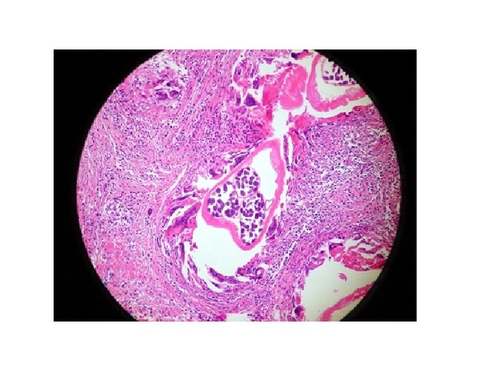
C-larva: spirally coiled within muscle fiber.

Hematoxylim - eosin s. Trichinella spiralis Encysted Larva Muscle Section

Wuchereria bancrofti Disease: Bancroftian filariasis or Elephantiasis Mosquito Culex intermediate host & vector.

Life cycle:

Filariae: A- Wucheraria bancrofti: sheathed microfilaria; short cephalic space long tail; aseptete nuclei.

Giemsa s. Wuchereria bancrofti Microfilaria in blood smear 210 – 320 X 7. 5 – 10 µm, Note : blood collected between the hour 10 – 2 at night, this microfilaria detected in blood after 6 mouth of infection

Adults of W. bancrofti. The male worm is on the left; the female is on the right.

Microfilariae of W. bancrofti in thick blood smears stained with Giemsa. Images courtesy of the Oregon State Public Health Laboratory.

B- Onchocerca volvulus: unsheathed microfilaria long cephalic space short tail Aseptete nuclei.

Ciemsa s. Onchcerca volvulus Microfilaria ( 280 – 330 X 6 – 9 µm )

Onchcerca volvulus -Skin Nodule Section with adult female and microfilariae In the surrounding fibrous connective tissues
- Slides: 49