Lab 34 Eye Structure Sheep Eye Dissection Lab

Lab 34: Eye Structure Sheep Eye Dissection

Lab 34: Eye Dissection Prep • • Get in lab groups (assigned by Mrs. M) Complete Part A/B Download needed Apps (Label box/Google Classroom) Assign roles for tomorrow (write on back) – Dissectors x 2 – Photographer x 1 – Facilitator+ Directions x 1 – Clean-up & Label Box: All participants

Objective After completing this exercise, you should be able to … 1. Identify the major structures of the eye 2. Describe the functions of the structures of an eye

Dissection Materials needed: • Textbook • Cellphone camera w/ Label Box & Google Classroom App installed • Sheep eye • Dissecting tray • Sharp scissors • Gloves • Probe

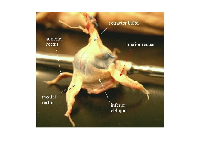
1. Trim away the fat and other connective tissues, but leave the extrinsic muscles and the optic nerve

2. Locate and observe the cornea, sclera, and iris

3. Use sharp scissors to make a coronal section of the eye • To do this, cut through the wall about 1 cm from the margin of the cornea and continue all the way around the eyeball

4. Gently separate the eyeball into anterior and posterior portions • Usually the jelly-like vitreous humor will remain with the posterior portion

5. Examine the anterior portion of the eye, and locate the cilliary body, which appears as a dark, circular structure • Note the iris and the lens

6. Use a dissecting needle to gently remove the lens, and examine it.

7. Examine the posterior portion of the eye. • Note the vitreous humor, which helps hold the lens in place Vitreous Humor

8. Carefully remove the vitreous humor and examine the retina • This will appear as a thin, nearly colorless to cream -colored membrane that detaches easily form the choroid coat

9. Locate the optic disc- the point where the retina is attached to the posterior wall of the eyeball and where the optic nerve originates aka “blind spot”

10. Note the iridescent area of the choroid coat beneath the retina • This colored surface in mammals with hooves is called the tapetum fibrosum it reflects light back through the retina • Thought to aid in night vision

11. Take any final pictures 12. Clean-up. All pans/utensils must be washed and dried. All gloves/eye material/dirty towels need to be disposed of in the proper trash bin.

Sheep Eye- Dissection Video 6 m 25 s http: //www. youtube. com/watch? v=G 2 Fqy. VDX 0 SM

Finish LAB 34…. 1. Complete Lab Report 34: Eye Structure with your lab group 2. Complete “Label Box” portion of the eye dissection • Submit to Google Classroom

Sheep Eye- Dissection Video 15 m http: //www. youtube. com/watch? v=DIz 78 Ey 9 fo. Q
- Slides: 19