LAB 3 THE MICROSCOPE AND THE CELL Objectives

LAB. 3 THE MICROSCOPE AND THE CELL Objectives 1. Identify and explain the functions of the primary parts of a compound microscope. 2. Use a compound (light) microscope to examine biological specimens. 3. Understand the differences between prokaryotic and eukaryotic cells and identify structure characteristic of each. 4. Understand the function of organelles in eukaryotic cells.

Part I The Microscope

1. Overview of Microscope The study of cytology of cells is concerned primarily with cellular structure and cell morphology, which is mainly dependent on microscopy techniques. Two types of microscopes: § (Compound) light microscope § Electron microscope A typical light microscope A electron microscope

2. Structure of the Light Microscope A microscope contains two main systemsilluminating system and imaging system. 1. illuminating system: focuses light on the specimen Components: Light source (light bulb): Intensity of light source can be adjusted. Condenser lens that focus light as a tight beam on the specimen slide. Condenser lens can be brought closer or farther from specimen on stage with the help of the condenser adjustment knob. Condenser iris diaphragm (similar to camera shtter): increase or reduce brightness.

Compound Light Microscope: The microscope above is called a compound light microscope. The microscope is called this due to the use of light to transmit the image to your eye through the use of lenses. It is called a compound microscope because it has more than one lens. Leeuwenhoek's early microscope was called a simple microscope because it only used one lens. This kind of simple microscope was much like a magnifiying glass that you might use to look at a insect or picture. This way of looking at things is limited and is what enticed people to create the compound microscope. The word Microscope is the combination of two words; "micro" meaning small and "scope" meaning view. How it Works The optical systems of the light microscope

2. Imaging system Oculars: contain lenses that magnify image 10 X Objective Lenses: 4 X: Low magnification objective (Scanning objective): 10 X: Medium magnification objective 40 X: High magnification objective 100 X: Oil immersion objective {ALWAYS start first with the scanning objective (4 X). You will NOT use the oil immersion objective in BIO 104 L} Body part: Arm, stage, Stage clip, Coarse and fine focus adjustment knobs. Use the coarse focus adjustment knob with 4 X, 10 X, and 40 X objectives. {ONLY use the fine focus adjustment with 40 X objective or higher. If you are not careful, you can ram the 40 X objective lenses on to the slide (scratches lens and breaks slide)}
 Arm Revolving nosepiece Objectives (4 X, 10 X, 40")
Oculars (10 X magnifying lenses) Arm Revolving nosepiece Objectives (4 X, 10 X, 40 X or 100 X magnifying lenses) Stage clip Coarse focus adjustment Stage Condenser (lightfocusing lens) Fine focus adjustment Condenser height adjustment (left side, not shown) X-Y stage Controls Iris diaphragm of condenser Base Substage lamp Light intensity control Fig. A typical compound light microscope Note: Always carry the microscope by the arm and base!

3. Important Concepts and Terms in Microscopy Magnification: Increase in apparent size of object (increases with higher objectives). Total magnifications = Objective magnification x Ocular magnification Sample: 4 X x 10 X x 40 X x 10 X =40 X 10 X = 100 X 10 X = 400 X 10 X = 1000 X Contrast: Level of difference between lightest and darkest part of image, affects ability to discern details. Without contrast, your cell would seem transparent (invisible). Most cells and organelles do not have any color, but the use of staining dyes can increase the contrast.

Field of view: Actual physical dimensions of what is viewed through lenses; low magnification gives larger field of view; high magnification produces smaller field of view. Depth of field: Actual thickness of object that is in sharp focus; low magnification objective has bigger depth of field; high magnification gives smaller depth of field. Parcentered: Ability of microscope to remain centered on image as you switch to higher objective lens from low objective lens. Parfocal: Ability of microscope to remain focused on image as you switch to higher objective lens. (A perfectly parfocal microscope would not require you to adjust the focus with the coarse or fine focus adjustment knob, as you switch to a higher objective).

Limit of resolution The minimum distance that a microscope can separate two points and still remain identifiable. Resolving Power The ability of a microscope to distinguish adjacent objects as separate entities.

4. The size of things Width of your thumb: 20 mm: Width of human cells: Width of mitochondria: Width of bacterial cell: Width of ribosome: Width of atom molecule: 10 -20 μm 20 nm 0. 2 nm
")
PART II: THE CELL (STRUCTURE AND FUNCTION)
 All living organisms belong to one of")
1. The 3 Domains of Life (Review) All living organisms belong to one of the three domains (categories) of life: Archaea, Bacteria and Eukarya (Eukaryotes). Organisms belonging to the Bacteria and Archaea domains are called prokaryotes. ARCHAEA LIFE DOMAIN BACTERIA PROKARYOA (prokaryotes) EUKARYA (eukaryotes) Archaea: is an unicellular organism and inhabits in extreme environmental conditions
 (has 4 kingdoms) Animalia Fungi Plantae Protista")
Prokaryotes Achaea Bacteria Eukarya (Eukaryotes) (has 4 kingdoms) Animalia Fungi Plantae Protista

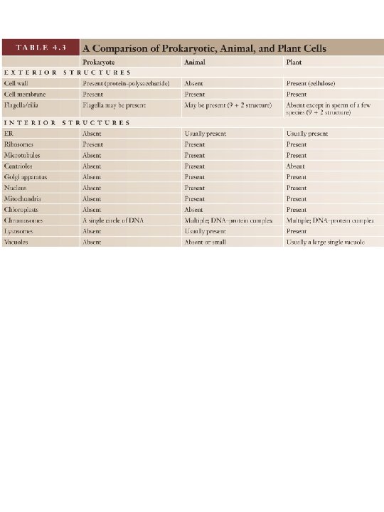
Features common to cells of all living organisms: 1. All cells have a plasma membrane (also called cytoplasmic membrane). 2. All cells are filled with a fluid called cytoplasm (cytosol). 3. All cells contain genetic information in the form of DNA in chromosomes. The expression of DNA produces RNA and proteins.
; multicellular (few) § They have cell")
2. Prokaryotic cells Common Characteristics: § Unicelluar (most); multicellular (few) § They have cell wall (surrounding cell membrane) § They do not have nucleus and organelles. § They are smaller than eukaryotes in size. § Contain only circular chromosome and circular plasmid DNA * § Can contain specialized structures for movement , such as flagella (whip-like tail for movement) or cilia (hair-like projections) § Simple shape: spiral-shaped, spherical, or rod -shaped. Fig. Shapes of bacteria cells.
 Cyanobacteria (also called blue-green algae)*: Cyanobacteria are the largest prokaryotes and they contain")
(1) Cyanobacteria (also called blue-green algae)*: Cyanobacteria are the largest prokaryotes and they contain light absorbing pigments (chlorophyll) and are capable of photosynthesis. Oscillatoria Anabaena Algae, green-algae, red-algae, and blue-green algae: § Algae belongs to eukaryotes, which could be unicellular or multicellular, such as Seaweeds. § Green-algae and red algae are two groups of Algae. § Blue-green algae is one group of bacteria
 Beneficial bacteria: Lactobacillus acidophilus is a probiotic (good bacteria). Humans used this")
* (2) Beneficial bacteria: Lactobacillus acidophilus is a probiotic (good bacteria). Humans used this bacterium to produce yogurt (they grow in milk, causing the milk to sour) because yogurt preserves better than milk. Lactobacillus acidophilus
 Pathogenic bacteria: Pathogenic bacteria cause disease. Bacillus anthracis is the bacteria that")
* (3) Pathogenic bacteria: Pathogenic bacteria cause disease. Bacillus anthracis is the bacteria that causes anthrax. (Don’t worry about manipulating the slides, the bacteria were all killed when the bacteria cells were stained to produce this slide). Bacillus anthracis

3. Eukaryotes: Eukaryotes are divided into four kingdoms: Animalia, Plantae, Fungi, and Protista. Fig. Four kingdoms of eukaryotes

Basic Characteristics of eukaryotes: § Can be multicellular organisms (all animals, all plants and most fungi) or unicellular organisms (protists, some fungi). § Some have cells wall (plant cells, fungi cells, and protistae cells) and some do not have cell wall (animal cells) § Have nucleus and other organelles. § Chromosome DNA are usually linear. § About 10 X larger than prokaryotic cells. § Can contain specialized structures for movement at their cell surface, such as flagella (whip-like tail for movement) or cilia (hair-like projections).

1. Animalia Kingdom § multicellular organisms § No cell wall § Most cells in an animal are usually arranged as tissues (layer of connected cells) Fig. Human inner cheek cells after staining their nuclei with methylene blue.

2. Plantae Kingdom § Plant cells are always surrounded by a cell wall § Multicellular structure (tissues); some unicellular. § Plants are capable of photosynthesis that is performed in chloroplasts. A chloroplast is a organelle containing pigments that can absorb visible light. § Plant cells can also contain other plastids (plant organelle). Amyloplasts are organelles that store starch in the cells of some plant tissues (such as potato). Amyloplasts can be observed after staining with Lugol’s solution (iodine solution).

chloroplasts in elodea plant cells. Onion cells Nucleus Amyloplasts in potato cells
")
Ulothrix (unicellular organism)
 and multicellular (mushrooms, most molds) organisms")
3. Fungi kingdom § Unicellular (yeasts, some molds) and multicellular (mushrooms, most molds) organisms that are not capable of movement or photosynthesis. § Fungi cells are usually surrounded by a cell wall 4. Protistae kingdom Protists consist of diverse eukaryotes that are essentially unicellular § Protists are unicellular organisms. § Some protists share characteristic of either animal and/or plant cells o Chloroplasts for photosynthesis o Capable of cellular movement o Surrounded by cell wall
. Amoebas")
Examples of protistae: Amoeba: moves by using cell extensions called pseudopods (false feet). Amoebas can elongate and retract pseudopods, and they are not only involved in cell movement but also serve in surrounding food particles. The food particles will be taken into the cell by phagocytosis and digested with the help of lysosomes. Contractile vacuoles are organelles that accumulate and expel water and cellular waste chemicals. Amoeba

Paramecium: moves by using hair-like ciliae at cell surface. Paramecia contain contractile vacuoles, organelles that accumulate and expel water and cellular waste chemicals. Unlike amoeba, paramecium has a distinct and permanent shape and certain areas of cytoplasm, (cell organelles), are specialized to carry out specific functions. Paramecium

How to determine the field of view when 10 X and 40 X objective lenses are used? 1. The ruler can be used to measure the diameter of the filed view when 4 X objective lens is used. After you have diameter, you figure out radius (diameter/2) and use following formula to calculate field of view (area of circle): Area of circle = 3. 14 x radius 2 (3. 14 = π) 2. However, the ruler cannot be used to measure the diameters of the field view when 10 x and 40 x lenses are used, because 10 x will give incorrect diameter and 40 x make space lines of the ruler out area of view. Therefore, these diameters must be calculated using following formula:

FOVlow x Maglow = FOVhig x Maghigh FOVlow = diameter of the field of view of the 4 x power objective Maglow = magnification objective, i. e. 4 x. of the low-power FOVhigh = diameter of the field of view of the 10 x (or 40 x) power objective (this is to be determined by calculation using the formula above. Maghigh = magnification of the high-power objective, i. e. 10 x or 40 x For example, if 3. 0 mm is the diameter of the field of view for a 4 x low-power objective, what is diameter of the field of view of the 10 x and 40 x high-power objectives, respectively?

Diameter for using 10 x objective lens: 3. 0 mm x 4 = FOVhigh x 10 FOVhigh = 3 x 4/10 = 1. 2 mm Diameter for using 40 x objective lens: 3. 0 mm x 4 = FOVhigh x 40 FOVhigh = 3 x 4/40 = 0. 3 mm Then use formula (Area of circle = 3. 14 x radius 2) to calculate area of circle (or called field of view)

For Quiz and Exam: In addition to studying theory, you must be able to distinguish by memory or by deductive logic, whether cells belong to prokaryotes or eukaryotes and whether they belong a particular kingdom of eukaryotes, based on their observable cell structures under the microscope. You must be able to recognize and identify and recognize organelles observable under the microscope.
- Slides: 33