Lab 3 Ascaris lumbricoides Life cycle of Ascaris

Lab. 3: Ascaris lumbricoides

Life cycle of Ascaris lumbricoides

Ascaris lumbricoides: Female A-Gross specimens: large size of worm Ø Female: cylindrical in shape, straight posterior end. Ø Male: smaller than female with curved posterior end. Ø Male

281 Male and female anterior end, with three lips Ascaris lumbricoides adult worm Note: white, brown redish or light brown or pink the posterior end of the male, curved with 2 spicules, the female with straight end


B-Egg: q Fertilized: • round in shape • with outer mammillated coat • thick transparent middle coat • thin membranous inner coat • single unembryonated egg. q Unfertilized: • elongated cylindrical in shape • with all other characters mentioned above.
 One cell stage")
Iodine s. Fertile, with larvae Semilunar space Vitelline layer (1 st) One cell stage 2 nd layer (thick) Decorticated egg: Its fertile ovum, but the outer coat is 3 rd layer outer coarse albuminous layer (regular) sometime lost Ascaris lumbricoides-Fertilized Egg In Iodine s. stool smear ( golden brown in colour ) 60 – 75 X 40 – 50 µm, is spherical or oval with semilunar space and regular albuminous layer

Ascaris lumbricoides- Fertilized Egg In Saline s. Decorticated egg: Its fertile ovum, but the outer coat is sometime lost

In Iodine s. In Saline s. elongated oval, no semilunar space with irrregular albuminous layer 88 – 94 X 40 – 50 µm Ascaris lumbricoides- Unfertilized Egg stool smear

Ascaris lumbricoides Larva in Lung Section Note : the larva also detected in sputum Hematoxylim - eosin s.

C. Cross section of worm: v Outer cuticular covering layer v Middle syncetial cellular layer with dorsal & ventral thickenings or cords in which dorsal ventral nerve cords pass. v lateral thickenings or cords in which lateral excretory vessels pass.

v. Inner musculer layer: divided by the Dorsal, ventral & lateral cords into 4 sector. v. Section through the intestine in the middle v. Coelomic cavity between muscles intestine, vwith male or female reproductive organs.

ovary Ascaris lumbreioides : cross section in female

Ascaris lumbreioides : cross section in female

Ascaris lumbreioides : cross section in male

Lab. 4: Hook worms A. Old world hook worm: Ancylostoma duodenale B. New world hook worms: Necator americanus

Life cycle of Hookworms

Ancylostoma duodenale: Female & Male

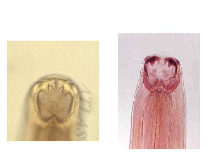
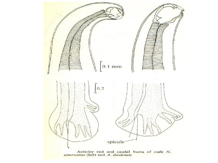
Ancylostoma duodenale: 1 -Buccal capsule: in male & female ü anterior end, enlarged, chitinized with: • 2 pairs of subequal ventral teeth-like thickenings, � 1 pair of dorsal small tooth-like thickenings.

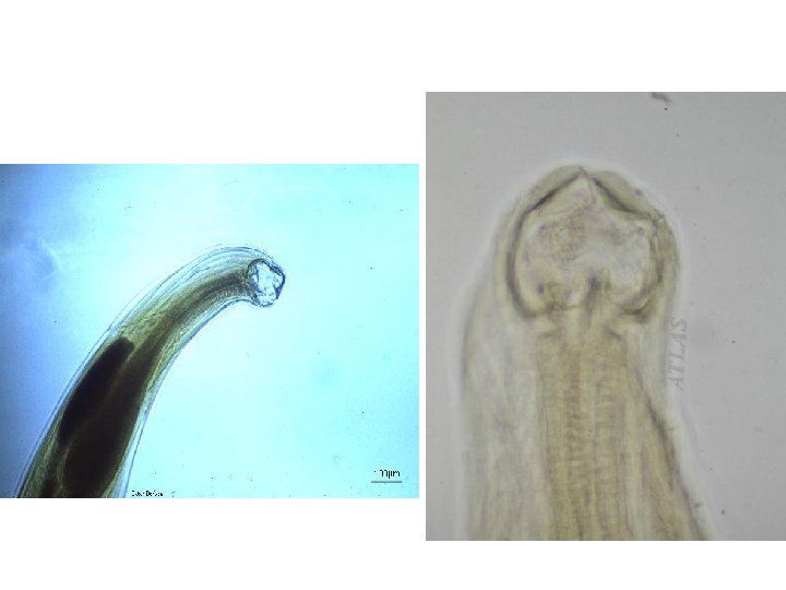
Ancylostoma duodenale: buccal capsule

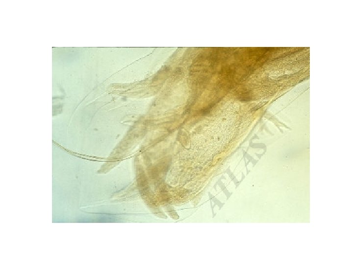
2 -Capulatory bursa in male: q Fan-like expansion of posterior part of cuticula, q With rib-like thickenings & 2 thin separated copulatory bursa. 2 separated spicules
 of A. duodenale")
posterior end in male (Bursa) of A. duodenale

Necator americanus: 1 -Buccal capsule: in male & female • anterior end, enlarged, • chitinized with 2 cutting plates on the ventral side.

Capulatory bursa in male: § Fan-like expansion of posterior part of cuticula, § with rib-like thickenings & 2 thin fused copulatory barbs.
 of N. americanus")
posterior end in male (Bursa) of N. americanus
 of")
Egg: ►Thin shelled transparent, ovoid & measures 6476 µm in early stages(4 -celled) of cleavage when laid in several hours may reach the early larval stage, then hatching in 24 -48 hrs. to L 1. 1 - celled 4 - celled

Eggs of hookworms in unstained wet mount

C- Rhabditoid larva: Mouth open Esophagus short with bulb.

Open mouth Bulb Rhabditoid larvae of hookworms

D- Filariform larva: more delicate closed mouth long esophagous

Bulb Closed mouth Filariform larvae of hookworms

Ancylostoma duodenale Filariform Larva in sputum
- Slides: 36