Lab 2 Trichuris trichiura Ascaris lumbricoides Trichuris trichiura

Lab 2: Trichuris trichiura & Ascaris lumbricoides

Trichuris trichiura Life cycle

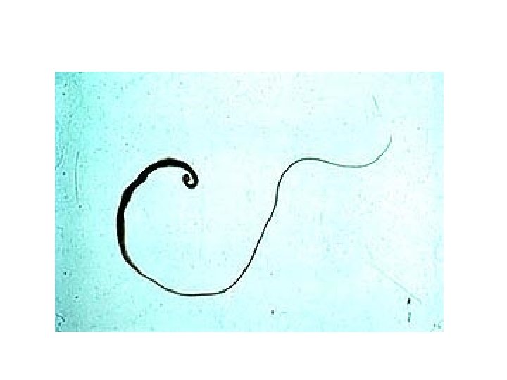
Trichuris trichiura: A-Female: vwhip-like body vwith thin anterior 2/3 body vthick posterior 1/3 body. vbead-like cells surrounding intestinal tract in the anterior end, called stichocytes.
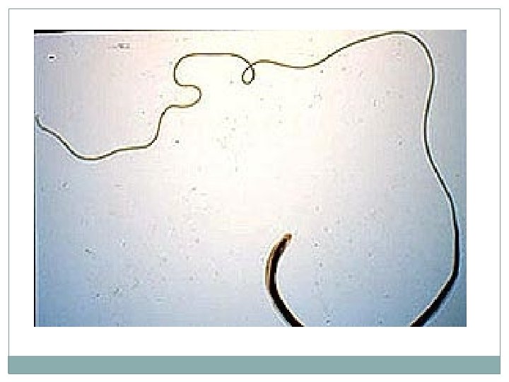

B-Male: body same as in female. posterior end strongly curved with one spicule.

Trichuris trichiura in the large intestine. Many worms are present, each with its anterior end embedded in the intestinal mucosa, resulting in the erythema. symptum, with havey infection ( prolapse of the rectum )

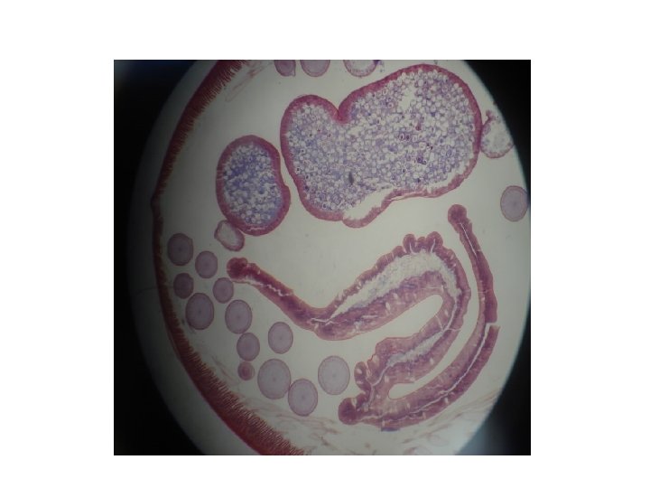
c. section in mucosa: small round section in anterior part of worm, in columnar epithelial cells of mucosa. larger rounded sections in posterior part of worm, in lumen of intestine.

D-Eggs: q. Barrel-shaped or lemon-shaped eggs q. With lateral plug like thickining. q. Egg single-celled, unembryonated.

Plug In Iodine s. In saline s. Trichuris trichiura Egg 50 X 25 µm Note : the egg barrel – shaped, with two polar plugs yellowish brown in colour ( bile – stained ) has a double shell, the outer one is bile – stained

Lab 3: Ascaris lumbricoides

Life cycle of Ascaris lumbricoides

Ascaris lumbricoides: Female A-Gross specimens: large size of worm Ø Female: cylindrical in shape, straight posterior end. Ø Male: smaller than female with curved posterior end. Ø Male

281 Male and female anterior end, with three lips Ascaris lumbricoides adult worm Note: white, brown redish or light brown or pink the posterior end of the male, curved with 2 spicules, the female with straight end

B-Egg: q Fertilized: • round in shape • with outer mammillated coat • thick transparent middle coat • thin membranous inner coat • single unembryonated egg. q Unfertilized: • elongated cylindrical in shape • with all other characters mentioned above.
 One cell stage")
Iodine s. Fertile, with larvae Semilunar space Vitelline layer (1 st) One cell stage 2 nd layer (thick) Decorticated egg: Its fertile ovum, but the outer coat is 3 rd layer outer coarse albuminous layer (regular) sometime lost Ascaris lumbricoides-Fertilized Egg In Iodine s. stool smear ( golden brown in colour ) 60 – 75 X 40 – 50 µm, is spherical or oval with semilunar space and regular albuminous layer

Ascaris lumbricoides- Fertilized Egg In Saline s. Decorticated egg: Its fertile ovum, but the outer coat is sometime lost

In Iodine s. In Saline s. elongated oval, no semilunar space with irrregular albuminous layer 88 – 94 X 40 – 50 µm Ascaris lumbricoides- Unfertilized Egg stool smear

C. Cross section of worm: v Outer cuticular covering layer v Middle syncetial cellular layer with dorsal & ventral thickenings or cords in which dorsal ventral nerve cords pass. v lateral thickenings or cords in which lateral excretory vessels pass.

v. Inner musculer layer: divided by the Dorsal, ventral & lateral cords into 4 sector. v. Section through the intestine in the middle v. Coelomic cavity between muscles intestine, vwith male or female reproductive organs.

ovary Ascaris lumbreioides : cross section in female
- Slides: 22