Lab 10 Electrophoresis Analytical biochemistry lab KAUbiochemistry dep
: Electrophoresis Analytical biochemistry lab KAU-biochemistry dep. L. Nouf Alshareef nf. shareef@hotmail. com")
Lab (10): Electrophoresis Analytical biochemistry lab KAU-biochemistry dep. L. Nouf Alshareef nf. shareef@hotmail. com

Gel electrophoresis • is a technique used to separate charged molecule (DNA, RNA and protein) under the influence of an electrical field. • Electrophoresis term refers to: the movement of particles through a porous matrix (gel) by electromotive force (EMF) according to their size (mass) and charge. • Used as analytical technique and preparative technique
 and charge:")
• Molecules move at different rates according to their weight (mass) and charge: 1 - Negatively charged molecules migrates toward cathode (+) electrode Positively charged molecule migrates toward anode (-) electrode 2 - Small size (low M. wt) molecules migrates faster Large size (high M. wt) molecules migrates slower

The rate of migration is also depends on: • Strength of electrical field • Sample: charge, size, shape and ionic strength • Medium (buffer): p. H, viscosity, temperature and ionic strength • Supporting material: Gel concentration Example: High voltage electrical field cause rapid movement but poor separation

Electrophoresis can be: Horizontal or slab gel Vertical Gel is poured in plat Gel poured between two glasses

Three major types of electrophoresis: • DNA electrophoresis, • 1 D protein electrophoresis • 2 D protein electrophoresis.

EQUIPMENTS AND MATERIALS
 • • • Supporting material (gel) Medium (buffer) Dyes Sample Marker")
Material (chemicals) • • • Supporting material (gel) Medium (buffer) Dyes Sample Marker
 Cellulose acetate Starch gel")
1 - Supporting medium: • • • Paper (filter paper) Cellulose acetate Starch gel Agarose gel Polyacrylamide gel electrophoresis (PAGE)

• Agarose and PAGE are commonly used. Agarose gel: • porous material that sample move though it. • Used in DNA and protein • Low range of conc. can prepared (0. 5 -3%) Polyacrylamide gel electrophoresis (PAGE): • Is also porous consists of two material: acrylamide + bisacrylamide (cross link) • Used in: DNA sequencing, protein, assessing M. wt of protein. • High range of conc. can prepared: (2 -20%) giving small pore size.

Polyacrylamide gel
 • Function: carry electric current. • Ionic")
2 - Buffer (ionic strength, p. H) • Function: carry electric current. • Ionic strength = concentration of ions in solution • When ionic strength of the buffer increased this lead to form sharp zones, but decrease the migration rate.

3 - Dyes Visualization Two types of dye are used: 1 - Tracking dye or loading dye: § Used to monitor the migration, help in sample loading § Bromophenol blue
 § Silver § Coomassie blue")
2 - Visualization dye: § Ethidium bromide (DNA, RNA) § Silver § Coomassie blue dye (protein). Ethidium bromide coonassie blue dye Silver staining

4 - Molecular weight size marker • mixture of molecules of known sizes may be protein (Da) or DNA (bp).

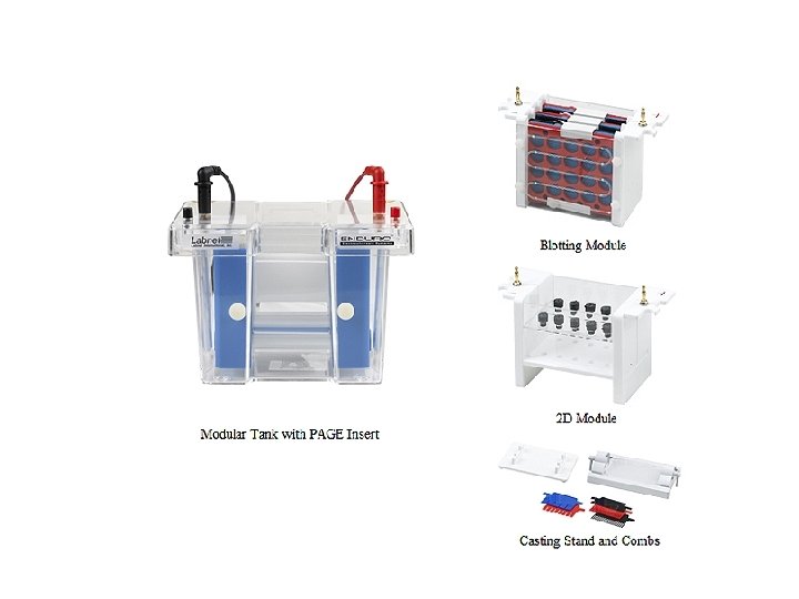
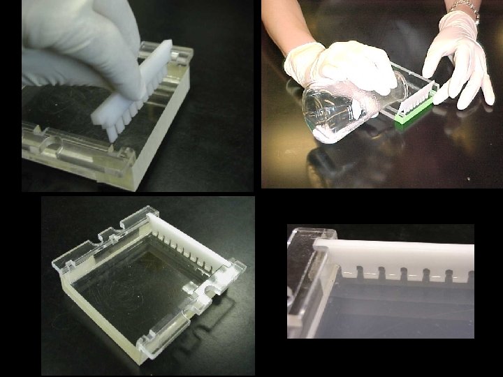
Equipments • • Casting tray Tank with cover Comb Power supply

Determination of Molecular Weight • • using PAGE of proteins or agarose gel for DNA known M. wt (marker) is used along with sample Run electrophoresis. plot a standard curve of distance migrated vs. log Mwt
, gamma globulin electrophoresis, immunoglobulin electrophoresis or Serum protein")
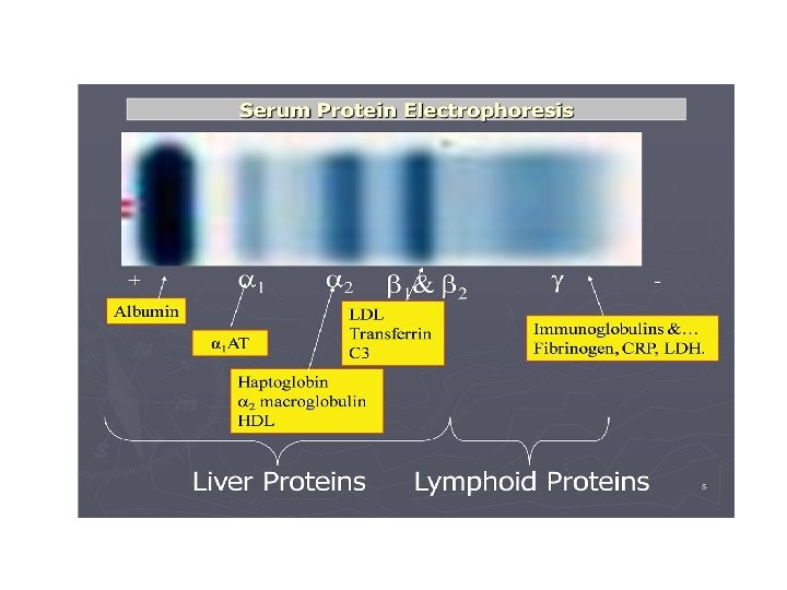
Lab practice: Immunoelectrophoresis • Immunoelectrophoresis (IEP), gamma globulin electrophoresis, immunoglobulin electrophoresis or Serum protein electrophoresis (SPEP) • Is screening test measures the major blood proteins. • Used to evaluate, diagnose, and monitor a variety of diseases. • Levels of blood proteins increase or decrease due to disease. • Serum proteins are separated into five fractions: albumin, a 1, a 2, b, and gamma proteins.

Serum Proteins • Proteins make up 6 -8% of blood. 50% serum albumin, 50% variety of serum globulins. • How to prepare serum: Blood withdraw >>>allows to clot>> > clear fluid called serum is separated out. • So, serum has same components of blood plasma without fibrinogen and other clotting factors.

• At p. H 8. 6 all proteins are negatively charged, but some more strongly than others. • serum proteins move toward the positive electrode. • The separated proteins appear as distinct bands. • They migrate in the order – – Albumin alpha beta globulins gamma globulins.
 2. Prepare")
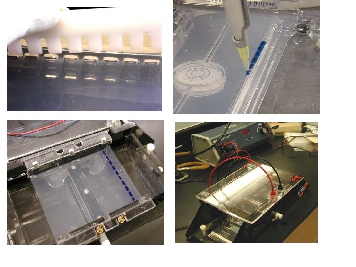
Procedure: 1. Prepare 1% agarose gel (1 g agarose +100 ml buffer) 2. Prepare sample by mixing 1 ml loading dye (BPB) + 5 ml serum 3. Load samples in gel wells (well should be in -ve electrode side) 4. Switch on power supply at 90 volt and run for 30 min. 5. Stain gel by soaking in commasie blue for 5 min 6. De-stain gel by soaking in de-staining solution for 10 -15 min 7. Identify the bands resluted.
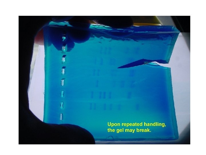

Result

Important Terms • • • Pouring Casting Loading Migration Running

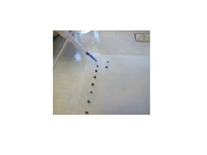
Sample wells

Sample loading

Loading with multi-channel pipette

Procedure preparation electrophoresis Experiment

1 - Agarose Gel Preparing : Before melting undissolved gel after melting dissolved gel
- Slides: 34