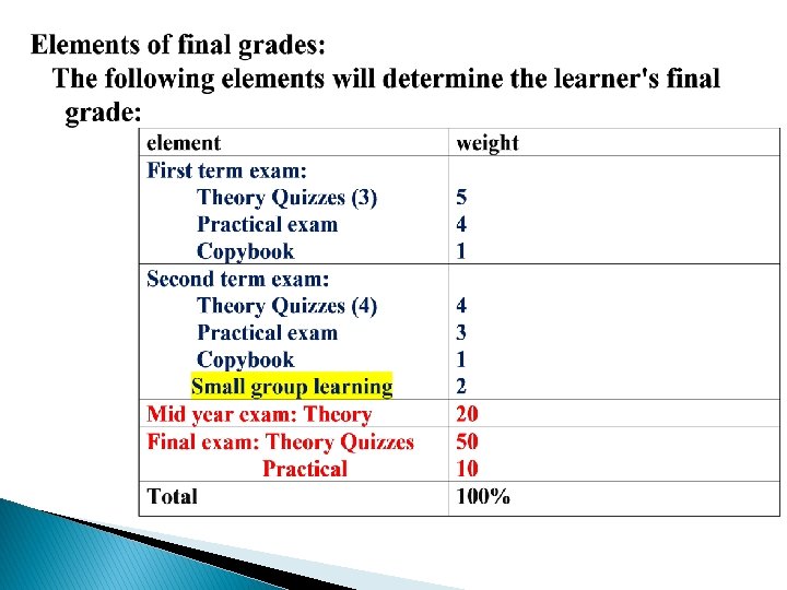
Lab 1 Introduction Enterobius vermicularis Credit hours 45

Lab. 1: Introduction + Enterobius vermicularis

Credit hours: 45 theoretical hours (2 hours in 1 st semester & 1 hour in the 2 nd semester) + 30 laboratories (2 hours for each lab. )

Nematode The intestinal nematodes Ascaris Hook worms Pin worm Whip worm The blood- and tissue dwelling nematodes The filaria Trichinella

General life cycle of Nematodes Adult ♂+♀ Egg L 4 L 1 L 3 L 2


Enterobius vermicularis :

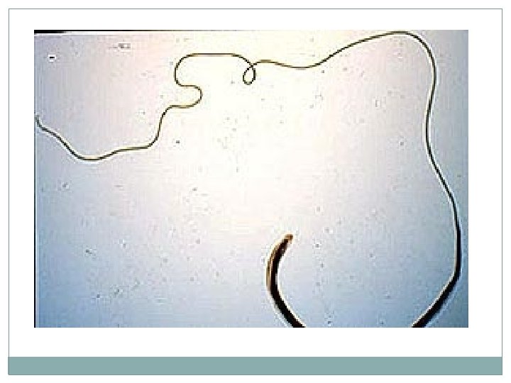
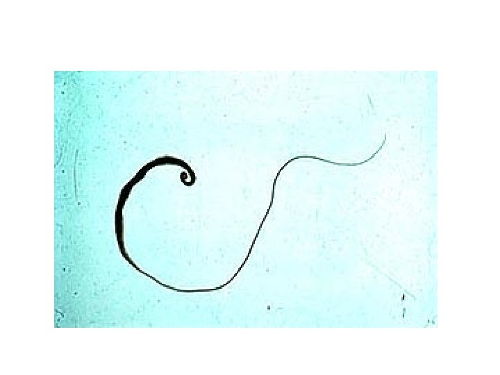
1. Enterobiusvermicularis : A. Male: v Anterior cuticular inflations, v Esophageal & esophageal bulb, v Curved posterior end (like an inverted question mark).

Enterobius vermicularis Adult Male posterior end is curved with one spicule, rarely seen in stool

Cervical alae Oesophagus Bulb Globular bulb Enterobius vermicularis Adult Anterior end Note : the cuticular cervical expansions ( alae ) and the double – bulb muscular oesophagus ( similar in both sexes )

Enterobius vermicularis B. Female: q. Anterior cuticular inflations, q. Esophageal &esophageal bulb, q. Paired gonads, q. Long pointed posterior end.

D. Ova: D-shaped eggs, with or without enclosed larva.

Eggs of E. vermicularis in a cellulose-tape preparation. Eggs of E. vermicularis in a wet mount. Egg in Fecal Concentrate X 400

Tape test for pinworms Flashlight Test: At night, the female adult worms deposit their eggs outside the rectum or anal area.
 The NIH swab. The cellophane is now")
Scotch tape technique ( graham swab ) The NIH swab. The cellophane is now usually replaced with sticky tape The techniques which used for detected the eggs of E. vermicularis from perianal skin, eggs are rarely recovered in stool Note : eggs can also be recovered from under the finger nails

C. Section in appendix: section of worms in lumen of appendix lateral alae.

Section of adult worm Lateral alae Enterobius vermicularis Adult in Appendix Section Note : cuticular lateral alae like spine in cross section of adult worm

Lab. 2: Trichuris trichiura

Trichuris trichiura Life cycle

Trichuris trichiura: A-Female: vwhip-like body vwith thin anterior 2/3 body vthick posterior 1/3 body. vbead-like cells surrounding intestinal tract in the anterior end, called stichocytes.

B-Male: body same as in female. posterior end strongly curved with one spicule.

Trichuris trichiura in the large intestine. Many worms are present, each with its anterior end embedded in the intestinal mucosa, resulting in the erythema. symptum, with havey infection ( prolapse of the rectum )

c. section in mucosa: small round section in anterior part of worm, in columnar epithelial cells of mucosa. larger rounded sections in posterior part of worm, in lumen of intestine.

D-Eggs: q. Barrel-shaped or lemon-shaped eggs q. With lateral plug like thickining. q. Egg single-celled, unembryonated.

Plug In Iodine s. In saline s. Trichuris trichiura Egg 50 X 25 µm Note : the egg barrel – shaped, with two polar plugs yellowish brown in colour ( bile – stained ) has a double shell, the outer one is bile – stained

Lab. 3: Ascaris lumbricoides

Life cycle of Ascaris lumbricoides

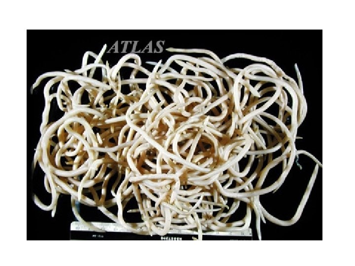
Ascaris lumbricoides: Female A-Gross specimens: large size of worm Ø Female: cylindrical in shape, straight posterior end. Ø Male: smaller than female with curved posterior end. Ø Male

281 Male and female anterior end, with three lips Ascaris lumbricoides adult worm Note: white, brown redish or light brown or pink the posterior end of the male, curved with 2 spicules, the female with straight end

B-Egg: q Fertilized: • round in shape • with outer mammillated coat • thick transparent middle coat • thin membranous inner coat • single unembryonated egg. q Unfertilized: • elongated cylindrical in shape • with all other characters mentioned above.
 One cell stage")
Iodine s. Fertile, with larvae Semilunar space Vitelline layer (1 st) One cell stage 2 nd layer (thick) Decorticated egg: Its fertile ovum, but the outer coat is 3 rd layer outer coarse albuminous layer (regular) sometime lost Ascaris lumbricoides-Fertilized Egg In Iodine s. stool smear ( golden brown in colour ) 60 – 75 X 40 – 50 µm, is spherical or oval with semilunar space and regular albuminous layer

Ascaris lumbricoides- Fertilized Egg In Saline s. Decorticated egg: Its fertile ovum, but the outer coat is sometime lost

In Iodine s. In Saline s. elongated oval, no semilunar space with irrregular albuminous layer 88 – 94 X 40 – 50 µm Ascaris lumbricoides- Unfertilized Egg stool smear

Ascaris lumbricoides Larva in Lung Section Note : the larva also detected in sputum Hematoxylim - eosin s.

C. Cross section of worm: v Outer cuticular covering layer v Middle syncetial cellular layer with dorsal & ventral thickenings or cords in which dorsal ventral nerve cords pass. v lateral thickenings or cords in which lateral excretory vessels pass.

v. Inner musculer layer: divided by the Dorsal, ventral & lateral cords into 4 sector. v. Section through the intestine in the middle v. Coelomic cavity between muscles intestine, vwith male or female reproductive organs.

ovary Ascaris lumbreioides : cross section in female

Ascaris lumbreioides : cross section in female

Ascaris lumbreioides : cross section in male
- Slides: 42