Kingdom Protista o This is a very large


-contain phycoerythrin, making them appear reddish in color. Most are multicellular")


,")


.")
- Slides: 43
Kingdom Protista o This is a very large, diverse Kingdom n All Eukaryotic n Ecologically important/Can cause disease o Traditionally thought of as having 3 sub-groups n Protozoa-Animal-like protists (heterotrophic) n Plant-like protists-Algae (autotrophic) n Fungus-like (slime molds)protists (decomposers) Modern scientists are close to splitting this into several Kingdoms n There are more subtle divisions than the ones described above n Many are unicellular, but some are multicellular n Not all are heterotrophic or autotrophic, but are “mixotrophic, ” and capable of combining Photosynthesis and heterotrophic nutrition n They have varied methods of reproduction and movement o
ac te ria A rc ha ea D ip lo m on Pa ad ra s ba sa Eu lid gl s en i ds C ili at es D in of la ge A pi l co late s m pl O om ex a yc et D es ia to m s B ro w n Fo a ra lga e m C hl inif e or ar ra a G ch la ni uc op op hy R ed hyt te e s al a l g g G ae re ae en al ga La e nd pl an Fu ts ng i C ho an of A la ni ge m lla al te s Lo s bo se C am el lu oe la ba r sl e Pl im as e m sl m im od ol e ia ds m l ol ds B Figure 29 -8 Bacteria Archaea Eukarya Chromalveolata Discicristata Excavata Alveolata Stramenopila Unikonta Rhizaria Plantae Opisthokonta Green plants Eight major lineages of eukaryotes (protist branches are in color) Amoebozoa
Figure 29 -11 THE ENDOSYMBIOSIS THEORY-formation of Protists Pyruvate and O 2 Aerobic bacterium ATP Anaerobic eukaryote 1. Eukaryotic cell 2. Bacterium lives 3. Eukaryote supplies surrounds and engulfs bacterium. within eukaryotic cell. bacterium with protection and carbon compounds. Bacterium supplies eukaryote with ATP.
Figure 29 -10 ORIGIN OF THE NUCLEAR ENVELOPE 1. Ancestor of the eukaryotes. Chromosomes Plasma membrane 2. Infoldings of plasma membrane surround the chromosomes. 3. Eukaryotic cell. Nucleus Endoplasmic reticulum
Figure 29 -19 Amoeboid motion via pseudopodia Swimming via flagella Swimming via cilia
Figure 29 -14 Pseudopodia engulf food Ciliary currents sweep food into gullet Cilia Gullet Protist Prey Pseudopodium Food items
Figure 29 -3 -Table 29 -1
Supergroup Excavates o This is a newly-proposed clade with an “excavated” feeding groove on one side of the cell They also have a unique cytoskeleton o 3 Divisions o n n n Diplomonads-have a modified mitochondria (mitosome) that lacks an electron transport chain o Ex: Giardia lamblia (intenstinal parasite) Parabasalids-reduced mitochondria called hydrogenosomes, releasing Hydrogen gas as a waste product of respiration o Ex: Trichomonas vaginalis (an STD) Euglenozoans-a Mixotroph that is predominantly autotrophic, but can become heterotrophic if deprived of light o Euglenoids (ex: the Euglena that we examine in lab)
Figure 29 -23 Giardia lamblia Two nuclei
Figure 29 -24 Trichomonas vaginalis Cluster of flagella Rod of microtubules
Figure 29 -26 Euglena gracilis Chloroplasts Eyespot Flagellum
Supergroup Chromalveolates o May have evolved by secondary endosymbiosis where a common ancestor engulfed a red alga (which itself evolved by primary endosymbiosis) o Large group with many subgroups n Alveolates have a membrane-bound sac under the plasma membrane whose function is unknown They include Dinoflagellates, Apicomplexans, and Cilliates n Stramenopiles are marine algae with numerous, fine flagella and includes Diatoms, Golden Algae, Brown Algae, and Oomycetes (Water molds)
Figure 29 -16 SECONDARY ENDOSYMBIOSIS Predatory protist Photosynthetic protist Nucleus Chloroplast Nucleus 1. Photosynthetic protist is engulfed. 2. Nucleus from photosynthetic protist is lost. 1 2 3 4 Organelle with four membranes
Alveolates o o o Dinoflagellates-Have cells that are reinforced by cellulose plates like armor n n n Many are photosynthetic, but have mixotrophic tendancies Cause Blooms like red tides in costal waters Ex: Pfiesteria in the Neuse River n Ex: Plasmodium which causes Malaria n n n Ex: Paramecium Have 2 nuclei Micronuclei divide via meiosis to become haploid and are exchanged during conjugation Macronuclei are produced from fused micronuclei Apicomplexans-All are parasitic of animals with a complex life cycle involving a vector Cilliates-So called for obvious reasons n
Figure 29 -18 Dinoflagellate cells
Figure 29 -3 Cell type that infects humans during a mosquito bite (present in mosquito saliva) (n) MITOSIS MIT O SIS in human (n) t wall in S Infectio n of gu SI Mosquito host TO MI Human host d oo bl s d re an of m n hu io n ct s i fe ell c In Infection of liver cells Male gamete (n) mosqu ito MEI follo OSIS w MIT ed by Oocyst FERTIL OSI IZATION S (2 n) Female gamete (n) Cell types that infect mosquitoes during a mosquito bite (present in human blood)
Figure 29 -15 Cilia Cell mouth Contractile vacuole Gullet Macronucleus and micronucleus Food vacuoles Anal pore
o Diatoms-Unicellular algae with a glass-like wall of silicon dioxide n n n Include approximately 100, 000 species Nanoengineers are studying their tests (shells) for manufacturing minute motors or medicine delivery systems Blooms can occur when nutrients are available, causing many to die and sink to the bottom of the ocean, where they’re less likely to be broken down by bacteria, suggesting that they can help remove Carbon from the atmosphere and keep it out of the atmosphere o Golden Algae-Contain large amounts of carotenoids, giving them a golden color o Brown Algae-Commonly called Seaweed which exhibits alternation of generations (complex life cycle)
Figure 29 -32 Actinoptychus heliopelta Glass tests
Figure 29 -20 b A life cycle dominated by diploid cells (species shown here is the diatom Cyclotella meneghiniana) Gametes (n) MEIOSIS MI TO SIS AT IO N SEXUAL REPRODUCTION (offspring produced by meiosis) IZ ASEXUAL REPRODUCTION (offspring produced by mitosis) IL Mature cell (2 n) FE RT MITOSIS (2 n) TH GROW Zygote (2 n)
Figure 29 -34 Durvillaea species Blade Stipe Holdfast
Figure 29 -21 Alternation of generations in which multicellular haploid and diploid forms look identical (here, Ectocarpus siliculosus) Male and female gametophytes are separate MITOSIS Meiosis occurs in specialized structures IS IOS ME Spores (n) MIT OSI S Gametes are produced by mitosis in specialized structures Gametophytes (n) M Sporophyte ITOS IS (2 n) Gametes (n) Egg Sperm Haploid Diploid Zygote FERTILIZATION (2 n) Zygote geminates and grows into sporophyte Alternation of generations in which multicellular haploid and diploid forms look different (here, Laminaria solidungula) Meiosis occurs in specialized structures S EIO IS M MITO SIS Spores (n) Male and female gametophytes are separate MIT OSI S Gametophytes (n) M Sperm IT O SI S Sporophyte (2 n) Zygote germinates and grows into sporophyte MITOSIS Zygote (2 n) N IZATIO FERTIL Egg
Figure 29 -33 Phytophthora infestans Structures that produce spores
o These have typically been known as the amoebas, however, we now know that amoebas are not monophyletic o Contains 2 main groups n Forams (Foraminforans) which have a calcium carbonate shell with pseudopods extending through the shell n Radiolarians have symmetrical internal skeletons made of silica with the pseudopods radiating from the central part of the cell
Figure 29 -36 Calcarina species Pseudopodia projecting from shell
n Red Algae (rhodophytes)-contain phycoerythrin, making them appear reddish in color. Most are multicellular o Also called “seaweed” o Used to make nori for sushi n Green Algae (viridplantae)-Many argue placing these in the plant kingdom because off their pigment composition and structure.
Figure 29 -38 Lithothamnion species
o Amoebozoans are split into: n Slime Molds o Cellular Slime Molds-These can actually be unicellular or o n n multicellular, depending on food availability (less food=multicellularity). They’r mostly haploid (only the zygote is diploid). Plasmodial Slime Molds-usually yellow or orange and form a mass called a plasmodium. It’s 1 multinucleate cell with lots of cytoplasm that streams to distribute nutrients. They actually pulse! Gymnamoebas-Found in soil and fresh water. Can eat bacteria or detritus Entamoebas-All parasitic and generally spread through contaminante drinking water and food (Ex: E. histolytica cause amebic dyssentry)
Figure 29 -40 Physarum polycephalum Single cell with many nuclei
Kingdom Fungi o Recall from Biology - fungi are a network of threadlike hyphae called mycelium (the mushroom/fuzzy mold is only the spore-delivery system, not the body of the fungus) o Cell walls made of chitin (a polysaccharide) o Heterotrophic - extracellular digestion with many varied enzymes and absorb nutrients o Multicellular (except certain yeasts) o Ecologically important group of organisms because they are decomposers.
Figure 31 -6 Both the reproductive structure and mycelium are composed of hyphae. Hyphae Reproductive structure Cell wall Septum Hyphae Mycelium Hyphae are usually broken into compartments by septa. Pore
Fungal Reproduction o Fungi reproduce using spores o Spores are produced via Sporangia (spore-delivery structrue ex: mushroom) in one of two ways: n Sexual-hyphae of adjacent organisms touch and fuse creating cells that are n+n (dikaryotic-2 nuclei). At some point, the nuclei fuse , forming diploid (2 n) spores. n Asexual-spores are produced via mitosis in the adult organism. Yeasts go through a process called “budding” to reproduce
Ecological importance of Fungi o Lichens-symbiotic relationship between a fungus and an algae or cyanobacteria. These provide food in extreme habitats, since they can withstand extremes. o Mycorrhizal Relationships-symbiotic relationship in which the hyphae of fungi become intertwined with plant roots. Fungus increases the surface area for absorption and decomposes organic material in the soil. The plant provides the fungus sugars. This also connects plants to one another for nutrient transfer. o Endophytes-fungi living inside of plants. The fungus is protected and fed, while protecting the plant from parasites o Animal/Fungus Symbiosesn cattle keep fungi in their guts to help them digest cellulose n Leaf-cutter ants farm fungus to digest the leaves for them. In return, the ants spread the fungus. The ants also culture bacteria that produce antibiotics to combat invasion of the colony by other fungi.
Figure 31 -20 Cross section of a lichen, showing three layers Asexual reproduction occurs when “mini-lichens” are produced Soredia Fungal layer Algal layer Fungal layer Substrate Asci produced by fungus Top view of lichens on a rock
Figure 31 -1 With normal mycorrhizal fungi Without
Fungal Pathogens o There are several animal diseases caused by fungi. Aspergillosis (lung infection), athlete’s foot, ringworm, jock itch, yeast infections (thrush) o Fungal infections in plants are often very destructivecorn smut, brown rot in fruit, blights, etc o Some fungi produce poisons called mycotoxins, which can destroy tissue and cause death
Fungus Stations/interactions, evolution o Read through the Fungal Activities. o Complete these in any order. o You may use notes on Edmodo, Campbell textbook, articles and online sources.
Uses of Fungi o Food and Beverages-yeasts are used to make alcoholic beverages, cheeses use molds to give them flavor, other species are used for other fermentation (soy sauce), and many mushrooms are edible o Medical uses-penicillins come from bread mold, yeasts are used in biomedical research to study problems with molecular genetics, and research into using fungi for controlling pest populations is ongoing
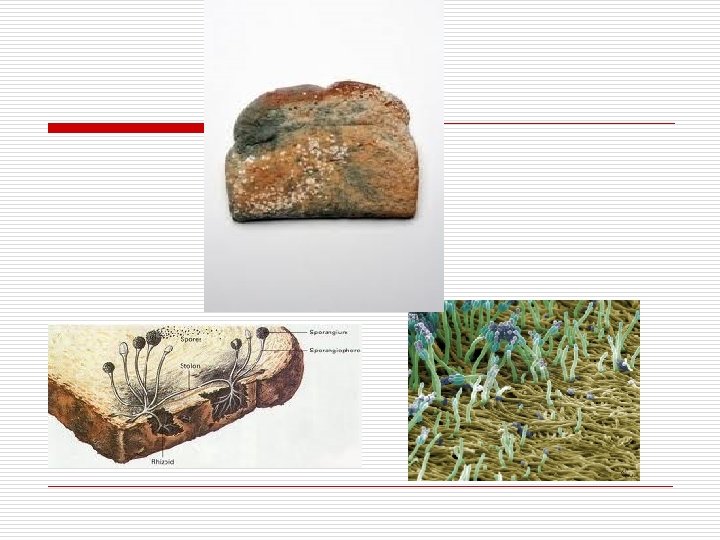
Fungal Origin and Phylogeny o o Probably originated from an aquatic, single-celled protist Split into 5 main Phyla: n n Chytrids-Have flagellate spores, inhabit ponds and damp soil, unicellular or only composed of a few cells, and a few species have alternation of generations Zygomycetes- (ex: Black Bread mold)Live in soil or decaying plants, and some form mycorrhizal relationships with plants, o Produce zygospores for sexual reproduction and often are self-sterile, but can reproduce when they contact an individual of a different mating type Includes Microspores-small parasitic spores of eukaryotic cells n n n o Glomeromycetes-these form most of the mycorrhizal relationships with plants. They have no septa in their hyphae, extend their hyphae into the cell walls of plants, but not the plasma membrane Ascomycetes-(ex: most yeasts, powdery mildews, pink, blue-green, and brown mold, also morels and truffles) Produce ascospores for sexual reproduction, and conidia for asexual reproduction. Are commonly called sac fungi b/c o the asci that hold the sexual spores (think about the Sordaria lab) Basidiomycetes- (ex: bracket fungi, mushrooms, puffballs) Commonly called the club fungi b/c they produce a club-shaped basidium for sexual reproduction. They produce spores outside the basidium as opposed to the ascomycetes that produce spores inside of the asci. Basidia are within the gills of the fruiting body
Figure 31 -7 Chytridiomycota make swimming gametes and spores. Gametes Zygomycota hyphae yoke together and form a zygosporangium. Basidiomycota form spores on basidia (little pedestals). Ascomycota form spores in asci (sacs). Zygosporangium Spores Ascus Spore Basidia Flagella Hyphae
Figure 31 -13 b Chytridiomycota include the only fungi in which alternation of generations occurs. FUSION O MIT SIS MITOSIS Gametes form in gametangia Gametophytic mycelium (n) Spore (n) Gametes (n) (plasmogamy and Zygote karyogamy occur (2 n) simultaneously) n IS MEIOS MIT OS IS Sporophytic mycelium (2 n) 2 n Spores form in sporangia
Figure 31 -13 R Zygomycota form yoked hyphae that produce a spore-forming structure (zygosporangium). PLASMOGA MY Coenocytic hyphae Zygosporangium (n + n) Hyphae fuse Spores (n) Zygote YOG MITOSIS MEIOSIS n KAR AMY n+n 2 n Basidiomycota have reproductive structures with many spore-producing basidia. 2 n MEIOSIS KARYOGAMY Spores (n) Basidium Mature sporeproducing body (n + n) Hyphae of different mating types fuse Heterokaryotic mycelium begins to grow Spores (n) germinate to form hyphae IS OS MIT PLASMOGAMY Ascomycota have reproductive structures with many spore-producing asci. IS IOS ME MITOSIS Spores (n) germinate to form hyphae Ascocarp (mature AMY spore-producing YOG KAR body) (n + n) MIT Spores (n) 2 n OSI S Ascus Eight spores formed Dikaryotic mycelium begins to grow MIT OS Structures containing many nuclei form Hyphae of different mating types make contact and fuse IS PLASMOGAMY