Kinesiology Biomechanistic processes Lecture 14 15 Kinesiology Kinesiology

Kinesiology: Bio-mechanistic processes Lecture 14 & 15

Kinesiology • Kinesiology is the study of the principles of mechanics and anatomy in relation to human movement. • Brings together anatomy, physiology, physics, and geometry as they relate to the human bodies movement.

Kinesiology • A sound understanding of kinesiology allows for the development of a rational evaluation, a precise diagnosis, and an effective treatment of musculoskeletal disorders, and allows for safe, appropriate exercise prescription

Kinesiology • Kinesiology enables the detection and correction of various imbalances that may relate to stress, nutrition, learning problems, minor injuries and other issues encountered in daily life.

Kinesiology • The study of Kinesiology borrows heavily from the sciences of anatomy, biomechanics, and physiology. • Anatomy – the science of the shape and structure of the human body and its parts. • Biomechanics – a discipline that uses principles of physics to quantitatively study how forces interact with the living body. • Physiology – the biologic study of living organism.

Biomechanics “Biomechanics is the study of the structure function of biological systems”. and Bio= Living Mechanics= Forces and effects This includes studies of the tissues including bone, cartilage, ligament, tendon, muscle, and nerve, at multiple scales ranging from the single cell to whole body.

The study of mechanics in the human body divided into 2 areas: Kinematics – study of the variables that describe or quantify motion • Displacement • Velocity • Acceleration Kinetics – study of the variables that cause or influence motion • Forces • Torques • Mass

Biomechanists use the principles of mechanics in the analysis of human movement to answer questions such as: 1. How can human performance be enhanced? 2. How can injuries be prevented? 3. How can rehabilitation from injury be expedited?

Need for study • By having an understanding of the principles of analysis in biomechanics and the biomechanical properties of the primary tissues of the musculoskeletal system, we will be able to understand the mechanics of normal movement at each region and to appreciate the effects of impairments on the pathomechanics of movement.

Neuromuscular Skeletal Biomechanics

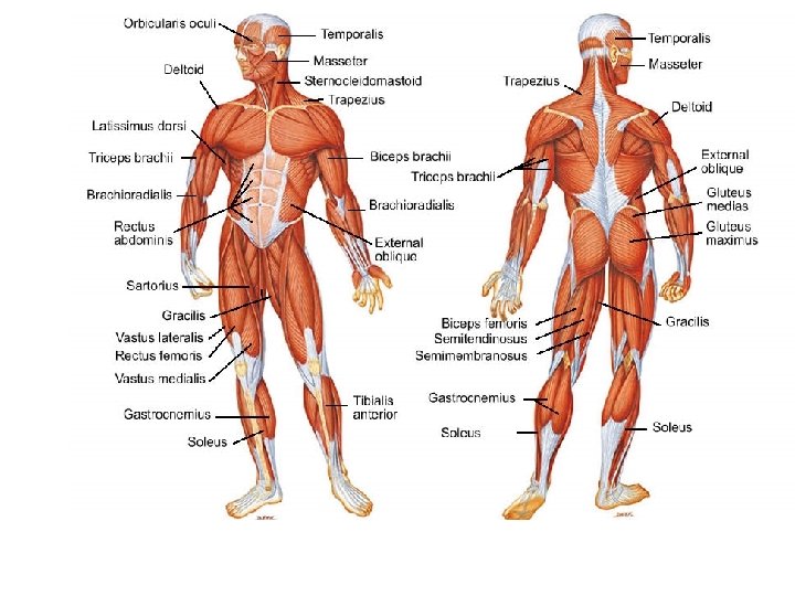
The Muscular System

Muscular System • Muscles provide the forces needed to make movement possible; they transmit their forces to tendons, whose forces in turn cause rotation of the bones about the joints.

Muscular System • Muscles, however, are not simple force generators: the force developed by a muscle depends not only on the level of neural excitation provided by the central nervous system (CNS), but also on the length and speed at which the muscle is contracting.

Muscular System • Muscles can shorten and pull but not push • Most muscles are arranged in opposing teams e. g. agonistic/antagonistic - as each team pulls, the other team relaxes and gets stretched • There are more than 640 muscles(320 in pairs). Nevertheless, exact number is difficult to define. • The muscles make up about 40% of the body mass.

• The longest muscle in the body is Sartorius. • The smallest muscle in the body is Stapedius. It is located deep in the ear. It is only 5 mm long and thinner than cotton thread. It is involved in hearing. • The biggest muscle in the body is Gluteus Maximus. It is located in the buttock. It pulls the leg backwards powerfully for walking and running.

Muscle Gross Structure. Muscles are molecular machines that convert chemical energy into force. Individual muscle fibers are connected together by three levels of collagenous tissue endomysium, which surrounds individual muscle fibers; perimysium, which collects bundles of fibers into fascicles; and epimysium, which encloses the entire muscle belly.

Structure of Muscle

Functions of Muscles • • Movement Maintenance of posture and muscle tone Heat production Protects the bones and internal organs.

Classification of Muscles Functionally • Voluntarily – can be moved at will • Involuntarily – can’t be moved intentionally Structurally • Striated – have stripes across the fiber • Smooth – no striations

Types of Muscles A. B. C. Skeletal Muscles Cardiac Muscles Smooth Muscles

A. Skeletal Muscle • Fibers are long and cylindrical. • Has many nuclei, striations and voluntary • Attached to skeleton by tendons and cause movements of bones at the joints • They do fatigue

Functions of Skeletal muscles A. Movements – muscles move bones by pulling not pushing 1. Synergists – any movement is generally accomplished by more than one muscle. 2. Agonist- most responsible for the movement. 3. Antagonist- muscles and muscle groups usually work in pairs. Biceps flex arm and its partner triceps extend arm. Two muscles are antagonists. 4. Levators - muscle that raise a body part.

Functions of Skeletal muscles B. Maintenance of posture or muscle tone – We are able to maintain our body position because of tonic contractions in our skeletal muscles. C. Heat Production – Contractions of muscles produce most of the heat required to maintain body temperature.

Structural Organization of Skeletal Muscle

Anatomy of a Skeletal Muscle Fiber • The skeletal muscle fiber is a cell. • The Sarcolemma is the plasma membrane. • It has multiple inward extensions which form a set of T Tubules (the T stands for transverse). • The Sarcoplasm is the cytoplasm & the Sarcoplasmic Reticulum is the endoplasmic reticulum. The Sarcoplasmic reticulum is responsible for controlling the release of Calcium ions. • Myofibrils are the cylindrical organelles found inside a muscle fiber. • Myofilaments are the filaments of a myofibril. • Myofilaments are organized into repeating units called Sarcomeres.

B. Cardiac Muscles • Cells are branched and appear fused with one another , has striations, each cell has a central nucleus and involuntary. • Found only in the heart. • Healthy cardiac muscle never fatigue.

C. Smooth Muscle • Fibers are thin and spindle shaped. No striations, single nuclei, involuntary and contracts slowly. They fatigue but slowly. • Found in circulatory (lining of blood vessels), in digestive, respiratory and urinary system.
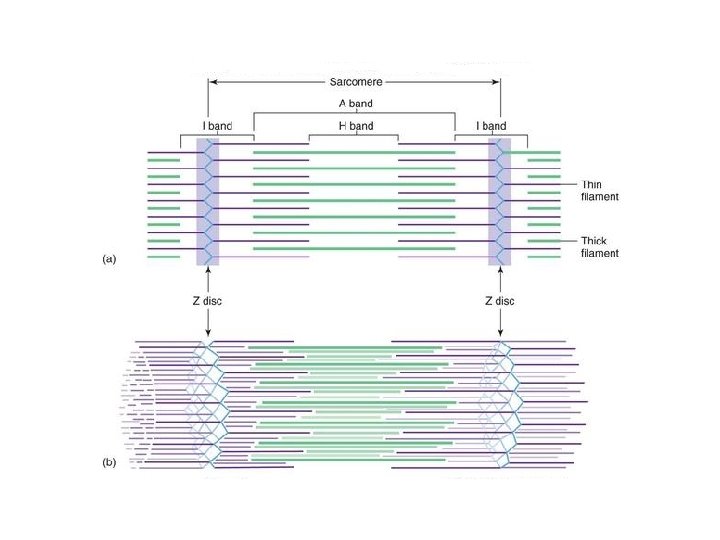
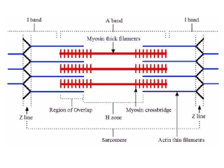
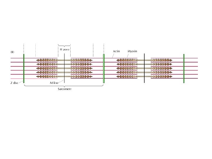
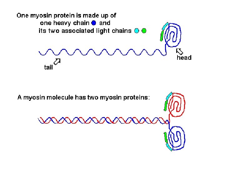


Microscopic Anatomy of Skeletal Muscle · Organization of the sarcomere · Thick filaments = myosin filaments · Composed of the protein myosin · Has ATPase enzymes

Microscopic Anatomy of Skeletal Muscle · Organization of the sarcomere · Thin filaments = actin filaments · Composed of the protein actin

Nerve Stimulus to Muscles · Neuromuscu lar junctions –association site of nerve and muscle

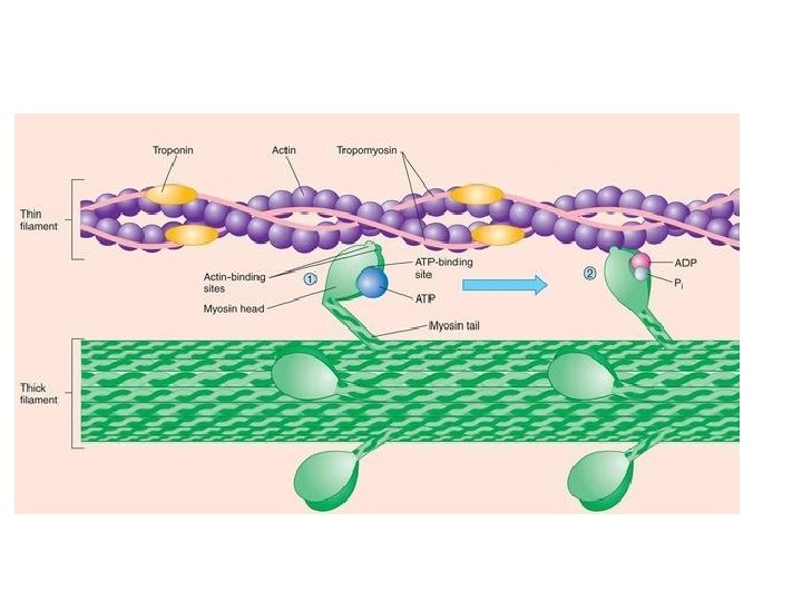
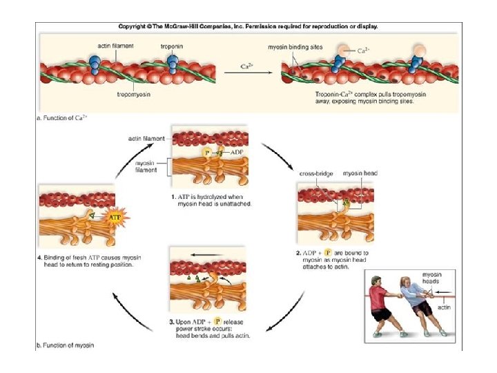
The Sliding Filament Theory of Muscle Contraction · Activation by nerve causes myosin heads (crossbridges) to attach to binding sites on the thin filament · Myosin heads then bind to the next site of the thin filament

The Sliding Filament Theory of Muscle Contraction · This continued action causes a sliding of the myosin along the actin · The result is that the muscle is shortened (contracted)

The Sliding Filament Theory
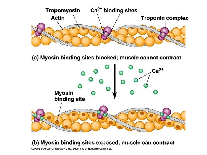

Calcium attaches to troponin/ tropomyosin; they roll away, exposing the active site on actin.

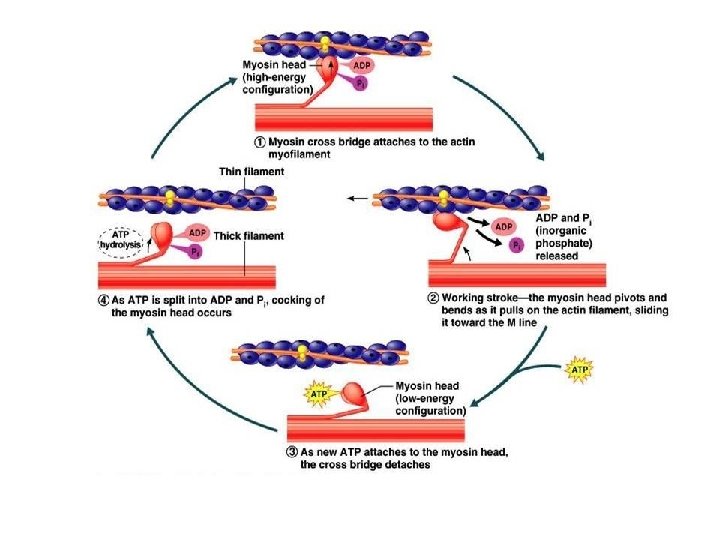
Myosin cross-bridges attach to active site on actin. After attachment, the cross-bridges pivot, pulling the thin filaments.

A fresh ATP replaces the ADP+Pi, allowing myosin and actin to detach. Energy from the splitting of the fresh ATP allows repositioning of the myosin head.

This leads back to Step 1, which continues the cycle as long as calcium ions are attached to troponin/tropomyosin.
- Slides: 47