
KIDS G I CENTER Neonatal Cholestasis Ictericia Neonatal



- Slides: 80
KIDS G. I. CENTER Neonatal Cholestasis (Ictericia Neonatal) Abordaje y manejo RUBEN GONZALEZ-VALLINA, MD, FAAP Director, Gastroenterology Out. Patient Initiatives rubengidoc@aol. com
BREAST MILK JAUNDICE Ø Early Onset ØVery common – 12 percent of breast-fed babies affected. ØOnset during the first three days of life. ØMay be related to caloric deprivation and dehydration.
BREAST MILK JAUNDICE Ø Late Onset Ø Occurs in approximately 0. 5 to 3 percent of healthy newborn. Ø Serum bilirubin rises rapidly after the fourth day of life. Ø Serum bilirubin peaks at the end of the second week of life. Ø Severe jaundice (bilirubin 15 mg per deciliter) occurs in 2 percent of breast-fed babies and 0. 3 percent of formula-fed babies.
BREAST MILK JAUNDICE Ø Ø Not associated with kernicterus. Cessation of breast-feeding for 24 to 48 hours results in a significant drop in serum bilirubin. Ø etiology [? ] Ø abnormal progesterone metabolite Inhibition of conjugating Ø mechanism ↑ levels of nonesterfied FA Ø ↑ absorption of bile from the GI tract
JAUNDICE – BREAST FEEDING • Bilirubin may to 20 mg/dl = in term infants • Need to interrupt feeding = extremely rare • Resolves spontaneously = without interruption • If bilirubin is too high = interrupt for up to 3 days / bilirubin will • Minimal rebound = after reinstitution of breast feeding
A breast-fed infant is found to have an indirect bilirubin level of 15 mg/d. L on day 15 of life. Of the following, the statement which most correctly describes hyperbilirubinemia as a result of ingestion of breast milk is: (A) Excess bilirubin production is the primary etiologic factor. (B) A bilirubin level >10 mg-d. L on the first day of life is typical (C) The condition will resolve spontaneously (D) Affected infants are at risk of kernicterus (E) The bilirubin is transmitted in the breast milk.
KERNICTERUS Early Signs: n Poor sucking n Lethargy Late Signs: Seizures n Fever n Direct bilirubin does not give kernicterus
JAUNDICE BREAST FEEDING VITAMIN K DEFICIENCY Vitamin K Content Ø Commercial formulas = 50 mcg/L Ø Human milk = <1 mcg/L
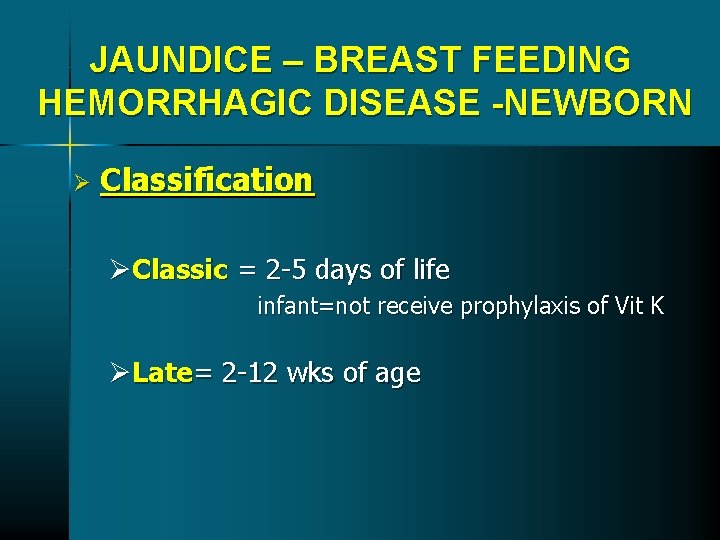
JAUNDICE – BREAST FEEDING HEMORRHAGIC DISEASE -NEWBORN Ø Classification ØClassic = 2 -5 days of life infant=not receive prophylaxis of Vit K ØLate= 2 -12 wks of age
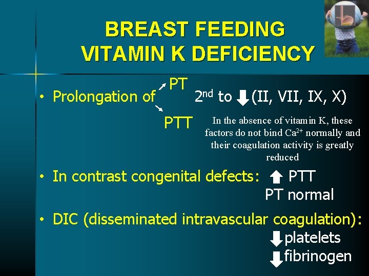
BREAST FEEDING VITAMIN K DEFICIENCY • Prolongation of PT PTT 2 nd to (II, VII, IX, X) In the absence of vitamin K, these factors do not bind Ca 2+ normally and their coagulation activity is greatly reduced • In contrast congenital defects: PTT PT normal • DIC (disseminated intravascular coagulation): platelets fibrinogen
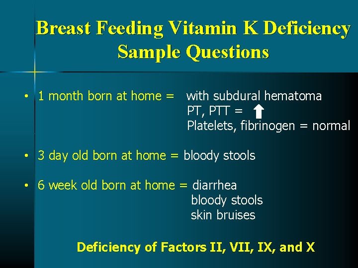
Breast Feeding Vitamin K Deficiency Sample Questions • 1 month born at home = with subdural hematoma PT, PTT = Platelets, fibrinogen = normal • 3 day old born at home = bloody stools • 6 week old born at home = diarrhea bloody stools skin bruises Deficiency of Factors II, VII, IX, and X
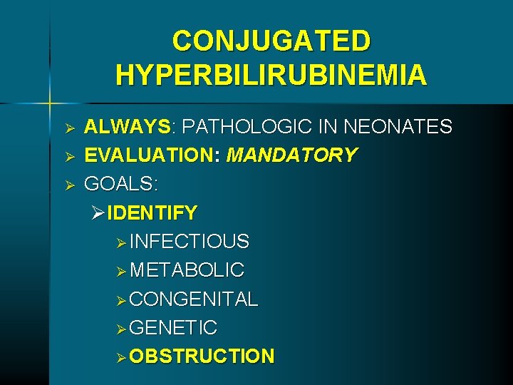
CONJUGATED HYPERBILIRUBINEMIA Ø Ø Ø ALWAYS: PATHOLOGIC IN NEONATES EVALUATION: MANDATORY GOALS: ØIDENTIFY Ø INFECTIOUS Ø METABOLIC Ø CONGENITAL Ø GENETIC Ø OBSTRUCTION
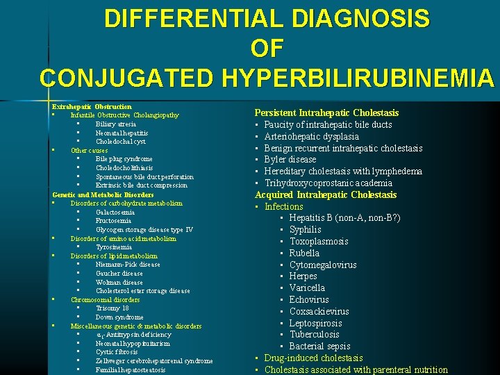
DIFFERENTIAL DIAGNOSIS OF CONJUGATED HYPERBILIRUBINEMIA Extrahepatic Obstruction • Infantile Obstructive Cholangiopathy • Biliary atresia • Neonatal hepatitis • Choledochal cyst • Other causes • Bile plug syndrome • Choledocholithiasis • Spontaneous bile duct perforation • Extrinsic bile duct compression Genetic and Metabolic Disorders • Disorders of carbohydrate metabolism • Galactosemia • Fructosemia • Glycogen storage disease type IV • Disorders of amino acid metabolism • Tyrosinemia • Disorders of lipid metabolism • Niemann-Pick disease • Gaucher disease • Wolman disease • Cholesterol ester storage disease • Chromosomal disorders • Trisomy 18 • Down syndrome • Miscellaneous genetic & metabolic disorders • α 1 -Antitrypsin deficiency • Neonatal hypopituitarism • Cystic fibrosis • Zellweger cerebrohepatorenal syndrome • Familial hepatosteatosis Persistent Intrahepatic Cholestasis • Paucity of intrahepatic bile ducts • Arteriohepatic dysplasia • Benign recurrent intrahepatic cholestasis • Byler disease • Hereditary cholestasis with lymphedema • Trihydroxycoprostanic academia Acquired Intrahepatic Cholestasis • Infections • Hepatitis B (non-A, non-B? ) • Syphilis • Toxoplasmosis • Rubella • Cytomegalovirus • Herpes • Varicella • Echovirus • Coxsackievirus • Leptospirosis • Tuberculosis • Bacterial sepsis • Drug-induced cholestasis • Cholestasis associated with parenteral nutrition
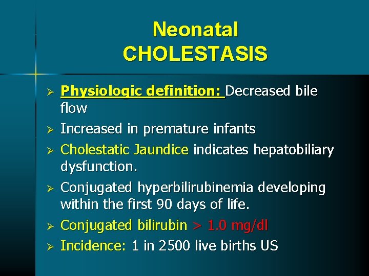
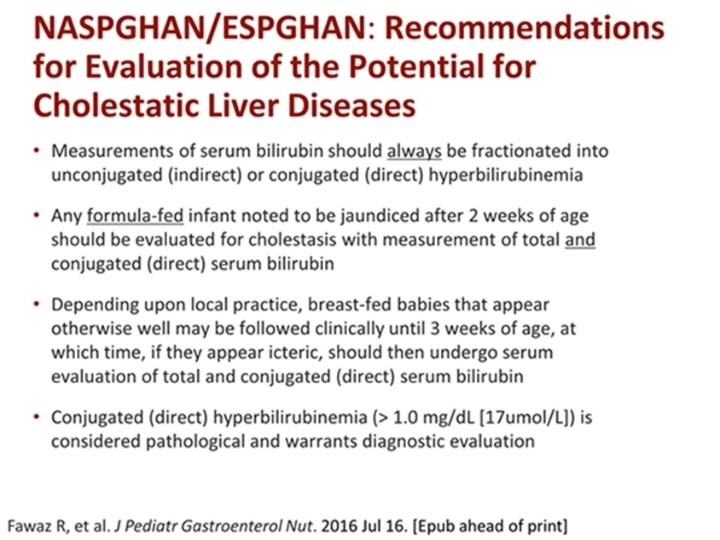
Neonatal CHOLESTASIS Ø Ø Ø Physiologic definition: Decreased bile flow Increased in premature infants Cholestatic Jaundice indicates hepatobiliary dysfunction. Conjugated hyperbilirubinemia developing within the first 90 days of life. Conjugated bilirubin > 1. 0 mg/dl Incidence: 1 in 2500 live births US
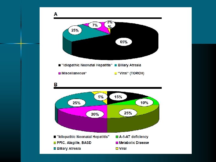
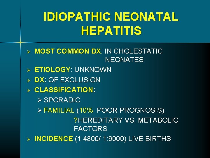
IDIOPATHIC NEONATAL HEPATITIS Ø Ø Ø MOST COMMON DX: IN CHOLESTATIC NEONATES ETIOLOGY: UNKNOWN DX: OF EXCLUSION CLASSIFICATION: Ø SPORADIC Ø FAMILIAL (10% POOR PROGNOSIS) ? HEREDITARY VS. METABOLIC FACTORS INCIDENCE (1: 4800/ 1: 9000) LIVE BIRTHS
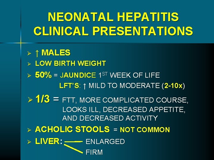
NEONATAL HEPATITIS CLINICAL PRESENTATIONS Ø ↑ MALES Ø LOW BIRTH WEIGHT Ø 50% = JAUNDICE 1 ST WEEK OF LIFE LFT’S: ↑ MILD TO MODERATE (2 -10 x) Ø 1/3 = FTT, MORE COMPLICATED COURSE, LOOKS ILL, DECREASED APPETITE, AND DECREASED ACTIVITY Ø Ø ACHOLIC STOOLS = NOT COMMON LIVER: ENLARGED FIRM
TREATMENT Ø Ø CONSERVATIVE PROGNOSIS : GOOD
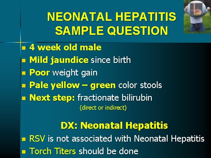
NEONATAL HEPATITIS SAMPLE QUESTION n n n 4 week old male Mild jaundice since birth Poor weight gain Pale yellow – green color stools Next step: fractionate bilirubin (direct or indirect) DX: Neonatal Hepatitis n n RSV is not associated with Neonatal Hepatitis Torch Titers should be done
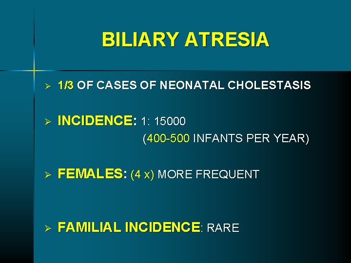
BILIARY ATRESIA Ø 1/3 OF CASES OF NEONATAL CHOLESTASIS Ø INCIDENCE: 1: 15000 (400 -500 INFANTS PER YEAR) Ø FEMALES: (4 x) MORE FREQUENT Ø FAMILIAL INCIDENCE: RARE
BILIARY ATRESIA Ø IS A DYNAMIC AND PROGRESSIVE PANDUCTULAR SCLEROTIC PROCESS ØETIOLOGY UNKNOWN REOVIRUS TYPE 3 (? )
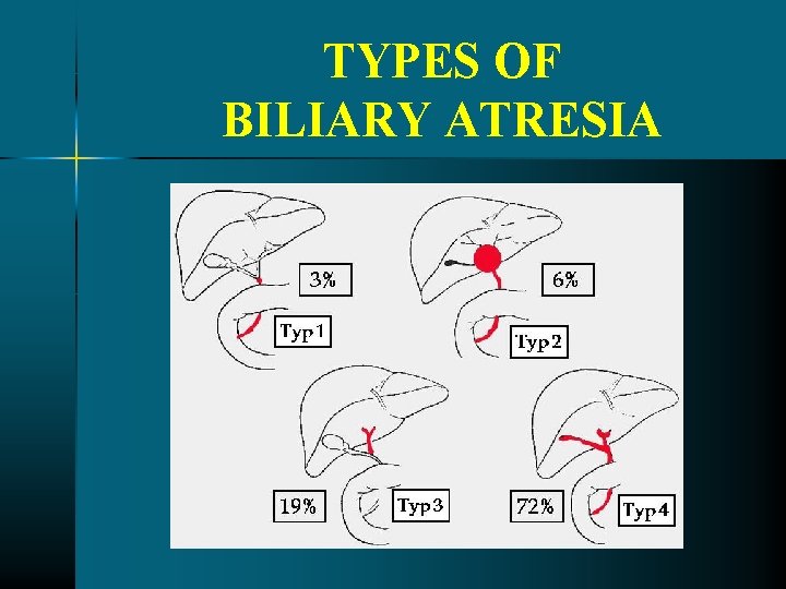
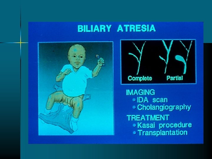
TYPES OF BILIARY ATRESIA
BILIARY ATRESIA CLINICAL PRESENTATIONS Ø Ø Ø HINT OBSTRUCTIVE JAUNDICE ØPATIENT LOOKS WELL PARENCHYMAL OR METABOLIC DISEASE ØPATIENT LOOKS ILL
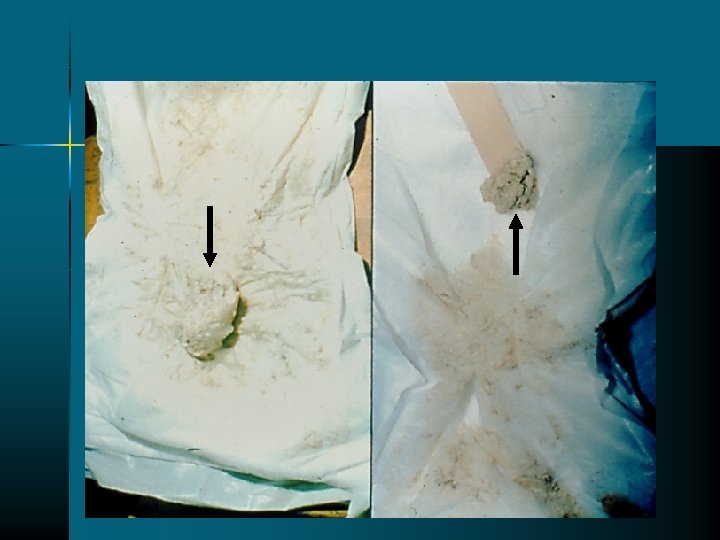
BILIARY ATRESIA CLINICAL PRESENTATIONS Ø JAUNDICE: 3 -5 WEEKS AFTER BIRTH Ø LFT’S: MILD ELEVATION UP TO 5 x (USUALLY IN THE 200 RANGE, 150 -300). Ø BILIRUBIN: 14 -15 RANGE, MOSTLY DIRECT Ø ALK PHOSPHATASE: MARKEDLY ELEVATED Ø HEPATOSPLENOMEGALY Ø ACHOLIC STOOLS: COMMON
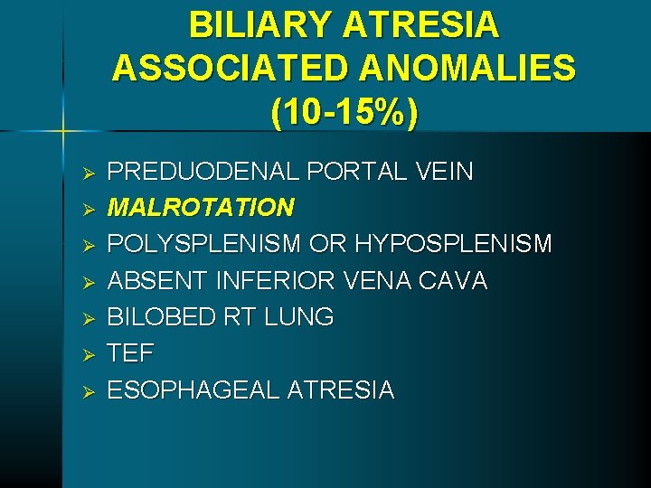
BILIARY ATRESIA ASSOCIATED ANOMALIES (10 -15%) Ø Ø Ø Ø PREDUODENAL PORTAL VEIN MALROTATION POLYSPLENISM OR HYPOSPLENISM ABSENT INFERIOR VENA CAVA BILOBED RT LUNG TEF ESOPHAGEAL ATRESIA
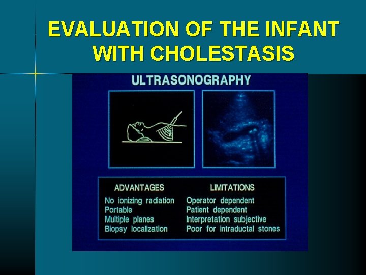
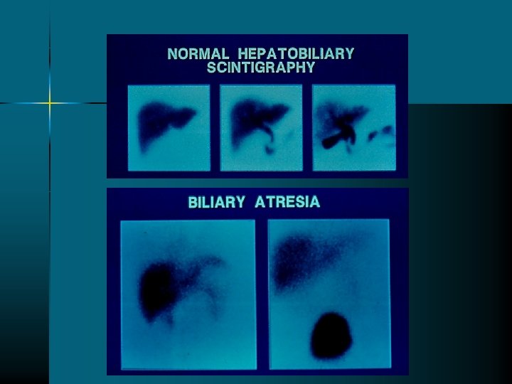
EVALUATION OF THE INFANT WITH CHOLESTASIS
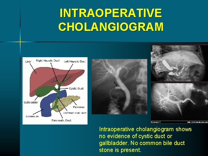
INTRAOPERATIVE CHOLANGIOGRAM Intraoperative cholangiogram shows no evidence of cystic duct or gallbladder. No common bile duct stone is present.
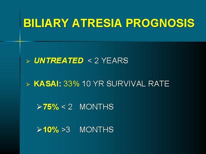
BILIARY ATRESIA PROGNOSIS Ø UNTREATED < 2 YEARS Ø KASAI: 33% 10 YR SURVIVAL RATE Ø 75% < 2 MONTHS Ø 10% >3 MONTHS
BILIARY ATRESIA SAMPLE QUESTIONS Ø Ø Ø 4 week old = female, male Looks well except that appear yellow Mild hepatosplenomegaly TB = 10, LFTS = 200 range Next Step: URGENT to differentiate between intra/extrahepatic obstruction and fractionate bilirubin (direct or indirect)
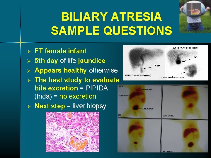
BILIARY ATRESIA SAMPLE QUESTIONS Ø Ø Ø FT female infant 5 th day of life jaundice Appears healthy otherwise The best study to evaluate bile excretion = PIPIDA (hida) = no excretion Next step = liver biopsy
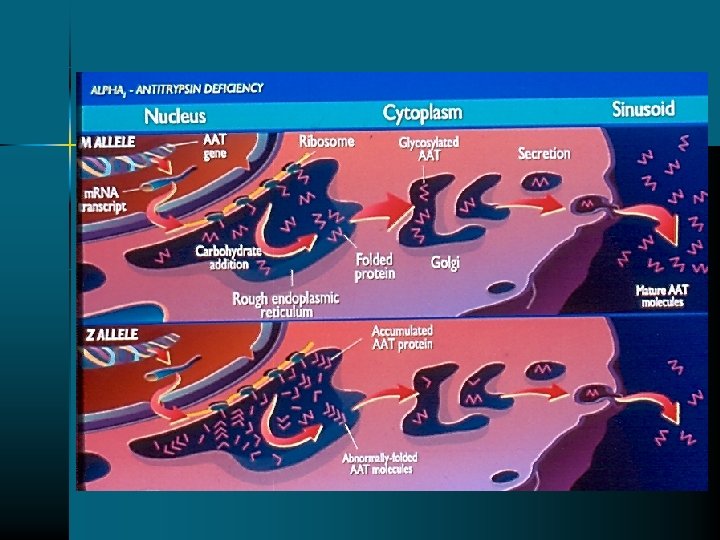
ALPHA 1 -ANTITRYPSIN Ø IS A GLYCOPROTEIN Ø PRODUCED BY HEPATOCYTES (2 GRAMS PER DAY) Ø SERINE – PROTEINASE INHIBITOR Ø INHIBITOR OF: NEUTROPHIL ELASTASE ACID PROTEASES OF ALVEOLAR MACROPHAGES
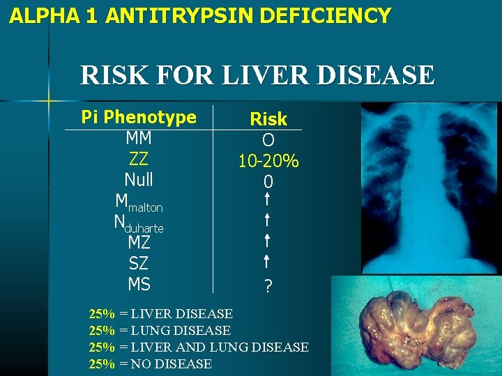
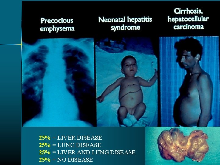
ALPHA 1 ANTITRYPSIN DEFICIENCY RISK FOR LIVER DISEASE Pi Phenotype MM ZZ Null Mmalton Nduharte MZ SZ MS Risk O 10 -20% 0 ? 25% = LIVER DISEASE 25% = LUNG DISEASE 25% = LIVER AND LUNG DISEASE 25% = NO DISEASE
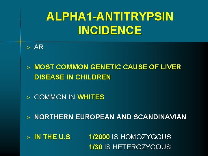
ALPHA 1 -ANTITRYPSIN INCIDENCE Ø AR Ø MOST COMMON GENETIC CAUSE OF LIVER DISEASE IN CHILDREN Ø COMMON IN WHITES Ø NORTHERN EUROPEAN AND SCANDINAVIAN Ø IN THE U. S. 1/2000 IS HOMOZYGOUS 1/30 IS HETEROZYGOUS
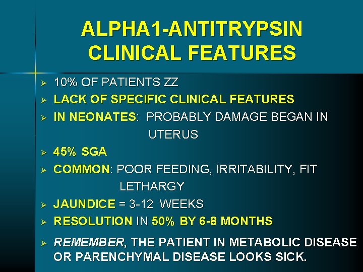
ALPHA 1 -ANTITRYPSIN CLINICAL FEATURES Ø Ø Ø Ø 10% OF PATIENTS ZZ LACK OF SPECIFIC CLINICAL FEATURES IN NEONATES: PROBABLY DAMAGE BEGAN IN UTERUS 45% SGA COMMON: POOR FEEDING, IRRITABILITY, FIT LETHARGY JAUNDICE = 3 -12 WEEKS RESOLUTION IN 50% BY 6 -8 MONTHS REMEMBER, THE PATIENT IN METABOLIC DISEASE OR PARENCHYMAL DISEASE LOOKS SICK.
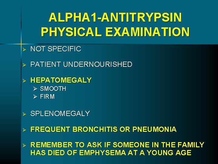
ALPHA 1 -ANTITRYPSIN PHYSICAL EXAMINATION Ø NOT SPECIFIC Ø PATIENT UNDERNOURISHED Ø HEPATOMEGALY Ø SMOOTH Ø FIRM Ø SPLENOMEGALY Ø FREQUENT BRONCHITIS OR PNEUMONIA Ø REMEMBER TO ASK IF SOMEONE IN THE FAMILY HAS DIED OF EMPHYSEMA AT A YOUNG AGE
25% = LIVER DISEASE 25% = LUNG DISEASE 25% = LIVER AND LUNG DISEASE 25% = NO DISEASE
ALPHA 1 ANTITRYPSIN DEFICIENCY SAMPLE QUESTIONS 10 year old with no PMH; presents with UGI bleeding • Hepatosplenomegaly • Ascites • platelets • Mild in LFTs, GGTP • PT Test of diagnosis? Alpha 1 -antitrypsin level and phenotype Always remember to get a phenotype because level could be normal, or in infections. It is an acute phase reactant.
ALPHA 1 ANTITRYPSIN DEFICIENCY SAMPLE QUESTIONS 9 month old with • • FH = + emphysema Hepatomegaly + icterus Looks ill Appetite Irritability lethargy Test for diagnosis? Alpha 1 -antitrypsin level and phenotype Always remember to get a phenotype because level could be normal in infections. It is an acute phase reactant.
ALPHA 1 ANTITRYPSIN DEFICIENCY SAMPLE QUESTIONS • serum alpha 1 antitrypsin level is associated with early cirrhosis • Most common manifestation in infancy • Hepatic cirrhosis
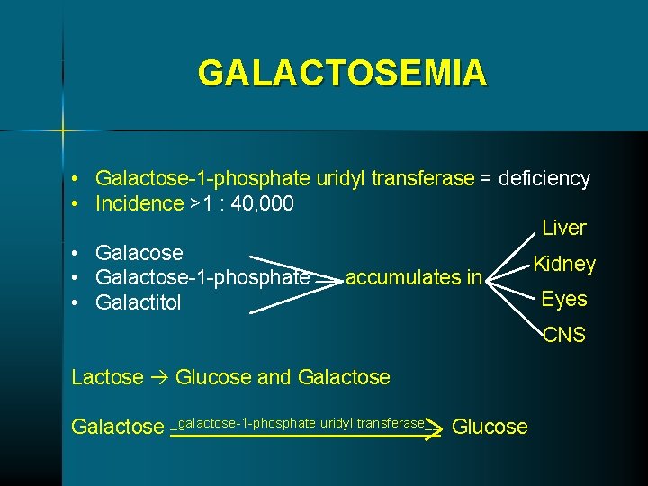
GALACTOSEMIA • Galactose-1 -phosphate uridyl transferase = deficiency • Incidence >1 : 40, 000 Liver • Galacose Kidney • Galactose-1 -phosphate accumulates in Eyes • Galactitol CNS Lactose Glucose and Galactose _galactose-1 -phosphate uridyl transferase__ Glucose
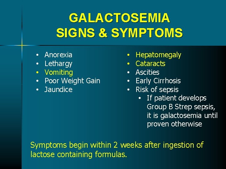
GALACTOSEMIA SIGNS & SYMPTOMS • • • Anorexia Lethargy Vomiting Poor Weight Gain Jaundice • • • Hepatomegaly Cataracts Ascities Early Cirrhosis Risk of sepsis • If patient develops Group B Strep sepsis, it is galactosemia until proven otherwise Symptoms begin within 2 weeks after ingestion of lactose containing formulas.
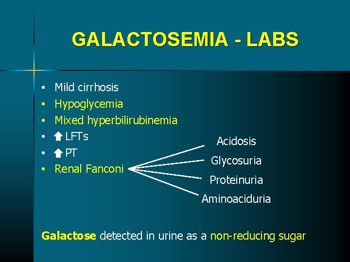
GALACTOSEMIA - LABS • • • Mild cirrhosis Hypoglycemia Mixed hyperbilirubinemia LFTs PT Renal Fanconi Acidosis Glycosuria Proteinuria Aminoaciduria Galactose detected in urine as a non-reducing sugar
GALACTOSEMIA DIAGNOSIS • Measure in RBC’s = galactose-1 - phosphate uridyl activity • Treatment = remove galactose from diet
GALACTOSEMIA SAMPLE QUESTIONS • 2 week old breast fed • appetite • Jaundice, hepatomegaly • Glucose oxidate test in urine = (-) • Check reducing substance in urine, order a Clinitest Galactose detected in urine as a non-reducing sugar
GALACTOSEMIA SAMPLE QUESTIONS 4 week old V + D; multiple cow’s milk formula changes • Appetite • Lethargic • Jaundice, hepatomegaly • Order: • Galactose-1 -phosphate uridyl transferase activity in RBCs • Diagnosis: • Galactosemia • Treatment: • Soy formula
GALACTOSEMIA SAMPLE QUESTIONS 2 -3 month breast fed; prolonged jaundice • V+D • Hepatomegaly, Hypotonia • Initial test = check reducing substance • Order a galactose-1 -phosphate uridyl transferase activity in RBCs
HEREDITARY FRUCTOSE INTOLERANCE Ø Ø Incidence: 1: 20, 000 AR Deficiency: Adolase B ØLiver ØKidney ØSmall Bowel Failure to Split Fructose 1 Phosphate
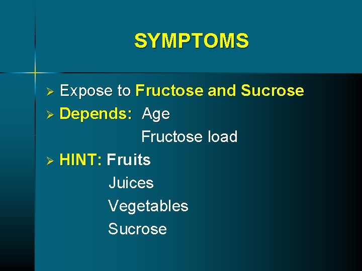
SYMPTOMS Expose to Fructose and Sucrose Ø Depends: Age Fructose load Ø HINT: HINT Fruits Juices Vegetables Sucrose Ø
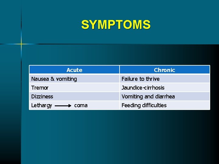
SYMPTOMS Acute Chronic Nausea & vomiting Failure to thrive Tremor Jaundice-cirrhosis Dizziness Vomiting and diarrhea Lethargy coma Feeding difficulties
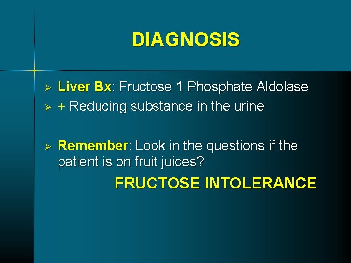
DIAGNOSIS Ø Ø Ø Liver Bx: Fructose 1 Phosphate Aldolase + Reducing substance in the urine Remember: Look in the questions if the patient is on fruit juices? FRUCTOSE INTOLERANCE
THANK YOU!!!!!!
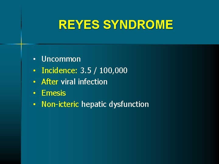
REYES SYNDROME • • • Uncommon Incidence: 3. 5 / 100, 000 After viral infection Emesis Non-icteric hepatic dysfunction
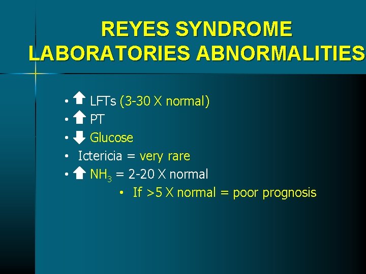
REYES SYNDROME LABORATORIES ABNORMALITIES • LFTs (3 -30 X normal) • PT • Glucose • Ictericia = very rare • NH 3 = 2 -20 X normal • If >5 X normal = poor prognosis
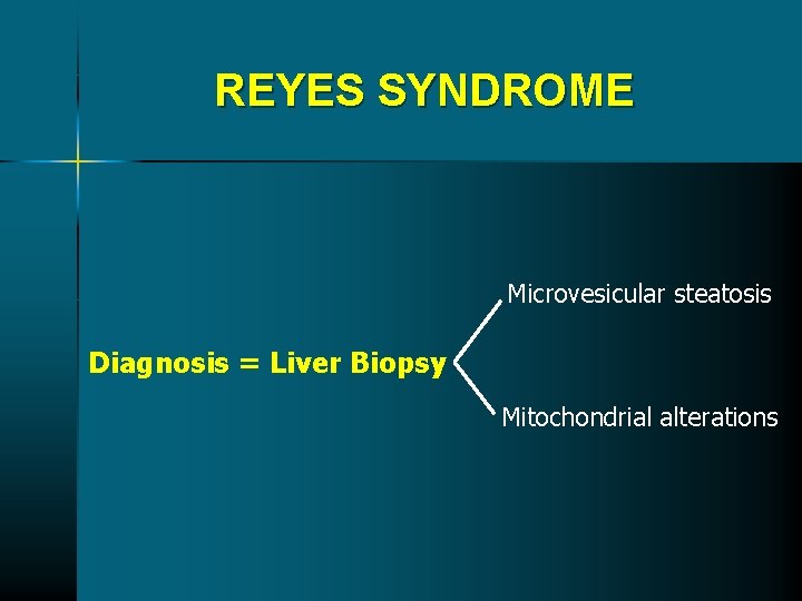
REYES SYNDROME Microvesicular steatosis Diagnosis = Liver Biopsy Mitochondrial alterations
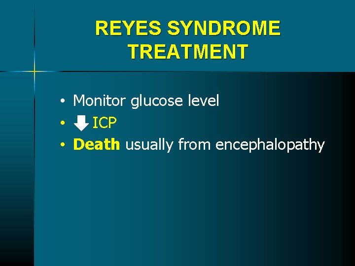
REYES SYNDROME TREATMENT • Monitor glucose level • ICP • Death usually from encephalopathy
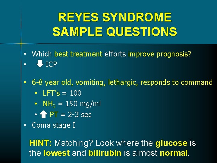
REYES SYNDROME SAMPLE QUESTIONS • Which best treatment efforts improve prognosis? • ICP • 6 -8 year old, vomiting, lethargic, responds to command • LFT’s = 100 • NH 3 = 150 mg/ml • PT = 2 -3 sec • Coma stage I HINT: Matching? Look where the glucose is the lowest and bilirubin is almost normal.
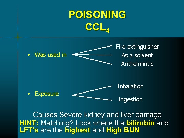
POISONING CCL 4 • Was used in Fire extinguisher As a solvent Anthelmintic Inhalation • Exposure Ingestion Causes Severe kidney and liver damage HINT: Matching? Look where the bilirubin and LFT’s are the highest and High BUN
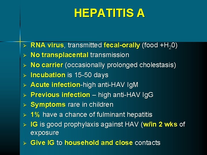
HEPATITIS A Ø Ø Ø Ø Ø RNA virus, transmitted fecal-orally (food +H 20) No transplacental transmission No carrier (occasionally prolonged cholestasis) Incubation is 15 -50 days Acute infection-high anti-HAV Ig. M Previous infection – high anti-HAV Ig. G Symptoms rare in children 1% have a chance of fulminant hepatitis IG is good prophylaxis against HAV (w/in 2 wks of exposure Give IG to household and close contacts
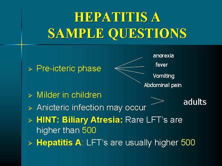
HEPATITIS A SAMPLE QUESTIONS anorexia Ø Pre-icteric phase fever Vomiting Abdominal pain Ø Ø Milder in children adults Anicteric infection may occur HINT: Biliary Atresia: Rare LFT’s are higher than 500 Hepatitis A: A LFT’s are usually higher 500
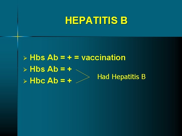
HEPATITIS B Hbs Ab = + = vaccination Ø Hbs Ab = + Had Hepatitis B Ø Hbc Ab = + Ø
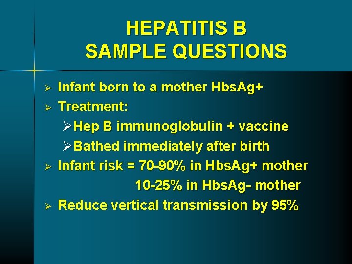
HEPATITIS B SAMPLE QUESTIONS Ø Ø Infant born to a mother Hbs. Ag+ Treatment: ØHep B immunoglobulin + vaccine ØBathed immediately after birth Infant risk = 70 -90% in Hbs. Ag+ mother 10 -25% in Hbs. Ag- mother Reduce vertical transmission by 95%
HEPATITIS B SAMPLE QUESTIONS Hepatitis Bs Ag = + Ø No need to be vaccinated Ø
CONSTIPATION Majority is due to functional or behavioral problem Ø Small percentage of children present with constipation have an organic cause Ø Some breast-fed babies will stool 1/5 -10 days Ø Functional fecal retention is the most common nonorganic cause HINT: Rectal examination: examination Rectal ampulla is full Ø
HIRSCHPRUNG DISEASE 1/5000 births Ø Absence of enteric ganglionic neurons that begin at the anus and then extend proximally Ø Increased association with Down syndrome Ø Evaluate any term infant that does not pass meconium within 48 hours of birth Ø Suction rectal biopsy for diagnosis, full-thickness biopsy needed for unclear cases Ø Treat with surgical resection HINT: Rectal Ampulla is empty KUB: No granular appearance or Ca 2+ in ABD = CF yes Ø
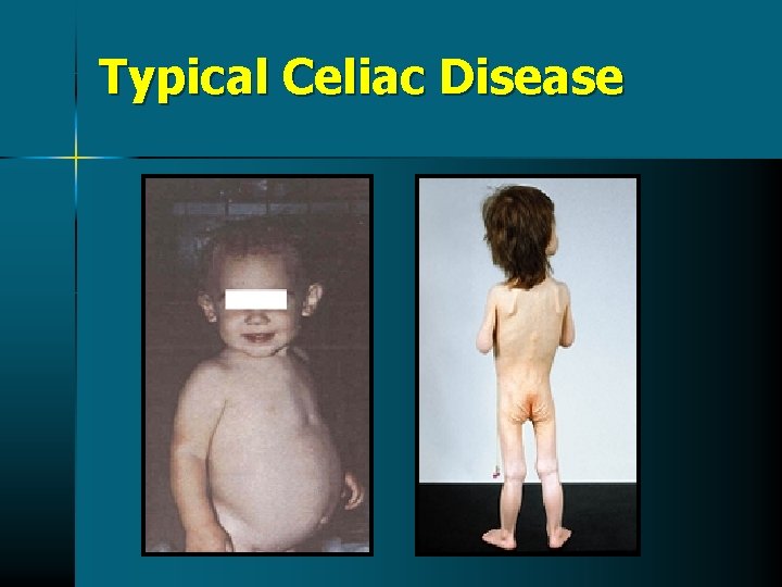
Typical Celiac Disease
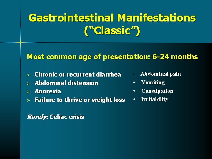
Gastrointestinal Manifestations (“Classic”) Most common age of presentation: 6 -24 months Ø Ø Chronic or recurrent diarrhea Abdominal distension Anorexia Failure to thrive or weight loss Rarely: Celiac crisis • Abdominal pain • Vomiting • Constipation • Irritability
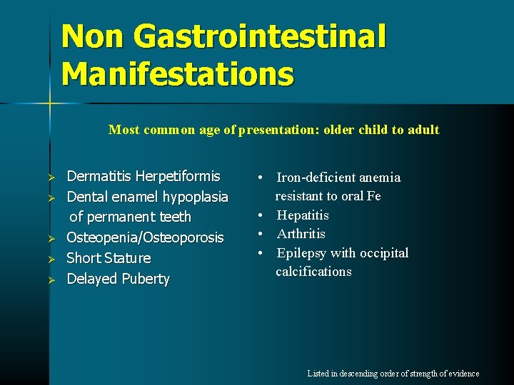
Non Gastrointestinal Manifestations Most common age of presentation: older child to adult Ø Ø Ø Dermatitis Herpetiformis Dental enamel hypoplasia of permanent teeth Osteopenia/Osteoporosis Short Stature Delayed Puberty • Iron-deficient anemia resistant to oral Fe • Hepatitis • Arthritis • Epilepsy with occipital calcifications Listed in descending order of strength of evidence
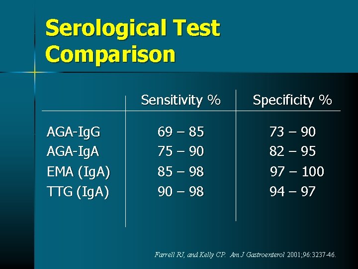
Serological Test Comparison AGA-Ig. G AGA-Ig. A EMA (Ig. A) TTG (Ig. A) Sensitivity % Specificity % 69 – 85 75 – 90 85 – 98 90 – 98 73 – 90 82 – 95 97 – 100 94 – 97 Farrell RJ, and Kelly CP. Am J Gastroenterol 2001; 96: 3237 -46.
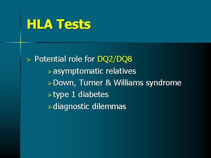
HLA Tests Ø Potential role for DQ 2/DQ 8 Ø asymptomatic relatives Ø Down, Turner & Williams syndrome Ø type 1 diabetes Ø diagnostic dilemmas
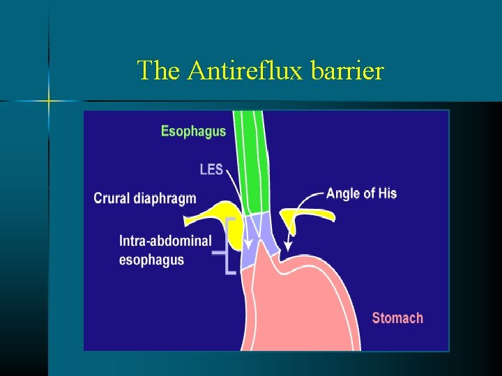
The Antireflux barrier
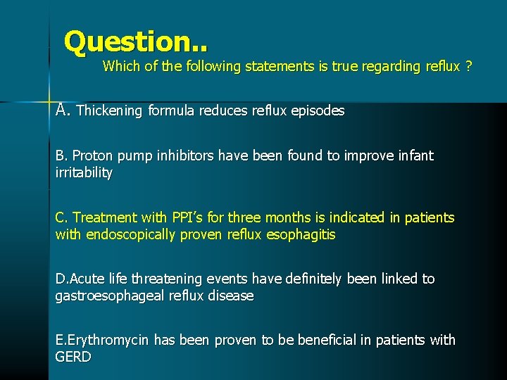
Question. . Which of the following statements is true regarding reflux ? A. Thickening formula reduces reflux episodes B. Proton pump inhibitors have been found to improve infant irritability C. Treatment with PPI’s for three months is indicated in patients with endoscopically proven reflux esophagitis D. Acute life threatening events have definitely been linked to gastroesophageal reflux disease E. Erythromycin has been proven to be beneficial in patients with GERD
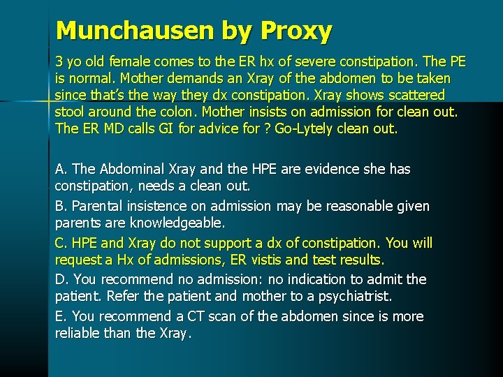
Munchausen by Proxy 3 yo old female comes to the ER hx of severe constipation. The PE is normal. Mother demands an Xray of the abdomen to be taken since that’s the way they dx constipation. Xray shows scattered stool around the colon. Mother insists on admission for clean out. The ER MD calls GI for advice for ? Go-Lytely clean out. A. The Abdominal Xray and the HPE are evidence she has constipation, needs a clean out. B. Parental insistence on admission may be reasonable given parents are knowledgeable. C. HPE and Xray do not support a dx of constipation. You will request a Hx of admissions, ER vistis and test results. D. You recommend no admission: no indication to admit the patient. Refer the patient and mother to a psychiatrist. E. You recommend a CT scan of the abdomen since is more reliable than the Xray.
KIDS G. I. CENTER RUBEN GONZALEZ-VALLINA, MD, FAAP