
Kidney Injury and Liver Disease in the ICU

- Slides: 29
Kidney Injury and Liver Disease in the ICU German T. Hernandez, MD, FASN, FACP Associate Professor of Medicine Division of Nephrology & Hypertension Paul L. Foster School of Medicine TTUHSC at El Paso
Learning Objectives • 1. Define the Hepatorenal Syndrome • 2. Discuss the use of emerging medical therapies in Hepatorenal Syndrome • 2. Recognize the abdominal compartment syndrome as cause of acute kidney injury
Acute Kidney Injury • Many Definitions: • Increase in serum creatinine ≥ 1. 5 x baseline within 7 days (RIFLE) – or • Increase in serum creatinine by 0. 3 mg/d. L or ≥ 1. 5 x baseline with 48 hrs (AKIN) Crit Care 2004; 8: B 204 Crit Care 2001; 11: R 31
Acute Kidney Injury: Classification • Prerenal AKI • Intrinsic AKI – Acute Tubular Necrosis (ATN) – Interstitial Nephritis – Glomerulonephritis – Vascular syndromes – Intra-tubular obstruction (crystals, myeloma casts) • Post-renal AKI
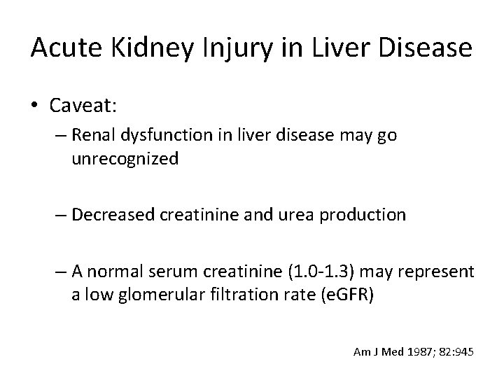
Acute Kidney Injury in Liver Disease • Caveat: – Renal dysfunction in liver disease may go unrecognized – Decreased creatinine and urea production – A normal serum creatinine (1. 0 -1. 3) may represent a low glomerular filtration rate (e. GFR) Am J Med 1987; 82: 945
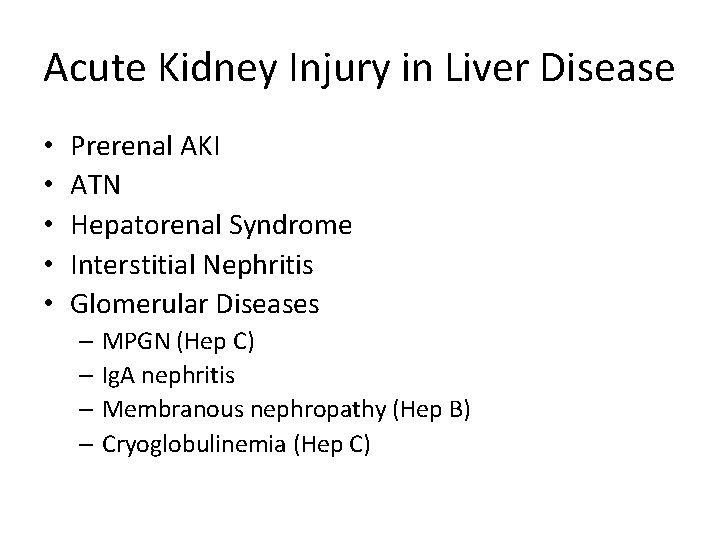
Acute Kidney Injury in Liver Disease • • • Prerenal AKI ATN Hepatorenal Syndrome Interstitial Nephritis Glomerular Diseases – MPGN (Hep C) – Ig. A nephritis – Membranous nephropathy (Hep B) – Cryoglobulinemia (Hep C)
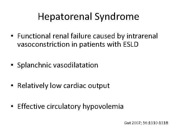
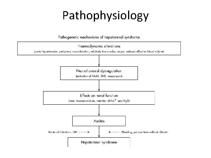
Hepatorenal Syndrome • Functional renal failure caused by intrarenal vasoconstriction in patients with ESLD • Splanchnic vasodilatation • Relatively low cardiac output • Effective circulatory hypovolemia Gut 2007; 56: 1310 -1318
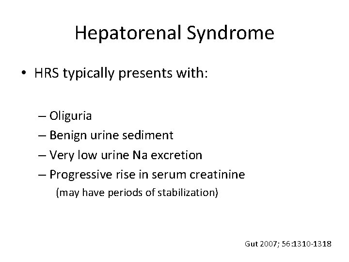
Hepatorenal Syndrome • HRS typically presents with: – Oliguria – Benign urine sediment – Very low urine Na excretion – Progressive rise in serum creatinine (may have periods of stabilization) Gut 2007; 56: 1310 -1318
Pathophysiology
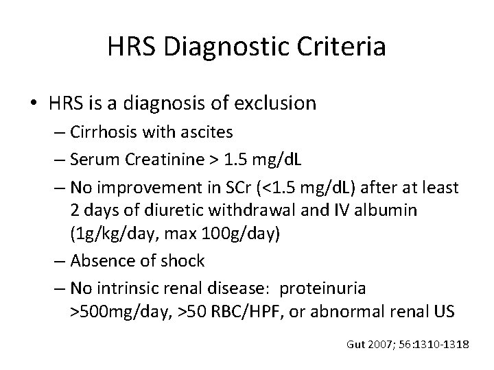
HRS Diagnostic Criteria • HRS is a diagnosis of exclusion – Cirrhosis with ascites – Serum Creatinine > 1. 5 mg/d. L – No improvement in SCr (<1. 5 mg/d. L) after at least 2 days of diuretic withdrawal and IV albumin (1 g/kg/day, max 100 g/day) – Absence of shock – No intrinsic renal disease: proteinuria >500 mg/day, >50 RBC/HPF, or abnormal renal US Gut 2007; 56: 1310 -1318
Hepatorenal Syndrome • Type-1 HRS – Rapid progression of kidney injury with a rise in SCr >2 x baseline in less than 2 weeks – Can develop spontaneously, but commonly follows: • SBP or other infection • GI bleeding Gut 2007; 56: 1310 -1318
Hepatorenal Syndrome • Type-2 HRS – Associated with diuretic-resistant ascites and less renal insufficiency than type-1 HRS Gut 2007; 56: 1310 -1318
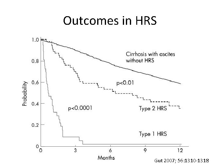
Outcomes in HRS Gut 2007; 56: 1310 -1318
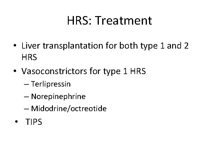
HRS: Treatment • Liver transplantation for both type 1 and 2 HRS • Vasoconstrictors for type 1 HRS – Terlipressin – Norepinephrine – Midodrine/octreotide • TIPS
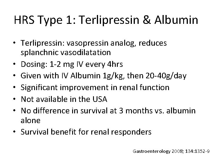
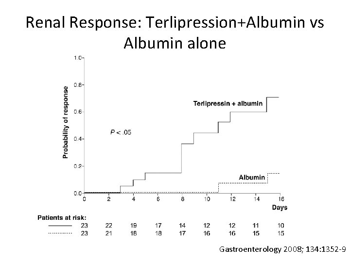
HRS Type 1: Terlipressin & Albumin • Terlipressin: vasopressin analog, reduces splanchnic vasodilatation • Dosing: 1 -2 mg IV every 4 hrs • Given with IV Albumin 1 g/kg, then 20 -40 g/day • Significant improvement in renal function • Not available in the USA • No difference in survival at 3 months vs. albumin alone • Survival benefit for renal responders Gastroenterology 2008; 134: 1352 -9
Renal Response: Terlipression+Albumin vs Albumin alone Gastroenterology 2008; 134: 1352 -9
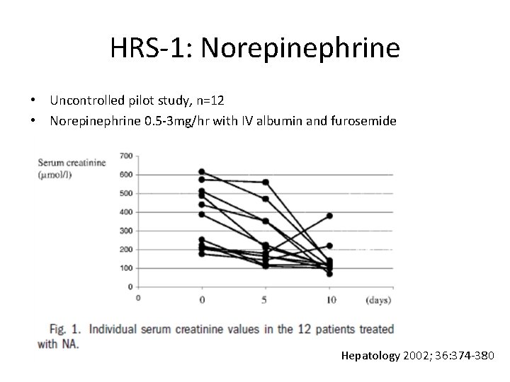
HRS-1: Norepinephrine • Uncontrolled pilot study, n=12 • Norepinephrine 0. 5 -3 mg/hr with IV albumin and furosemide Hepatology 2002; 36: 374 -380
HRS-1: Midodrine & Octreotide • Midodrine- selective alpha-1 adrenergic agonist – Causes increase in peripheral vascular resistance • Octreotide-analogue of somatostatin – Inhibits endogenous vasodilator release, thereby reducing splanchnic vasodilatation • The combination is thought to improve renal and systemic hemodynamics
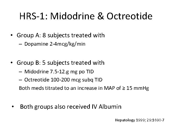
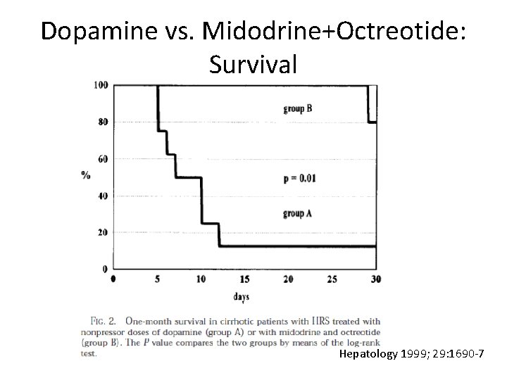
HRS-1: Midodrine & Octreotide • Group A: 8 subjects treated with – Dopamine 2 -4 mcg/kg/min • Group B: 5 subjects treated with – Midodrine 7. 5 -12. g mg po TID – Octreotide 100 -200 mcg subq TID Both meds titrated to an increase in MAP of ≥ 15 mm. Hg • Both groups also received IV Albumin Hepatology 1999; 29: 1690 -7
Dopamine vs. Midodrine+Octreotide Hepatology 1999; 29: 1690 -7
Dopamine vs. Midodrine+Octreotide: Survival Hepatology 1999; 29: 1690 -7
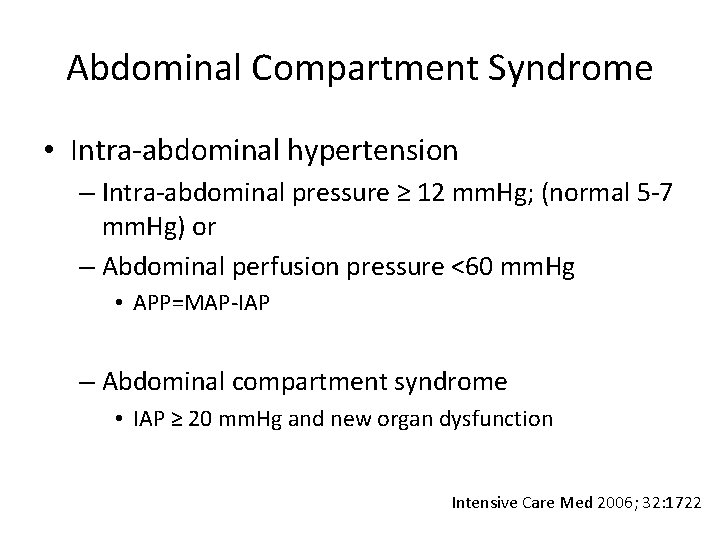
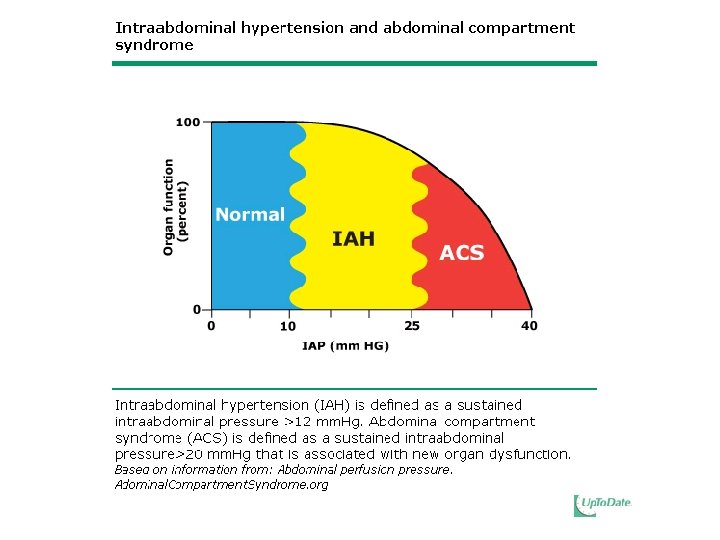
Abdominal Compartment Syndrome • Intra-abdominal hypertension – Intra-abdominal pressure ≥ 12 mm. Hg; (normal 5 -7 mm. Hg) or – Abdominal perfusion pressure <60 mm. Hg • APP=MAP-IAP – Abdominal compartment syndrome • IAP ≥ 20 mm. Hg and new organ dysfunction Intensive Care Med 2006; 32: 1722
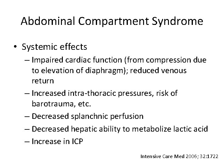
Abdominal Compartment Syndrome • Systemic effects – Impaired cardiac function (from compression due to elevation of diaphragm); reduced venous return – Increased intra-thoracic pressures, risk of barotrauma, etc. – Decreased splanchnic perfusion – Decreased hepatic ability to metabolize lactic acid – Increase in ICP Intensive Care Med 2006; 32: 1722
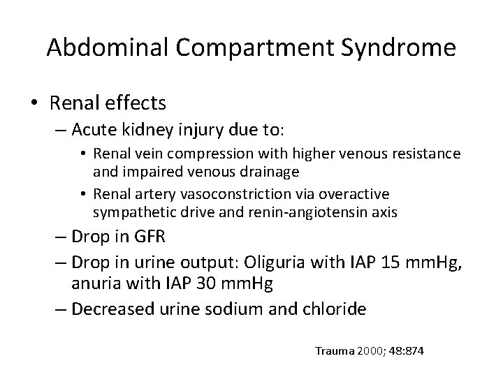
Abdominal Compartment Syndrome • Renal effects – Acute kidney injury due to: • Renal vein compression with higher venous resistance and impaired venous drainage • Renal artery vasoconstriction via overactive sympathetic drive and renin-angiotensin axis – Drop in GFR – Drop in urine output: Oliguria with IAP 15 mm. Hg, anuria with IAP 30 mm. Hg – Decreased urine sodium and chloride Trauma 2000; 48: 874
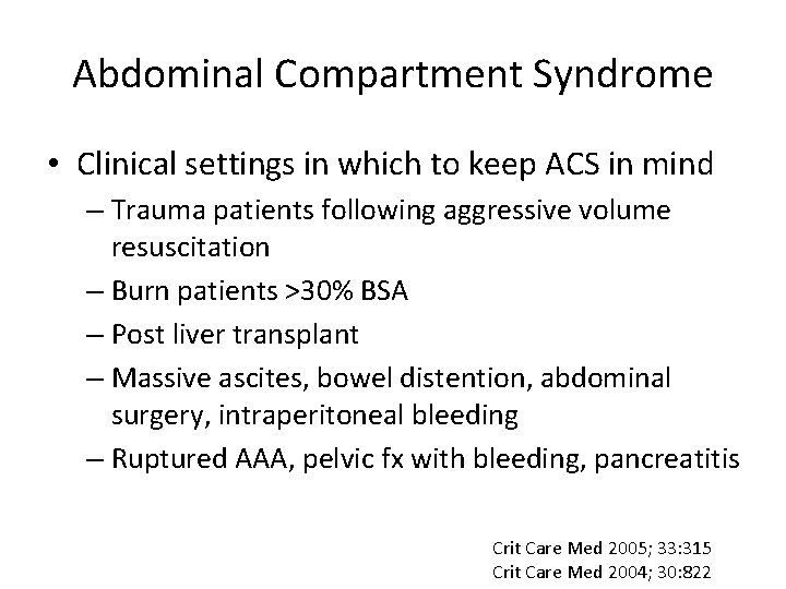
Abdominal Compartment Syndrome • Clinical settings in which to keep ACS in mind – Trauma patients following aggressive volume resuscitation – Burn patients >30% BSA – Post liver transplant – Massive ascites, bowel distention, abdominal surgery, intraperitoneal bleeding – Ruptured AAA, pelvic fx with bleeding, pancreatitis Crit Care Med 2005; 33: 315 Crit Care Med 2004; 30: 822
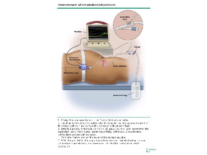
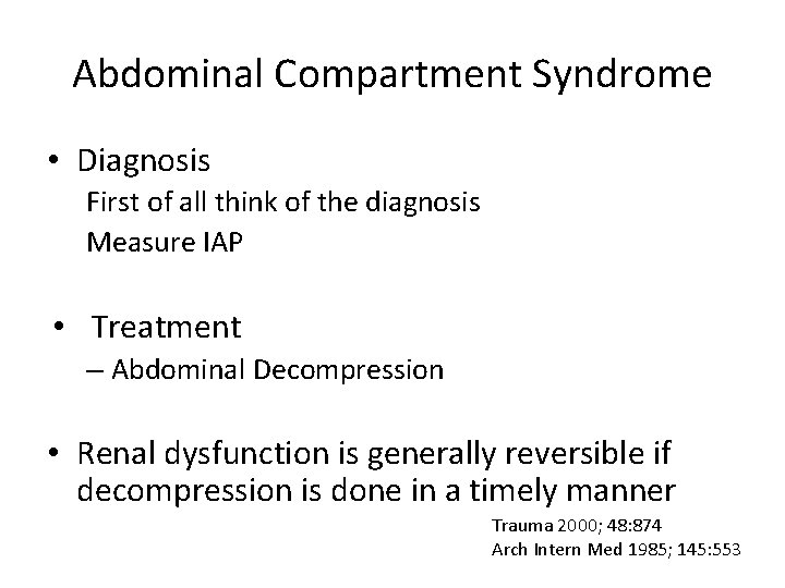
Abdominal Compartment Syndrome • Diagnosis First of all think of the diagnosis Measure IAP • Treatment – Abdominal Decompression • Renal dysfunction is generally reversible if decompression is done in a timely manner Trauma 2000; 48: 874 Arch Intern Med 1985; 145: 553
The End • Thank you for your attention