Kidney By Dr Abdel Aziz M Hussein Lecturer


Kidney By Dr. Abdel Aziz M. Hussein Lecturer of Medical Physiology

• The role of the kidney in homeostasis may include which of the following : • a) Regulation of extracellular fluid composition • b) Regulation of red blood cell formation. • c) Secretion of certain hormones , such as angiotensin II, prostaglandins and kinins • d) Secretion of erythropoietin

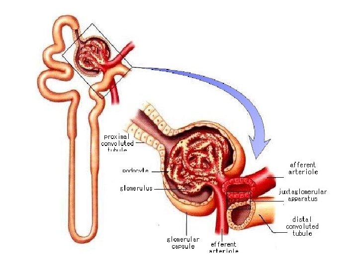
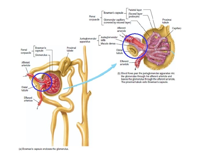
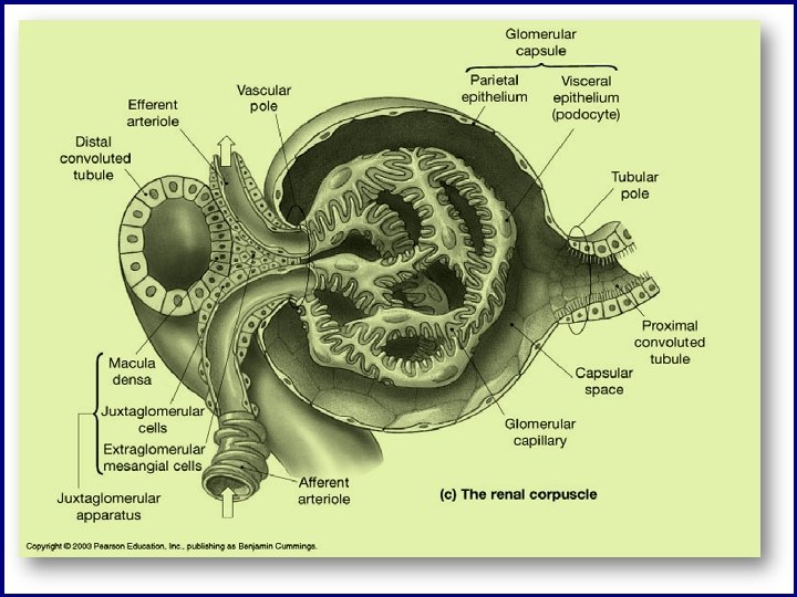
Juxta-glomerular Apparatus Def. , : • It is a combination of vascular, tubular and interstitial cells Site: • Vascular pole of Bowman's capsule Tubular pole Vascular pole
 or Lacis cells In")
Juxta-glomerular Apparatus Components: Macula Densa JG cells (E. G. mesangial) or Lacis cells In transitional zone between thick ALH and DCT In media of afferent arterioles Continuous with intraglomerular cells Densely crowded tubular cells Modified smooth ms cells with epitheloid appearance Interstitial cells between JG and MD cells

Juxta-glomerular Apparatus

Macula Densa Function • Monitor Na. Cl concentration in DCT (stimulated by low Na. Cl) Afferent ↓ ↑GFR V. C. Efferent Angiotensin II
 Synthesis, store and release of renin • B) acts")
Juxta-glomerular cell Function • A) Synthesis, store and release of renin • B) acts as Baroreceptors (detect tension in wall of afferent arterioles) JG cells ↓ wall tension Renin ↓ Renal Blood Flow
 Function • Form functional syncitium with macula densa")
Extra-glomerular Mesangial cell or Lacis (Polkisson) Function • Form functional syncitium with macula densa and JG cells

Functions of Juxtaglomerular Apparatus

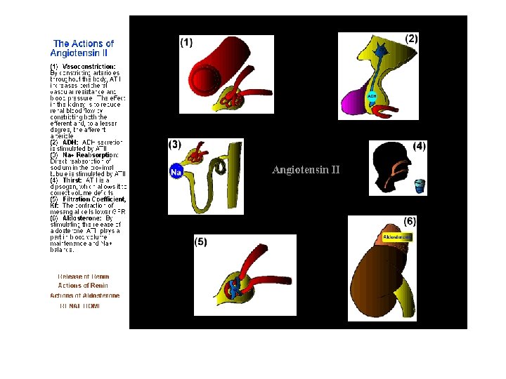
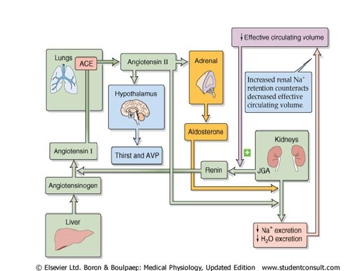
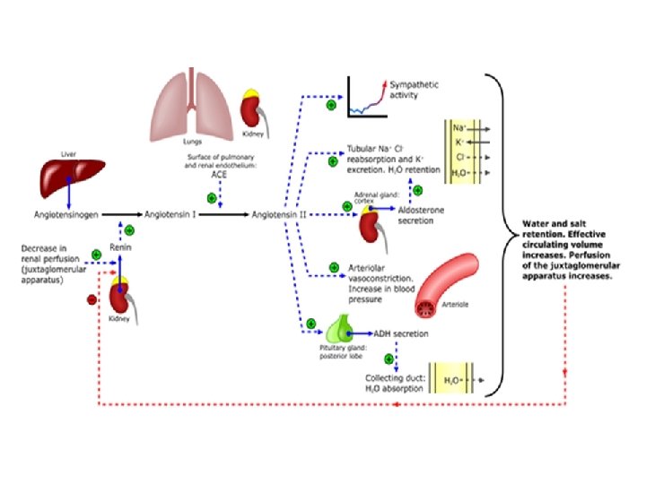
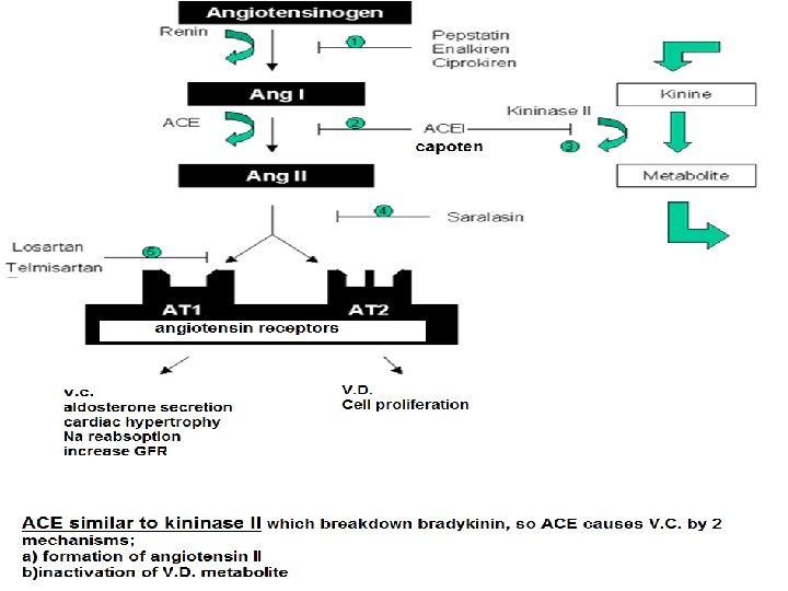
Functions of JGA • The only function of JGA is synthesis and secretion of renin Actions of Renin: V. C. Thirst and salt appetite Stimulates Na reabsorption from PT ACE Angiotensin I Renin Angiotensinogen Stimulates ACTH, ADH, etc…. Increase force of myocardial contraction Angiotensin II • Small dose (V. C. of efferent) • Large dose (generalized V. C. ) Stimulates Na and H 2 O reabsorption from intestine Aldosterone

Control of Renin Secretion
 Stimuli for Renin Release: they are 3 stimuli 1.")
Control of Renin Secretion A) Stimuli for Renin Release: they are 3 stimuli 1. Mechanical stimuli 1. Acute hypotension • 2. Renal artery stenosis • 3. Decrease in blood volume e. g. • in acute hypovoalemia and some chronic disease 2. Chemical stimuli 1. Macula densa mechanism • 2. Chronic K depletion • 3. Nervous stimuli Sympathetic stimulation via B 1 • receptors

1. Mechanical stimuli in the form of decreased wall tension 3. Nervous stimuli via B 1 receptor 2. Chemical stimuli as in Macula densa mechanism

Macula Densa Mechanism 1. Decrease of GFR 2. Decrease flow through renal tubules 3. Decrease Na. Cl at Macula densa 4. Macula densa secrete PGI 2 5. PGI 2 stimulate JG cells to secrete renin angiotensin II → V. C. of efferent arteriole → ↑ GFR

Macula Densa Mechanism 1. Increase of GFR 2. Increase flow through renal tubules 3. Increase Na. Cl at Macula densa 4. Macula densa secrete adenosine 5. Adenosine V. C. of afferent arterioles ↓GFR

Decreased Wall Tension Renin Decreased Ca → increase c. AMP Decreased wall tension Decrease of ABP ↓ Renal Blood Flow

Increased Sympathetic activity Decrease of renin Decrease of ABP
 Inhibitors of Renin: include 1. 2. 3. 4. 5.")
Control of Renin Secretion B) Inhibitors of Renin: include 1. 2. 3. 4. 5. 6. Angiotensin III Atrial natriuretic peptide (ANP) ADH Hyperkalemia Hypernatremia Do not forget: 4 A 2 H

 8% of total O")
Renal O 2 Consumption • Is characterized by; • 1) 8% of total O 2 consumption i. e. 1820 ml /min for both kidneys or 6 ml/min/100 gm kidney tissue • 2) Arteriovenous O difference is low i. e. 1. 5 ml O 2/ 100 ml blood compared to 4 -5 ml /100 ml in other tissues 19 ml % 17. 5 ml %
 Renal O 2 consumption is a flow dependent")
Renal O 2 Consumption • 3) Renal O 2 consumption is a flow dependent i. e. increase of RBF → increase of renal O 2 consumption • 4) Most of O 2 consumption is used for tubular reabsorption especially Na Increase of renal O 2 consumption Increase of RBF

Renal Blood Flow

Renal Blood Flow Def. : • It is the fraction of CO that supplies both kidneys i. e. renal fraction RBF

Renal Blood Flow Value: • ¼ CO or 1200 ml/min or 4 ml/ 1 gm kidney tissues Significance: • Is high to ensure high GFR NOT to supply excess O 2 for excess metabolism
 10% supply non-functioning structures of kidney • a) capsule")
Renal Blood Flow Distribution: A) 10% supply non-functioning structures of kidney • a) capsule • b) pelvis • c) perinephric fats B) 90% functioning structures • Cortex → 88% - 89% • Medulla → 1 - 2%

Renal Blood Flow 4 -5 ml/ min 1 gm Cortex 0. 7 - 1 ml /min/ 1 gm Outer Medulla 0. 2 – 0. 25 ml /min/ 1 gm Inner Medulla

Low Medullary RBF High viscosity of medullary blood flow High length of vasa recta Small number of vessels

THANKS
- Slides: 37