Kidney and Ureters Agenesis Horseshoe kidneys congenital cysts


 that")


")


")
- Slides: 39
Kidney and Ureters : Agenesis, Horseshoe kidneys, congenital cysts, Megaureter, Ectopic ureter, Ureterocele Dr Amit Gupta Associate Professor Dept Of Surgery
Bilateral Renal Agenesis • Bilateral Renal Agenesis was first recognized in 1671 by Wolfstrigel • Can occur secondary to a defect of the wolffian duct, ureteric bud, or metanephric blastema • Bilateral agenesis occurs in 1 of every 4000 births • Male predominance
• 40% of the affected infants are stillborn • Children who are born alive do not survive beyond 48 hours because of respiratory distress associated with pulmonary hypoplasia • The adrenal glands are usually normally positioned • Characteristic Potter facies and presence of oligohydramnios are pathognomonic • Complete ureteral atresia is observed in slightly more than 50% of affected individuals
Potter's facial appearance
Diagnosis • The characteristic Potter facies and the presence of oligohydramnios are pathognomonic and should alert for this severe urinary malformation. • Amnion nodosum—small white, keratinized nodules found on the surface of the amniotic sac—may also suggest this anomaly • Anuria after the first 24 hours without distention of the bladder should suggest renal agenesis
Diagnosis • BRA has been detected in higher proportion in cryptophthalmos or Frazer's syndrome, Klinefelter's syndrome , Kallmann's syndrome, esophageal atresia. • Renal ultrasonography confirm the presence or absence of urine within these structures. • Absence of uptake of the radionuclide in the renal fossa above background activity confirms the diagnosis of BRA. • Umbilical artery catheterization and an aortogram defines the absence of renal arteries and kidneys.
Unilateral Renal Agenesis • There are no tell tale signs (as with BRA) that suggest an absent kidney. • Diagnosis not suspected unless careful examination of the external and internal genitalia uncovers an abnormality that is associated with renal agenesis or an imaging study is done. • Unilateral agenesis occurs once in 1100 births • Males predominate in a ratio of 1. 8: 1
• More frequent on the left side • Ipsilateral ureter is completely absent in about half of the patients • Structures derived from the müllerian or wolffian duct are most often anomalous • Anomalies of other organ systems involve the cardiovascular (30%), gastrointestinal (25%), and musculoskeletal (14%) systems
Unilateral renal agenesis to be associated with other urologic abnormalities in 48% of patients – Primary vesicoureteral reflux (28%) – Obstructive megaureter (11%) – Ureteropelvic junction obstruction (3%)
Diagnosis • No specific symptoms heralding an absent kidney • The diagnosis should be suspected during a physical examination when the vas deferens or body and tail of the epididymis is missing or hypoplastic vagina is associated with a unicornuate or bicornuate uterus • Radionuclide imaging • No clear-cut evidence that patients with a solitary kidney have an increased susceptibility to other diseases
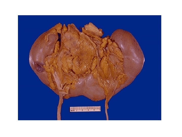
Horseshoe kidneys • Most common of all renal fusion anomalies • Occurs in 0. 25% of the population • fusion occurs before the kidneys have rotated on their long axis
The lower poles of the two kidneys touch and fuse as they cross the iliac arteries
• In 95% of patients, the kidneys join at the lower pole; in a small number, an isthmus connects both upper poles instead • Calyces normal in number, are atypical in orientation • Ureter may insert high on the renal pelvis and lie laterally • Blood supply to the horseshoe kidney can be quite variable
Arteriogram showing a multiplicity of arteries supplying kidney arising from aorta and common iliac arteries
• UPJ obstruction, causing hydronephrosis, occurs in one third of individuals • 60% patients remain asymptomatic for aprox. 10 years
Associated Anomalies
Diagnosis • Excretory urogram
Prognosis • 13% have persistent urinary infection or pain • 17% develop recurrent calculi • Renal carcinoma has been reported within a horseshoe kidney in 123 patients • Incidence of Wilms' tumor in horseshoe kidneys is more than twice
Congenital cysts • Kidney is one of the MC sites in body for cysts • Arise from the nephrons and collecting ducts after they have formed
Cystic Diseases of the Kidney
• Multicystic refers to a dysplastic entity • Polycystic most inherited, all without dysplasia and all with nephrons throughout the kidney • Many of the polycystic kidney disease entities progress to renal failure
‘Snowstorm’ appearance of infantile polycystic disease
Ectopic Kidney • Kidney not located in usual position • 1 in 1, 000 births, but only about one in 10 of these are ever diagnosed; up to 10% bilateral Most common: – Horseshoe Kidney – Unilateral renal agenesis – Pelvic kidney (Left kidney more likely to be abnormal)
Ectopic Kidney • Function is generally normal initially • Abnormal position leads to obstruction in 50% of ectopic kidneys • Increased risk UTI, kidney stones, VUR • Frequently associated with abnormalities of other organ systems (uterine, cardiac, skeletal)
Ectopic Kidney Locations
Ectopic Kidney (simple renal ectopia)
Mega ureter • Ureters wider than 7 to 8 mm • Normal ureteral diameter is rarely greater than 5 mm • Primary MGU is 2 -4 times more common in boys than girls • Slight predilection (1. 6 to 4. 5 times) for the left side • Bilateral in approximately 25% of patients • In 10% to 15% of children contralateral kidney may be absent or dysplastic
Three major classifications of megaureter based on primary and secondary causes
Pathophysiology • Distal end of the ureter, as it becomes intramural and subsequently submucosal, rearranges the muscular layers in its wall. • All layers become longitudinally oriented • Ureteral adventitia fuses to the bladder trigone by attachment to Waldeyer's sheath • Sympathetic and parasympathetic innervation to the distal ureter and UVJ area is believed to modulate primarily ureteral peristalsis
Diagnosis Ultrasound • Distinguishes MGU from UPJ obstruction based on the presence or absence of a dilated ureter VCUG • to rule out reflux Renal scintigraphy • Provides objective, reproducible parameters of function and obstruction
Whitaker's perfusion test & ureteral opening pressure • To evaluate obstruction, but their invasiveness and requirement for anaesthesia are drawbacks in children Magnetic resonance urography
Magnetic resonance urogram showing obstruction at the right ureterovesical junction
Management Primary Refluxing Megaureter • Medical management is often the initial approach • Surgery – Endoscopic subureteric injection, is recommended for persistent high-grade reflux in older children • Reconstructive surgery of a dilated ureter – distal ureterostomy for unilateral reflux – vesicostomy for bilateral disease
• Secondary Refluxing or Obstructive Megaureter – Management of secondary MGUs is initially directed at their root cause • Primary “Dilated” Nonrefluxing Megaureter: Nonobstructive versus Obstructive – Expectant management is preferred – Antibiotic suppression & radiologic surveillance is appropriate in most cases – Surgical correction
• Surgical Options – Plication or infolding for moderately dilated ureter Complications Persistent reflux and obstruction Postoperative VUR
Ectopic ureter • Ureter whose orifice terminates anywhere other than the normal trigonal position • Lateral ectopia : an orifice more cranial and lateral than normal • Caudal ectopia : orifice is more medial and distal than the normal position
• 80% are associated with a duplicated collecting system • Females : – More than 80% are duplicated – Urethra and vestibule are the most common sites • Males: – most ectopic ureters drain single systems – posterior urethra is the most common site • Drainage into the genital tract involves the seminal vesicle three times more often than the ejaculatory duct and vas deferens combined
Ureterocele (outpouching of ureter as it enters bladder)