KEY KNOWLEDGE The functions responsible for short term
 responses to physical activity in")
KEY KNOWLEDGE The functions responsible for short term (acute) responses to physical activity in the cardiovascular, respiratory and muscular systems. KEY SKILLS Participate in data collection, analyse and report on the acute responses occurring at the cardiovascular, respiratory and muscular systems in response to exercise. © Cengage Learning Australia 2011

© Cengage Learning Australia 2011

Acute responses to Exercise Acute responses are the short term changes that happen to the body from the start of exercise until the end of recovery • The type a extent of the response is dependent on the intensity, duration and type of exercise being undertaken • The acute responses take place in the three body systems: respiratory, cardiovascular and muscular systems. • These systems work together to supply more energy / ATP and oxygen to working muscles and then again to remove any waste products so that the body can keep up with the exercise demand. © Cengage Learning Australia 2011
 ventilation")
Acute Respiratory Responses 1. ↑ Ventilation (air breathed in and out per minute) ventilation (L/min) = tidal volume (L) x respiratory rate (breaths/min) 2. ↑Tidal volume (amount of inspired and expired air per breath) 3. ↑ Respiratory rate (breaths per minute) N. B. When asked for two respiratory acute responses, do not write Ventilation & tidal volume, or ventilation & respiratory rate…as they are part of the same thing. You can write tidal volume and respiratory rate q Why would ventilation increase immediately before the start of exercise? © Cengage Learning Australia 2011

Ventilation during submaximal and maximal exercise q Why does ventilation need to increase when you exercise? q Describe the changes in ventilation during submaximal exercise. q How and why does this differ during maximal exercise?

4. ↑ Gas exchange/diffusion Diffusion is the movement of gases from a high to a low concentration. Gases such as oxygen and carbon dioxide always move from areas of high concentration to areas of low concentration. Lungs • Oxygen concentration is high so it moves from the alveoli into the blood stream to be taken to muscles • Carbon dioxide concentration in the blood stream is high so it moves into the alveoli to be exhaled. • The rate of diffusion is increased by: • an increased diffusion gradient (through higher ventilation rates) • an increased surface area of the alveoli © Cengage Learning Australia 2011
 – NOT A RESPIRATORY RESPONSE •")
Diffusion at the Muscles (opposite concentrations to lungs) – NOT A RESPIRATORY RESPONSE • Oxygen concentration is low (as being used by the muscle) so it moves from the blood stream to be taken in by the muscles. • Carbon dioxide concentration in the muscles is high (as it is being produced by the aerobic system) so it moves into the blood stream to be transported to the lungs and exhaled.

q Explain how the four respiratory acute responses benefit the performer when they exercise.

Acute cardiovascular responses 1. ↑ stroke volume (ml of blood pumped out of the left ventricle per beat) 2. ↑ heart rate (beats per minute) 3. ↑ cardiac output (litres per minute) = SV x HR N. B. When asked for two cardiovascular acute responses, do not write cardiac output & heart rate, or cardiovascular & stroke volume…as they are part of the same thing in the equation. You can write stroke volume and heart rate © Cengage Learning Australia 2011

© Cengage Learning Australia 2011

During submaximal exercise: • Heart rate and stroke volume both increase to cause an increase in cardiac output. During maximal exercise: • Stroke volume plateaus during submaximal – why? • Heart rate continues to increase during maximal exercise which causes further increase in cardiac output.
 5. ↑ venous return to heart (assisted by")
4. ↑ blood pressure (mainly systolic) 5. ↑ venous return to heart (assisted by muscle pump, respiratory pump and venoconstriction – still approx 4% but 5 times as much blood compared to rest) 6. Redistribution of blood flow - ↑ blood directed towards working muscles (vasoconstriction of arterioles supplying inactive muscles reduces blood flow here and vasodilation of arterioles supplying muscles increases blood flow here) © Cengage Learning Australia 2011
 Heat is a by product of the aerobic")
6. ↓ blood volume (plasma loss) Heat is a by product of the aerobic energy system. When our muscles contract, heat is produced. Heat cannot accumulate in the body because that would cause our core body temperature to rise. So… Blood is redistributed to the surface of the skin (so less blood is going to the muscles) – via vasodilation/vasoconstriction Once at the surface of the skin, the water from plasma transfers through the surface of the skin and evaporates off the surface, causing an evaporative cooling effect This lowers muscle and body temperature q What are the negative impacts of this process on performance? q What strategies can be used to reduce these impacts?
 This is the difference in")
8. ↑ Arteriovenous oxygen difference (a-v. O 2 diff) This is the difference in oxygen concentration between the arterioles and the venules. [Artery Arteriole Capillaries (diffusion) Venules Veins] The AVO 2 difference indicates how much oxygen has been taken into the muscle. q How is oxygen transported in the blood? q How is oxygen carried from the blood and into the muscle? q Where does oxygen go to inside the muscle and what is it used for? © Cengage Learning Australia 2011
and")
Oxygen uptake during exercise There is a linear relationship between oxygen uptake (VO 2)and exercise intensity. i. e. as exercise intensity increases, so does oxygen uptake – until the body reaches it’s VO 2 max is the maximal volume of oxygen that the body can take in (respiratory), deliver (cardiovascular) and use by the muscles (muscular) per minute. q How can we test VO 2 max? q What would you expect oxygen uptake to look like during: q Submaximal exercise? q Maximal exercise?

Oxygen Uptake during submaximal exercise q How would the oxygen uptake curve differ during maximal exercise? Sketch what it would look like.

EPOC Excess Post-exercise Oxygen Consumption is the amount of oxygen you are taking in above resting levels at the end of exercise. Oxygen being delivered to the muscle > Oxygen demand. It is also the same as oxygen debt (repaying back the volume of oxygen owed at the start/during exercise) The extra oxygen is used to return the body back to pre-exercising conditions (see table below of what happens). The duration and volume of oxygen is dependent on the exercise intensity and duration. The larger the oxygen deficit (due to a high exercise intensity) the greater the EPOC.

What happens during the fast and slow phases of EPOC?

Acute Muscular Responses to Exercise 1. ↑ motor unit/fibre recruitment In order for movement to occur, our motor units need to be recruited. A motor unit is a motor neuron and all of the muscle fibres connected to it. Electrical impulses travel from our brain, down the spinal chord to the muscle. If the impulse is strong enough, all of the muscle fibres associated with the motor unit will contract (all or nothing principle). © Cengage Learning Australia 2011
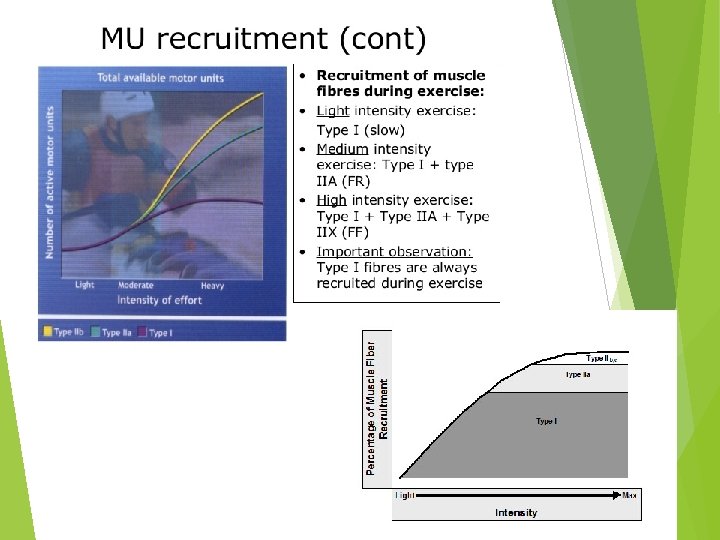

q Compare the number of muscle fibres that would be recruited for a 100 m run compared to a 800 m run. q Compare the type of muscle fibres that would be recruited for a steeple chase compared to the shot put. 2. ↓ fuel stores/energy substrates Energy substrates are the fuels that are used by the ATP-PC, Anaerobic Glycolysis and Aerobic systems. (PC, ATP, glycogen, triglycerides). They deplete when we exercise. Depletion depends on the exercise intensity, duration and whether the athlete can refuel during an event. q Compare the type and rate of energy substrate depletion during an AFL game, a 50 m swim and an iron man.
 q How is lactate")
3. ↑ lactate production (then ↑ H+ if not removed) q How is lactate produced? q What happens if lactate accumulates inside the muscle? Three ways that lactate can be broken down by the body. It can be: (i) Re-converted to pyruvate for immediate oxidation in the mitochondria. (ii) Transported out of the cell into the blood. Most blood lactate is oxidised by other muscles (particularly cardiac muscle and slow twitch muscle fibres). (ii) Transported to the liver to be converted to glucose or glycogen.

Lactate Inflection Point LIP: the last point where lactate entry, into, and removal from, the blood is balanced. It is defined as the final exercise intensity or oxygen uptake value at which blood lactate concentration is relatively stable during a maximal aerobic test. The LIP of an individual represents the maximal intensity at which blood lactate is in steady state (i. e. production = removal) q Describe the relationship between lactate production and exercise intensity.

q Describe the differences in lactate production between the beginner and advanced runner. q Circle the lactate inflection point for the two runners. q Explain why an advanced runner would have a higher lactate inflection point compared to the beginner. q How does having a high/delayed lactate inflection point benefit a performer?

4. ↑ muscle temperature As a by product of aerobic glycolysis
- Slides: 25