KCP731 SC 11 08587 M4 Soft tissue neck


 • • • Round-cell pattern Epithelioid")


 have attempted")
 RMS; (b, d)")


- Slides: 20
KCP-731
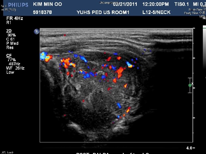
SC 11 -08587 • M/4 Soft tissue, neck, fine needle aspiration • 내원 한달 전부터 갑자기 커진 left posterior neck mass를 주소로 내원. Follow up neck sonography에서 크기가 증가되어 aspiration 시행
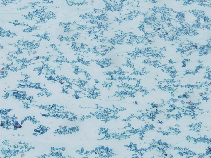
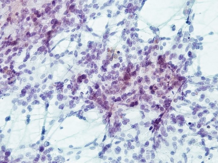
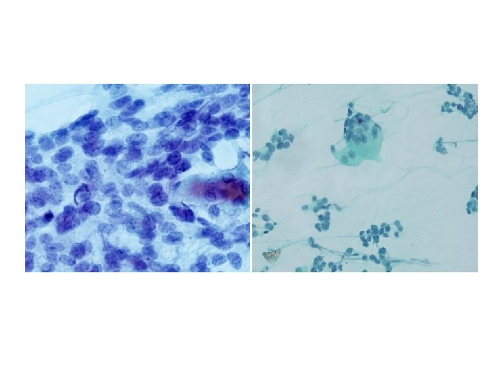
Cytologic features • • High cellularity Clustering of tumor cells Lacy pattern with partly mucoid background Monotonous cells Round to ovoid nuclei Mitotic figures, apoptosis Intranuclear inclusion Multinucleated giant cells
Differential diagnosis in cytologic specimen (head and neck) • • • Round-cell pattern Epithelioid pattern (thyroid, salivary gland, metastatic undifferentiated carcinoma) Giant cell pattern (GCT, histiocytic tumor) Anaplastic pattern (ALCL) Spindle cell pattern Mixed inflammtory pattern (Hodgkin lymphoma, granulocytic sarcoma) RMS Non-Hodgkin Lymphoma Ewing/PNET Neuroblastoma Chance clusters Embryonal (mixoid material/cytoplasmic vacuole) Coarse nuclear chromatin Convoluted nuclei Multinucleated cells Plasmacytoid cells High cellular smear Noncohesive cell population Naked nuclei in Pap smear Brisk mitoses and apoptosis Cyoplasmic vacuole Pale nucear chromatin Rosettes Neuropil (rare) Speckled nuclear chromatin Rosettes Neuropils Ganglion cells Calcification
Desmin Myogenin CD 99
Final diagnosis Rhabdomyosarcoma, most likely alveolar type
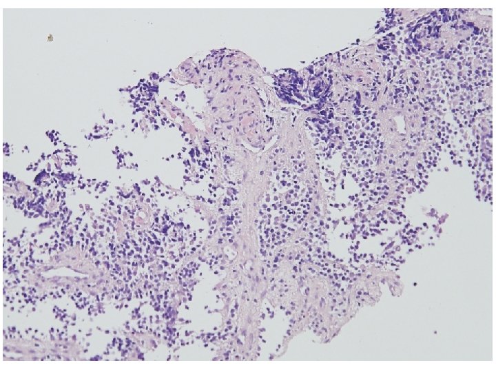
Rhabdomyosarcoma, alveolar type § Older mean ages than do the other type of RMS (adolescents) § Extremities and trunk § Worst prognosis § Histologic finding § § § Discohesive tumor cells in fibrous septa around alveoli/ spaces Multinucleated giant cells Solid variant (alveolar spaces x) § PAX 7 -FKHR –favorable course
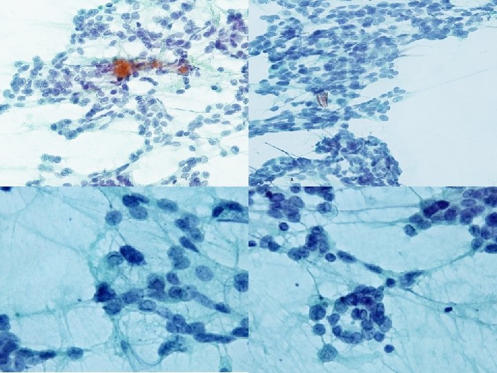
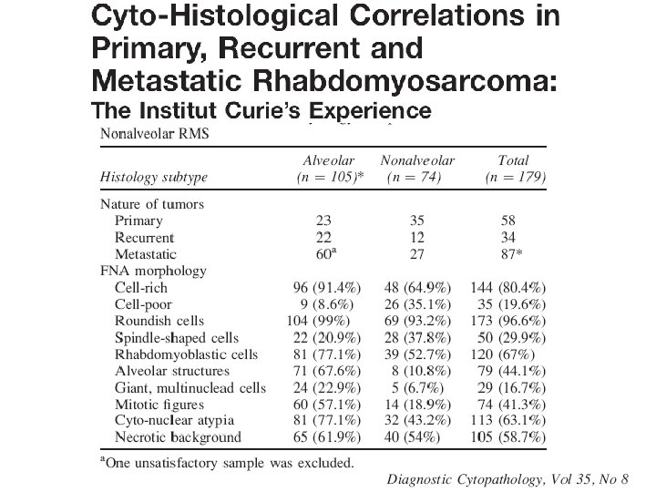
Cytologic morphology of RMS • A limited number of studies (250 cases) have attempted to characterize the cytologic morphology of RMS. • Embryonal subtype of RMS – Predominantly composed of primitive round cells with rounded nuclei – Spindle-shaped cells, large, tadpole or ribbon-shaped cells – Anisocytosis – Intranuclear or cytoplasmic inclusion • Alveolar subtype of RMS – Completely dissociated cells or clustering – Small and lymphocyte-like, presenting fine granular chromatin – Binucleated or multinucleated cells
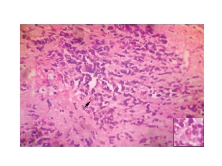
Figure 5. Morphological similarity between RMS, Ewing/PNET and synovial sarcoma. (a) RMS; (b, d) Ewing/PNET; (c) Synovial sarcoma
Cytologic features of round cell tumor KCP 731 RMS Ewing/ PNET ++ ++ +/- + Rhabdomyoblastic cell +/- ++ - Alveolar strucuture +/- +/- - Giant, cells +/- ++ ++ + Cyto-nuclear atypia + ++ +/- ++ + Necrotic background - ++ + +/- + Roundish cells Spindle cell multinucleated Mitotic figure Rhabdoid Synovial tumor sarcoma ++; Frequent, +; rare, +/- occasionally seen, - ; absent.
17 alveolar RMS 14 embryonal RMS 7 botryoid RMS Single cell predominent Spindle-, tadpole-, ribbon shape cells Occasional ill defined cell clusters Mucoid background Some cases Eccentric nuclei, dense cytoplasm Dense cytoplasm present Cytoplasmic vacuole present Rare Binuclear cells, multinuclear cells, hypercellularity Rare
Final diagnosis Rhabdomyosarcoma, most likely alveolar type