Kawasaki disease formerly known as mucocutaneous lymph node


")
 � Cardiac involvement is the most important manifestation of Kawasaki disease.")

 acute febrile phase Kawasaki disease is generally divided into three clinical")


 3 4 5 � Normocytic anemia is common. The platelet count is")


- Slides: 33
Kawasaki disease formerly known as �mucocutaneous lymph node syndrome Involve the skin , lymph node and eyes infantile PAN
DEFINITION is an acute febrile vasculitis of childhood first described by Dr. Tomisaku Kawasaki in Japan in 1967.
Introduction � Approximately 20% of untreated patients develop coronary artery abnormalities including aneurysms, with the potential for the development of coronary thrombosis, stenosis, myocardial infarction, and sudden death. � Whereas less than 5% of those treated with IVIG will develop cx � Kawasaki disease has replaced acute rheumatic fever as the leading cause of acquired heart disease in children in the United States and Japan.
ETIOLOGY The cause of the illness remains unknown � features strongly support an infectious origin; 1. age group affected, 2. periodic epidemics with a wavelike geographic spread of illness during the epidemic 3. the self-limited nature of the illness, and the 4. clinical features of fever, rash, enanthem, conjunctival injection, and cervical adenopathy. genetic causes of the disease become likely
EPIDEMIOLOGY �The incidence of Kawasaki disease in Asian children is substantially higher than in other racial groups �the illness occurs worldwide in all ethnic groups. � 80% of patients are younger than 5 yr, and only occasionally are teenagers and adults affected. #If the disease present at less than 1 year old patients >>It is a sign of worse prognosis
CLINICAL MANIFESTATIONS � The acute stage begins with the onset of fever and lasts 1 to 2 weeks. This is followed by the subacute stage, which includes desquamation of the extremities and the development of coronary aneurysms. The third stage is convalescence, during which the findings resolve Fever > ﻣﻬﻢ �high spiking (to 104°F or higher), �remittent, and unresponsive to antibiotics. �duration of fever is generally 1 -2 wk without treatment but may persist for 3 -4 wk. �Prolonged fever has been shown to be a risk factor for the development of coronary artery disease.
CLINICAL MANIFESTATIONS ﻣﻬﻢ �Persistant fever more than 5 days Plus at least 4 principal features � bilateral bulbar conjunctival injection, usually without exudate. >exudative conjunctival injection occur in cases of measles and bacterial infection � erythema of the oral and pharyngeal mucosa. � Change in lips and oral cavity : "strawberry" tongue, dry, cracked lips. � Change in extrimties: erythema and swelling of the hands and feet and peeling. � Polymorphous exanthem (maculopapular, erythema multiforme, or scarlatiniform) with accentuation in the groin area. � and nonsuppurative cervical lymphadenopathy, usually unilateral, with a node size of 1. 5 cm or greater in diameter.
bilateral bulbar conjunctival injection + scarlatiniform exanthem
strawberry tongue
Finger Desquamation � One to 3 wk after the onset of illness � Desquamation(periangual) of the fingers and toes may progress to involve the entire hand foot. � Perineal desquamation is common
CLINICAL MANIFESTATION (CONT) � Cardiac involvement is the most important manifestation of Kawasaki disease. � Myocarditis manifested by tachycardia, , and decreased ejection fraction and decreased ventricular function occurs in at least 50% of patients. � Pericarditis with a small pericardial effusion is common during the acute illness. � Coronary artery aneurysms generally develop during the 2 nd 3 rd wk of illness and can be detected by two-dimensional echocardiography. Valvular regurgitation and systemic artery aneurysms may occur but are uncommon. Giant coronary artery aneurysms) 8 mm internal diameter) pose the greatest risk for rupture, thrombosis or stenosis, and myocardial infarction.
Coronary artery aneurysm
CLINICAL MANIFESTATION (CONT) acute febrile phase Kawasaki disease is generally divided into three clinical phases. � The acute febrile phase, which usually lasts for 1 -2 wk, is characterized by fever and the other acute signs of illness.
SUBACUTE PHASE when fever and other acute signs have abated, but irritability, anorexia, and conjunctival injection may persist. is associated with �Desquamation. �Thrombocytosis. �the development of coronary aneurysms. �and the highest risk of sudden death. This phase generally lasts until about the 4 th wk.
Convalescent stage when all clinical signs of illness have disappeared and continues until the erythrocyte sedimentation rate (ESR) returns to normal, at approximately 6 -8 wk after the onset of illness.
PATHOGENESIS �severe vasculitis of all blood vessels �medium-sized arteries �predilection for the coronary arteries.
LABORATORY FINDINGS 1. 2. No specific diagnostic test An elevated ESR, C-reactive protein, and other acute phase reactants are almost universally present in the acute phase of illness and may persist for 4 -6 wk.
Lab findings(CONT) 3 4 5 � Normocytic anemia is common. The platelet count is generally normal in the 1 st wk of illness and rapidly rises by the 2 nd-3 rd wk of illness and may exceed 1, 000/mm 3. Antinuclear antibody and rheumatoid factor are not detectable. Sterile pyuria, mild elevations of the hepatic transaminases, and cerebrospinal fluid pleocytosis may be present. Low albumin, hyponatremia , thrombocytosis after one week , high GGT and transaminases
ECHOCARDIOGRAPHY �Two-dimensional echocardiography is the most useful test to monitor the potential development of coronary artery abnormalities and should be performed by a pediatric cardiologist. �The test should be performed at diagnosis and again after 2 -3 wk of illness. �If results of both of these are normal, a repeat study is performed 6 -8 wk after onset of illness.
FOLLOW UP For patients who develop coronary artery abnormalities; more frequent � echocardiographic studies � and potentially angiography may be indicated. Treatment of such patients should be determined in consultation with a pediatric cardiologist.
Predictor of poor prognosis �Young age , , male gender �Persistant fever and poor response to ttx �Labs abnormality
D DX MEASELES exudative conjunctivitis. Koplik spots. rash that begins on the face behind the ears. and a low white blood cell count and ESR.
Drug Rxn �periorbital edema �oral lesions � low ESR, may help to distinguish these reactions from Kawasaki disease.
Toxic shock syndrome hypotension renal involvement elevated creatine phosphokinase focus of staphylococcal infection. which are features of this illness but not of Kawasaki disease � �
scarlet fever A common clinical problem is the differentiation of from Kawasaki disease in a child who is a group A streptococcal carrier. Because patients with scarlet fever have a rapid clinical response to penicillin therapy, treatment with this therapy for 24 -48 hr with clinical reassessment at that time generally clarifies the diagnosis.
COMPLICATIONS AND PROGNOSIS �Recovery is complete and without apparent long- term effects for patients who do not develop coronary disease. � Recurrent illness occurs in only 1 -3% of cases � Overall, 50% of coronary artery aneurysms resolve echocardiographically by 1 -2 yr after the illness. � Giant aneurysms are unlikely to resolve and most often lead to thrombosis or stenosis. .
Treatment of Kawasaki Disease Acute Stage �Intravenous immune globulin 2 g/kg over 10 -12 hr. � aspirin 80 -100 mg/ kg/24 hr divided every 6 hr orally until 14 th illness day. (As anti-inflammatory drug)
Convalescent Stage � Aspirin 3 -5 mg/kg once daily orally until 6 -8 wk after illness onset. (As antithrombotic drug) Long-Term Therapy for Those with Coronary Abnormalities � Aspirin 3 -5 mg/kg once daily, orally � ± dipyridamole 4 -6 mg/kg/24 hr divided in two or three doses orally (some experts add warfarin for those patients at particularly high risk of thrombosis.
ASPIRIN antithrombotic Aspirin is decreased from anti-inflammatory to antithrombotic doses (3 -5 mg/kg/24 hr as a single dose) on the 14 th illness day or when a patient has been afebrile for at least 3 -4 days. Aspirin is continued for its antithrombotic effect until 6 -8 wk after onset, when the ESR has normalized, in patients who have not developed abnormalities detected by echocardiography.
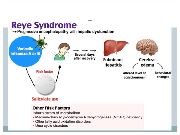
ASPIRIN--- PRECAUTIONS � Patients receiving long-term aspirin therapy are candidates for influenza vaccine to reduce the risk of Reye syndrome. The risk of Reye syndrome in children who take salicylates and who receive varicella vaccine is believed to be much lower than with wild-type varicella. � Different antiplt can be used in 6 week of vericella vaccine � IVIG and live vaccine � Physicians must weigh the relative risk of vaccine in children on long-term aspirin therapy against the risk of natural varicella infection Reye syndrome> encephalopathy + hepatitis
THANK YOU