Kamran fazel MD FCCM 1 AKI Acute Kidney


 has now replaced")


 Risk: 1. 5 x")


. Recognizes")


 suggests that AKI accounts")


")


.")


 is the")


")


- Slides: 83
Kamran fazel MD, FCCM 1
AKI- Acute Kidney Injury
HISTORY DEFINITION EPIDEMIOLOGY ETIOLOGY SUBTYPE PROGNOSIS BIOMARKER RISK FACTOR EVALUATION MANAGEMENT&GUIDELINE
1 -HISTORY
Acute Kidney Injury 2 nd Century AD: Galen surmises urine formed from kidneys 330 -1453 AD: Byzantine physicians describe oliguria as symptom of AKI, as well as detailed urine findings in AKI; also, the transition to polyuric phase as late finding in AKI is recognized 330 -1453 AD: likely precursors to ATN described: Aetius: “. . the reasons for the destruction of the kidney are the toxic influence of remedies and poisons, and external pressure” Nonus: “…hematuria results from poisonous drugs and serpent venom…” (Eftychiadis AC, Am J Nephrol 1997)
Acute Kidney Injury 1827: English physician Richard Bright describes microscopic hematuria, oliguria, and edema in acute and chronic renal inflammatory states, gives eponymic definition for acute/chronic GN.
Acute Kidney Injury WWI & WWII: Post-traumatic oliguria seen in combatants, crush syndrome evolves as an AKI dx 1950 -1960’s: AKI found retrospectively in ~20% of post-op open heart/aortic surgery
Acute Kidney Injury WW I: observations of thirst and oliguria in combat victims led to relationship between blunt trauma and AKI (Better, OS 1997) WWII: Spanish surgeon Joseph Trueta observes same in Spanish Civil War, WWII combatants Induces renal cortical vasospasm experimentally (Trueta, et al. , 1947) WWII: Bywaters and Beall link myoglobin to AKI in crush syndrome during London Blitz (1940)
2 -Definition of AKI
UK Renal Association 5 th Edition, 2011 Acute kidney injury (AKI) has now replaced the term acute renal failure and an universal definition and staging system has been proposed to allow earlier detection and management of AKI. The new terminology enables healthcare professionals to consider the disease as a spectrum of injury. This spectrum extends from less severe forms of injury to more advanced injury when acute kidney failure may require renal replacement therapy (RRT)
Clinically AKI is characterised by a rapid reduction in kidney function resulting in a failure to maintain fluid, electrolyte and acid-base homoeostasis.
Definition of AKI There are more than 35 definitions of AKI (formerly acute renal failure) in literature! Mehta R, Chertow G: Acute renal failure definitions and classification: Time for change? Journal of American Society of Nephrology 2003; 14: 2178 -2187. �
Acute Kidney Injury 2001 : Acute Dialysis Quality Initiative (ADQI) Risk: 1. 5 x inc in SCr, GFR dec 25%, UOP<0. 5 ml/kg/h x 6 h Injury: 2 x inc SCr, GFR dec 50%, UOP<0. 5 ml/kg/h x 12 h Failure: 3 x inc SCr, GFR dec 75%, UOP<0. 5/kg/h x 24 h Also anuria x 12 hr Loss: complete loss (inc need for RRT) > 4 wks ESRD: complete loss (inc need for RRT) > 3 months 2007: Acute Kidney Injury Network (AKIN) Modified RIFLE to include ΔSCr o. 3 mg/d. L from baseline, within 48 hr, based on 80% mortality risk
Definition of AKI As per the Acute Kidney Injury Network: An abrupt (within 48 hrs) reduction in kidney function defined as an increase in serum creatinine level of 0. 3 mg/dl OR An increase in serum creatinine ≥ 50% OR Urine output is < 0. 5 ml/kg/hr for >6 consecutive hours
Definition of AKI RIFLE classification AKIN classification
RIFLE classification Bellomo R, Ronco C, Kellum J, et al. : Acute renal failure-definition, outcome measures, animal models, fluid therapy and information technology needs: The Second International Consensus Conference of the Acute Dialysis Initiative (ADQI) Group. Critical Care 2004; 8: R 204 -R 212.
AKIN classification Modification of the RIFLE classification by Acute Kidney Injury Network (AKIN). Recognizes that small changes in serum creatinine (>0. 3 mg/dl) adversely impact clinical outcome. Uses serum creatinine, urinary output and time. Coca S, Peixoto A, Garg A, et al. : The prognostic importance of a small acute decrement in kidney function in hospitalized patients: a systematic review and meta-analysis. American Journal of Kidney Diseases 2007; 50: 712720.
AKIN classification AKIN Serum Creatinine stage Criteria Urinary Output Time Criteria 1 Cr ≥ 0. 3 mg/d. L or ≥ 150 -200% from baseline < 0. 5 m. L/kg/hr > 6 hrs 2 Cr to > 200 -300% from baseline < 0. 5 m. L/kg/hr > 12 hrs 3 Cr to > 300% from < 0. 5 baseline or Cr ≥ 4 mg/d. L m. L/kg/hr with an acute rise of at or anuria least 0. 5 mg/d. L X 24 hrs X 12 hrs *Patients needing RRT are classified stage 3 despite the stage they were before starting RRT Mehta R, Kellum J, Shah S, et al. : Acute kidney Injury Network: Report of an Initiative to improve outcomes Acute Kidney Injury. Critical Care 2007; 11: R 31. in
3 -Epidemiology
Epidemiology AKI occurs in ≈ 7% of hospitalized patients. 36 – 67% of critically ill patients (depending on the definition). 5 -6% of ICU patients with AKI require RRT. Nash K, Hafeez A, Hou S: Hospital-acquired renal insufficiency. American Journal of Kidney Diseases 2002; 39: 930 -936. Hoste E, Clermont G, Kersten A, et al. : RIFLE criteria for acute kidney injury are associated with hospital mortality in critically ill patients: A cohort analysis. Critical Care 2006; 10: R 73. Osterman M, Chang R: Acute Kidney Injury in the Intensive Care Unit according to RIFLE. Critical Care Medicine 2007; 35: 1837 -1843.
Data from the Intensive Care National Audit Research Centre (ICNARC) suggests that AKI accounts for nearly 10 percent of all ICU bed days.
4 -Etiology
Etiology Hemodynamic 30% Parenchymal 65% Acute tubular necrosis 55% Acute glomerulonephritis 5% Vasculopathy 3% Acute interstitial nephritis 2% Obstruction 5%
Common causes of AKI in ICU Sepsis Major surgery Low cardiac output Hypovolemia Medications (20%) Uchino S, Kellum J, Bellomo R, et al. : Acute renal failure in critically ill patients: A multinational, multicenter study. JAMA 2005; 294: 813 -818.
Nephrotoxins NSAIDs Aminoglycosides Amphotericin Penicillins Acyclovir Cytotoxics Radiocontrast dye Dennen P, Douglas I, Anderson R, : Acute Kidney Injury in the Intensive Care Unit: An update and primer for the Intensivist. Critical Care Medicine 2010; 38: 261 -275.
5 -Subtype
Acute Kidney Injury Subtype AKI PRERENAL INTRINSIC POSTRENAL
Acute Kidney Injury PRERENAL Volume loss/Sequestration Impaired Cardiac Output Hypotension (and potentially hypo-oncotic states) Net result: glomerular hypoperfusion
Acute Kidney Injury RENAL/INTRINSIC Vascular disorders: small vessel large vessel Glomerulonephritis Interstitial disorders: Inflammation intercalative processes Tubular necrosis: Ischemia Toxin Pigmenturia
Acute Kidney Injury POSTRENAL Intrarenal Crystals Proteins Extrarenal Pelvis/Ureter Bladder/Urethra
6 -PROGNOSIS
Mortality according to RIFLE Mortality increases proportionately with increasing severity of AKI (using RIFLE). AKI requiring RRT is an independent risk factor for inhospital mortality. Mortality in pts with AKI requiring RRT 50 -70%. Even small changes in serum creatinine are associated with increased mortality. Hoste E, Clermont G, Kersten A, et al. : RIFLE criteria for acute kidney injury are associated with hospital mortality in critically ill patients: A cohort analysis. Critical Care 2006; 10: R 73. Chertow G, Levy E, Hammermeister K, et al. : Independent association between acute renal failure and mortality following cardiac surgery. American Journal of Medicine 1998; 104: 343 -348. Uchino S, Kellum J, Bellomo R, et al. : Acute renal failure in critically ill patients: A multinational, multicenter study. JAMA 2005; 294: 813818. Coca S, Peixoto A, Garg A, et al. : The prognostic importance of a small acute decrement in kidney function in hospitalized patients: a systematic review and meta-analysis. American Journal of Kidney Diseases 2007; 50: 712 -720.
Acute kidney injury has a poor prognosis with the mortality ranging from 10%-80% Patients who present with uncomplicated AKI, have a mortality rate of up to 10%. In contrast, patients presenting with AKI and multiorgan failure have been reported to have mortality rates of over 50%. If renal replacement therapy is required the mortality rate rises further to as high as 80%
Non-Oliguric vs. Anuric Oliguric renal failure. Functionally, urine output less than that required to maintain solute balance (can’t excrete all solute taken in). Defined as urine output < 400 ml/24 hr. Anuric renal failure. Defined as urine output < 100 ml/24 hr. Less common – suggests complete obstruction, major vascular catastrophy, or more commonly severe ATN.
Non-Oliguric vs. Anuric Classifying by urine output may help establish a cause. Oliguria – more common with obstruction, prerenal azotemia Nonoliguric – intrarenal causes – nephrotoxic ATN, acute GN, AIN. More importantly, assists in prognosis. Significantly higher mortality with oliguric renal failure. 80% vs. 25% mortality in Oliguric vs. non-oliguric ARF Nonoliguric renal failure may also suggest greater liklihood of recovery of function.
7 -BIOMARKER
BIOMARKER RESEARCH IN DEFINITIONS AND GOALS a characteristic that is objectively measured and evaluated as an indicator of normal biological processes, pathogenic processes, or pharmacologic responses to a therapeutic intervention a biomarker is “any substance, structure or process that can be measured in the body or its products and influence or predict the incidence or outcome of disease”
What is GFR? How is it Calculated? The Glomerular Filtration Rate (GFR) is the volume of fluid filtered from glomerular capillaries into the Bowman’s capsule per unit time
Suspect AKI in a sick patient with a modest rise in their creatinine Large acute drop in GFR with oligoanuria GFR falls rapidly to near zero - only shown by oliguria Slow rise in Cr until eventually a new steady state is reached Only a small early rise in Cr: not easy to recognise as AKI
Limitations to Serum Creatinine as a Reflection of GFR The serum creatinine concentration does not increase above the normal range until the GFR declines below 50 m. L/min, and large declines in GFR may occur above this level without a concomitant increase in the serum creatinine value.
Limitations to Serum Creatinine as a Reflection of GFR In a cachectic patient with very low muscle mass, creatinine generation may be so feeble that the serum creatinine level remains “normal” (<0. 9 mg/d. L) even in the presence of a GFR less than 25 m. L/min.
Serum creatinine is a useful marker of stable renal function, but it is unreliable when GFR is rapidly changing.
Because it may take up to 48 hours for GFR to return to baseline, in the postoperative period the serum creatinine value may still increase for a few days while GFR is actually recovering.
Urine flow rate is an unreliable marker of acute renal failure and may vary from anuric (zero flow), to oliguric (urinary flow rate <15 m. L/hr), to nonoliguric (15 -80 m. L/hr), to polyuric (>80 m. L/hr).
Indices of Tubular Injury �β 2 -Microglobulin �Urinary N-Acetyl-β-D-glucosaminidase �Neutrophil gelatinase-associated lipocalin (NGAL)
8 -Risk Factors
Risk Factors for AKI Age > 75 yrs Chronic kidney disease (CKD, e. GFR < 60 mls/min/1. 73 m 2) Cardiac failure Diabetes mellitus Hypovolemia Nephrotoxic medication Atherosclerotic peripheral vascular disease Liver disease Sepsis
Risk Factors for Ischemic Tubular Injury Volume depletion Aminoglycosides Radiocontrast NSAIDs, Cox-2 inhibitors Sepsis Rhabdomyolysis Preexisting renal disease HTN Diabetes mellitus Age > 50 Cirrhosis
Radiocontrast-Induced Acute Renal Failure �Induces renal vasoconstriction and direct cytotoxicity via oxygen free radical formation �Risk factors: �Renal insufficiency �Advanced age �Hypotension - Diabetes - > 125 ml contrast �Usually non-oliguric ARF; irreversible ARF rare
Prevention of Radiocontrast Nephropathy Intervention Strength of Evidence Clarity of Risk -Benefit Grade of Recommendation Volume expansion with normal saline Good Clear A: Intervention is always indicated and acceptable Volume expansion with sodium bicarbonate Fair Clear B: Intervention may be effective and is acceptable Iso-osmolar contrast Fair Clear B: Intervention may be effective and is acceptable Theophylline Fair Unclear C: May be considered; minimal or no relative impact N-acetylcysteine Good Unclear C: May be considered; minimal or no relative impact Hemofiltration Fair Unclear I: Insufficient evidence to recommend for or against Fenoldopam Good Unclear D: Not useful Hemodialysis Good Unclear D: Not useful
Prevention of Contrast-Induced Nephropathy Avoid use of intravenous contrast in high risk patients if at all possible. Use pre-procedure volume expansion using isotonic saline (? bicarbonate). NAC Avoid concomitant use of nephrotoxic medications if possible. Use low volume low- or iso-osmolar contrast Dennen P, Douglas I, Anderson R, : Acute Kidney Injury in the Intensive Care Unit: An update and primer for the Intensivist. Critical Care Medicine 2010; 38: 261 -275.
Prevention of AKI in ICU Recognition of underlying risk factors Diabetes CKD Age HTN Cardiac/liver dysfunction Maintenance of renal perfusion Avoidance of hyperglycemia Avoidance of nephrotoxins Dennen P, Douglas I, Anderson R, : Acute Kidney Injury in the Intensive Care Unit: An update and primer for the Intensivist. Critical Care Medicine 2010; 38: 261 -275.
Prevention of AKI in hepatic dysfunction Intravenous albumin significantly reduces the incidence of AKI and mortality in patients with cirrhosis and SBP. Albumin decreases the incidence of AKI after large volume paracentesis. Albumin and terlipressin decrease mortality in HRS. Sort P, Navasa M, Arroyo V, et al. : Effect of intravenous albumin on renal impairment and mortality in patients with cirrhosis and spontaneous bacterial peritonitis. New England Journal of Medicine 1999; 341: 403 -409. Gines P, Tito L, Arroyo V, et al. : Randomised comparative study of therapeutic paracentesis with and without intravenous albumin in cirrhosis. Gastroenterology 1988; 94: 1493 -1502. Gluud L, Kjaer M, Christensen E: Terlipressin for hepatorenal syndrome. Cochrane Database Systematic Reviews 2006; CD 005162.
KDIGO Clinical Practice Guideline for Acute Kidney Injury � 4. 4. 3: We suggest using oral NAC, together with i. v. isotonic crystalloids, in patients at increased risk of CI-AKI. � 4. 4. 4: We suggest not using theophylline to prevent CI-AKI. � 4. 4. 5: We recommend not using fenoldopam to prevent CIAKI. � 4. 5. 1: We suggest not using prophylactic intermittent hemodialysis (IHD) or hemofiltration (HF) for contrastmediaremoval in patients at increased risk for CI-AKI. VOL 2 | SUPPLEMENT 1 | MARCH 2012
9 -EVALUATION
Baseline Set of Laboratories to Consider Biochemistry urea and electrolytes Hematology CBC Urinalysis (+/- microscopy, eosinophils) Urinary Biochemistry electrolytes, urea, osmolality Microbiology urine/blood culture when/if infection suspected Imaging renal ultrasound CXR, abdominal x-ray ECG
Acute Kidney Injury LABORATORY DATA Creatinine; also BUN/Cr ratio CBC: anemia, thrombocytopenia HCO 3ˉ: anion gap, lactic acid, ketones K CPK/LDH/Uric acid/liver panel Serologies: Complement ESR, RF, ANA, ANCA, Anti. GBM Electrophoresis Toxicology studies
Evaluation of Renal Failure Is the renal failure acute or chronic? laboratory values do not discriminate between acute vs. chronic oliguria supports a diagnosis of acute renal failure Clues to chronic disease Pre-existing illness – DM, HTN, age, vascular disease. Uremic symptoms – fatigue, nausea, anorexia, pruritis, altered taste sensation, hiccups. Small, echogenic kidneys by ultrasound.
Diagnostic Evaluation of Renal Failure 10015% 80 Cumulative 60% Correct Diagnosis 40 - 25% 60% 200 Hx, PE, Labs Therapeutic Trials Renal Biopsy
Renal Biopsy-When? n n Exclude pre- and post-renal failure, and clinical findings are not typical for ATN Extra-renal manifestations that suggest a systemic disorder Heavy proteinuria RBC casts
AKI �Physical Exam. �Assessing volume status. � Is the patient intravascularly volume depleted? � Neck veins – JVP � Peripheral edema or lack of. � Orthostatic vitals. �Not always straightforward. �Pt. may be edematous (low albumin) or have significant right sided heart disease.
�BUN/Creatinine ratio. �> 20: 1 – suggest prerenal or obstruction. �Can be elevated by anything leading to increased urea production/absorption. GI bleed � TPN � Steroids � Drugs – Tigecycline. � �Creatinine in anephric state typically only rises 1 mg/dl/day. �If greater – should be concerned for rhabdomyolysis
AKI: Diagnostic studies-urine Urinalysis for sediment, casts Response to volume repletion with return to baseline SCr 24 -72 hr c/w prerenal event Urine Na; FENa (%) = UNa x SCr x 100 SNa x UCr FENa < 1%: Prerenal FENa 1 -2%: Mixed FENa > 2%: ATN Hansel’s stain
Classic Lab Findings in AKI Causes UNa Fe. Na *Fe. Urea BUN/Cr Prerenal <10 <1% <35% >20 Renal >20 >2% >50% <15 Postrenal >40 >4% >15
Urine Patterns in Renal Disease Urinary Pattern Renal Disease Hematuria with red cell casts, heavy proteinuria, or lipiduria Glomerular disease or vasculitis Granular and epithelial casts with free epithelial cells Acute Tubular Necrosis Pyuria with white cell and granular casts and no/mild proteinuria Tubular or interstitial disease or obstruction Hematuria and pyuria with no or variable Acute interstitial nephritis, glomerular casts(excluding red cell casts) disease, vasculitis, obstruction, renal infarction Pyuria alone Usually infections, sterile pyuria suggests TB
10 -MANAGEMENT&GUIDELINE
5 Key Steps in Evaluating Acute Renal Failure I. Obtain a thorough history and physical; review the chart in detail II. Do everything you can to accurately assess volume status III. Always order a renal ultrasound IV. Look at the urine V. Review urinary indices
Management Importantly, manangement of AKI is varied and depends on the cause. Given no effective pharmaceutical options, management of AKI is primarily supportive. Prerenal azotemia is usually responsive to isotonic fluid repletion Managament of ATN includes discontinuation of nephrotoxic agents, optimization of hemodynamics, continued monitoring of renal function (acid/base status, electrolyte abnormalities). Postrenal causes warrant removal of the obstruction.
Management of AKI in ICU Maintain renal perfusion Correct metabolic derangements Provide adequate nutrition ? Role of diuretics
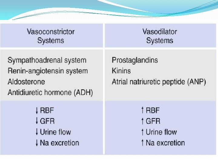
Maintaining renal perfusion �Human kidney has a compromised ability to autoregulate in AKI. �Maintaining haemodynamic stability and avoiding volume depletion are a priority in AKI. Kelleher S, Robinette J, Conger J: Sympathetic nervous system in the loss of autoregulation in acute renal failure. American Journal of Physiology 1984; 246: F 379 -386.
Maintaining renal perfusion �Current studies do not include patients with established AKI. �The individual BP target depends on age, comorbidities (HTN) and the current acute illness. �A generally accepted target remains MAP ≥ 65. Bourgoin A, Leone M, Delmas A, et al. : Increasing mean arterial pressure in patients with septic shock: Effects on oxygen variables and renal function. Critical Care Medicine 2005; 33: 780 -786.
Volume resuscitation – which fluid? SAFE study – no statistical difference between volume resuscitation with saline or albumin in survival rates or need for RRT. Post – hoc analysis – albumin was associated with increased mortality in traumatic brain injury subgroup and improved survival in septic shock patients. Finfer S, Bellomo R, Boyce N, et al. : A comparison of albumin and saline for fluid resuscitation in the intensive care unit. New England Journal of Medicine 2004; 350: 2247 -2256.
Which inotrope/vasopressor? There is no evidence that from a renal protection standpoint, there is a vasopressor agent of choice to improve kidney outcome. Dennen P, Douglas I, Anderson R, : Acute Kidney Injury in the Intensive Care Unit: An update and primer for the Intensivist. Critical Care Medicine 2010; 38: 261 -275.
KDIGO Clinical Practice Guideline for Acute Kidney Injury 3. 5. 1: We recommend not using lowdose dopamine to prevent or treat AKI. 3. 5. 2: We suggest not using fenoldopam to prevent or treat AKI. VOL 2 | SUPPLEMENT 1 | MARCH 2012
Renal vasodilators? “Renal” dose dopamine doesn’t reduce the incidence of AKI, the need for RRT or improve outcomes in AKI. It may worsen renal perfusion in critically ill adults with AKI. Side effects of dopamine include increased myocardial oxygen demand, increased incidence of atrial fibrillation and negative immuno-modulating effects. Lauschke A, Teichgraber U, Frei U, et al. : “Low-dose” dopamine worsens renal perfusion in patients with acute renal failure. Kidney 2006; 69: 1669 -1674. Argalious M, Motta P, Khandwala F, et al. : “Renal dose” dopamine is associated with the risk of new onset atrial fibrillation after cardiac surgery. Critical Care Medicine 2005; 33: 1327 -1332.
KDIGO Clinical Practice Guideline for Acute Kidney Injury 3. 4. 1: We recommend not using diuretics to prevent. AKI. 3. 4. 2: We suggest not using diuretics to treat AKI, exceptin the management of volume overload VOL 2 | SUPPLEMENT 1 | MARCH 2012.
Management Cont. Things to do for patients with AKI Renally dose medications Avoid nephrotoxins Monitor I/Os Serial assessment of serum creatinine Renal Replacement Therapy (i. e dialysis) is the central component of care for patients with severe AKI The generally accepted indications for renal replacement therapy in the setting of AKI include: Acidosis Electrolyte disturbance Ingestion/Intoxication Volume Overload Overt Uremia
Acute Kidney Injury INDICATIONS FOR RENAL REPLACEMENT THERAPY �Consensus generally includes: 1. 2. 3. 4. 5. 6. Refractory volume overload Severe metabolic acidosis; HCO 3 may be variable, but declining level of factor; also falling p. H to 7. 1 -7. 2 Hyperkalemia, with levels > 6. 5, or documented rapid rise refractory to medical therapy Major uremic target organ manifestations i. e. pericarditis, progressive neuropathy, seizure Platelet dysfunction, bleeding diasthesis AKI in setting of dialyzable drug/toxin
Guideline 8. 6 – AKI : Vascular access for RRT We recommend that subclavian access should be avoided in patients at risk of progressing to CKD stage 4 or 5 due to the risks of compromising future, permanent vascular access.
Guideline 8. 7 – AKI : Vascular access for RRT We suggest that non-dominant arm upper limb vasculature should be preserved as a contingency for future permanent access.
When to call nephrology Any known dialysis patient admitted Any known renal transplant patient admitted Any case of AKI where cause not clear Worsening AKI Emergency dialysis indications Suspect glomerulonephritis