K 812 18 Sept 07 Cardiac Contractile Element


 m. RNA analyses by reverse transcription (RT)-polymerase chain reaction (PCR)")


 and")


![When [Ca] = Kd then [Tn. C] = [Tn. C·Ca] Kd = [Ca] at](https://slidetodoc.com/presentation_image/bc4dc0af2ebc7d433a4df287ce23c10f/image-38.jpg "When [Ca] = Kd then [Tn. C] = [Tn. C·Ca] Kd = [Ca] at")


- Slides: 45
K 812 18 Sept 07 Cardiac Contractile Element 18 Sept 07 1
Myosin Isoforms 18 Sept 07 2
18 Sept 07 3
Coordinated regulation of cardiac MHC gene via antisense transcription 18 Sept 07 Haddad, F. et al. Am J Physiol Heart Circ Physiol 290: H 2351 -H 2361 2006 Copyright © 2006 American Physiological Society 4
Myosin heavy chain (MHC) m. RNA analyses by reverse transcription (RT)-polymerase chain reaction (PCR) using competitive PCR 18 Sept 07 Haddad, F. et al. Am J Physiol Heart Circ Physiol 290: H 2351 -H 2361 2006 5
Antisense beta-MHC RNA expression in NC and Ab. Con heart in 3 regions of the myocardium 18 Sept 07 Haddad, F. et al. Am J Physiol Heart Circ Physiol 290: H 2351 -H 2361 2006 6
Fig. 2. b-MHC pre-m. RNA expression in NC and Ab. Con heart Haddad, F. et al. Am J Physiol Heart Circ Physiol 290: H 2351 -H 2361 2006 Copyright © 2006 American Physiological Society
Fig. 3. b-MHC m. RNA expression in NC and Ab. Con heart Haddad, F. et al. Am J Physiol Heart Circ Physiol 290: H 2351 -H 2361 2006 Copyright © 2006 American Physiological Society
Fig. 4. Antisense b-MHC RNA expression in NC and Ab. Con heart in 3 regions of the myocardium Haddad, F. et al. Am J Physiol Heart Circ Physiol 290: H 2351 -H 2361 2006 Copyright © 2006 American Physiological Society
Fig. 5. Quantitative b-MHC RNA analyses in the apex region using real-time PCR and SYBRgreen fluorescence Haddad, F. et al. Am J Physiol Heart Circ Physiol 290: H 2351 -H 2361 2006 Copyright © 2006 American Physiological Society
Fig. 6. a-pre-m. RNA expression in NC and Ab. Con heart Haddad, F. et al. Am J Physiol Heart Circ Physiol 290: H 2351 -H 2361 2006; doi: 10. 1152/ajpheart. 01111. 2005 Copyright © 2006 American Physiological Society
Fig. 7. a-MHC m. RNA expression in NC and Ab. Con hearts Haddad, F. et al. Am J Physiol Heart Circ Physiol 290: H 2351 -H 2361 2006; doi: 10. 1152/ajpheart. 01111. 2005 Copyright © 2006 American Physiological Society
Titin 18 Sept 07 13
18 Sept 07 14
Layout of titin in cardiac sarcomere 18 Sept 07 Granzier, H. L. et al. Circ Res 2004; 94: 284 -295 15
Mechanism of passive and restoring force generation 18 Sept 07 Granzier, H. L. et al. Circ Res 2004; 94: 284 -295 16
Exon-intron structure of human titin gene 18 Sept 07 Granzier, H. L. et al. Circ Res 2004; 94: 284 -295 17
18 Sept 07 18
Passive tension/sarcomere length relations of cardiac myocytes that predominately express N 2 B titin and those that predominately express N 2 BA titin Granzier, H. L. et al. Circ Res 2004; 94: 284 -295
Contribution of titin and collagen to passive myocardial tension of mouse LV (MLV) and dog LV (DLV ) Granzier, H. L. et al. Circ Res 2004; 94: 284 -295
Titin summary • Very large molecule which spans half a sarcomere from M- to Z-line • Single gene which encodes for a protein of ~4. 2 MDa • Many isoforms exist which are the product of alternative splicing – each has different physical properties • Cardiac muscle expresses two forms: a short one -N 2 B (~2. 9 MDa) and a longer one N 2 BA (~3. 3 MDa) • Two cardiac forms have different physical properties – N 2 B is stiffer – and differential distributions • N 2 B isoform can undergo PKA phosphorylation making it less stiff
Myosin binding protein C My. BP C
Schematic structure of the cardiac sarcomere de Tombe, P. P. Circ Res 2006; 98: 1234 -1236
Rabbit psoas fibers labeled with goat antiserum to rabbit myosin binding protein C Flashman, E. et al. Circ Res 2004; 94: 1279 -1289
Harris et al. Hypertrophic cardiomyopathy in cardiac myosin binding protein-C KO mice Circulation Research 90: 594, 2002
Figure 2. c. My. BP-C m. RNA transcript expression in knockout mice. A, RT-PCR of RNA using primers to exons 1 to 2 (a), 3 to 5 (b), and 25 to 34 (c) resulted in PCR products of expected size when c. DNA from wild-type and heterozygous mice was amplified. c. DNA from c. My. BP-C-/- failed to yield product using primers to exons 3 to 5. Positive controls using cloned c. My. BP-C c. DNA and negative controls lacking reverse transcriptase (-RT) or DNA (H 2 O) are also shown. B, Northern blot using total RNA ( 3 µg/lane) was initially hybridized with a probe to exons 25 to 34 (3' probe). The blot was stripped and re-hybridized with a probe to exons 1 to 2 (5' probe).
Figure 3. c. My. BP-C protein expression in knockout mice. Left, Representative SDS PAGE of rat cardiac (RC) and rat skeletal (RS) myofibrils, and total cell homogenates or myofibrils. Right, Western blots.
Figure 4. Cardiac hypertrophy in c. My. BP-C knockout mice. A, Representative cross sections of hearts from adult (12 weeks) female littermate stained with Masson’s trichrome. Scale bar=1 mm. B, Heart (wet weight)-to-body weight ratios C, Relative expression of BNP, ANF, and a-skeletal actin RNA normalized to GAPDH D, Representative SDS-PAGE showing upregulation of ß-myosin heavy chain in littermate female c. My. BP-C-/- mice (16 weeks).
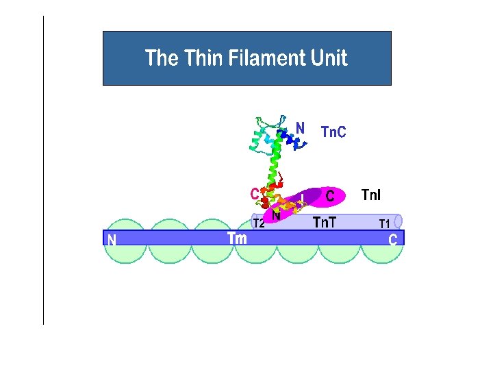
Thin filament
18 Sept 07 31
18 Sept 07 32
18 Sept 07 33
cardiac troponin C 161 amino acids 4 E-F hand Ca sites Sites III and IV –high affinity Ca/Mg sites Site I – dysfunctional Site II - low affinity Ca site – regulatory 18 Sept 07 35
EF hand Ca 2+ coordination V R C-terminus M M C V Helix D L F E D F D Ca 2+ V Loop D E T G S G D V Helix C E D I N-terminus 18 Sept 07 36
18 Sept 07 37
When [Ca] = Kd then [Tn. C] = [Tn. C·Ca] Kd = [Ca] at 50% [Tn. C·Ca]max 18 Sept 07 38
Non equilibrium states Rate of formation Rate of dissociation When rates of formation and dissociation are equal: 18 Sept 07 39
E M M V Helix C Helix B I K D G L D T K G D S E E E S V I G Non-functional Site II Ca 2+ C T G V V F D L E G E D F F D L A I V D Helix A F M Helix D M A N-terminus 18 Sept 07 C-terminus 41
comparison of Tn. C Site I sequences X Y Z-Y–X -Z Mammals – fast skeletal DADGGGDISVKELGTV Mammals – cardiac LGAEDGCISTKELGKV Fish QDAEDGCISTKELGKV 18 Sept 07 – cardiac 42
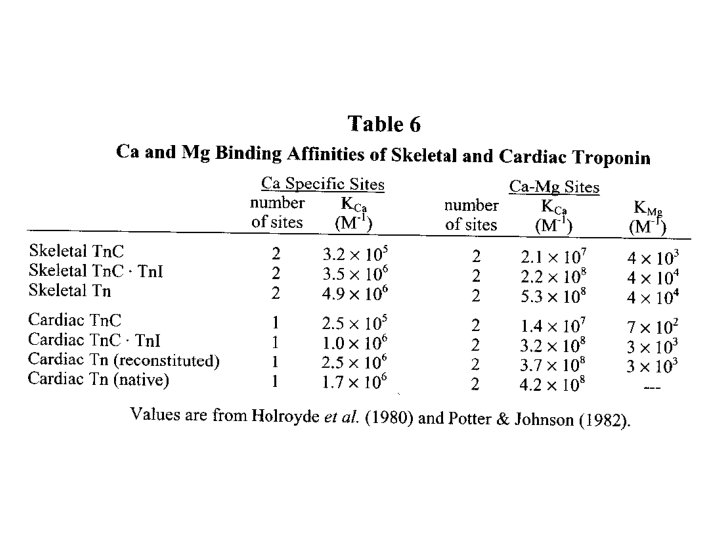
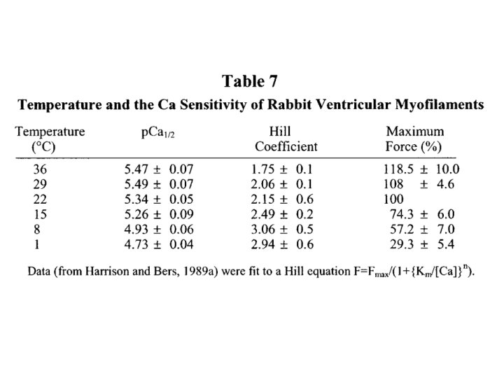
Bers – Table 8