Joints of the Vertebral Column Anatomy 1 Vertebrae

Joints of the Vertebral Column Anatomy 1

• Vertebrae from C 2 to S 1 – 3 Joints • Anterior joint • 2 posterior joints • Craniovertebral joints – Atlanto-occipital joint – Atlanto-axial joint

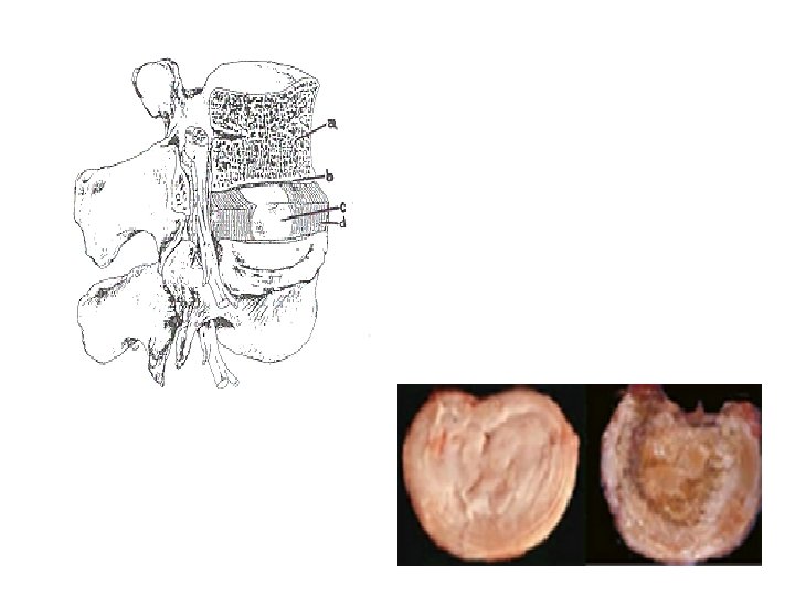
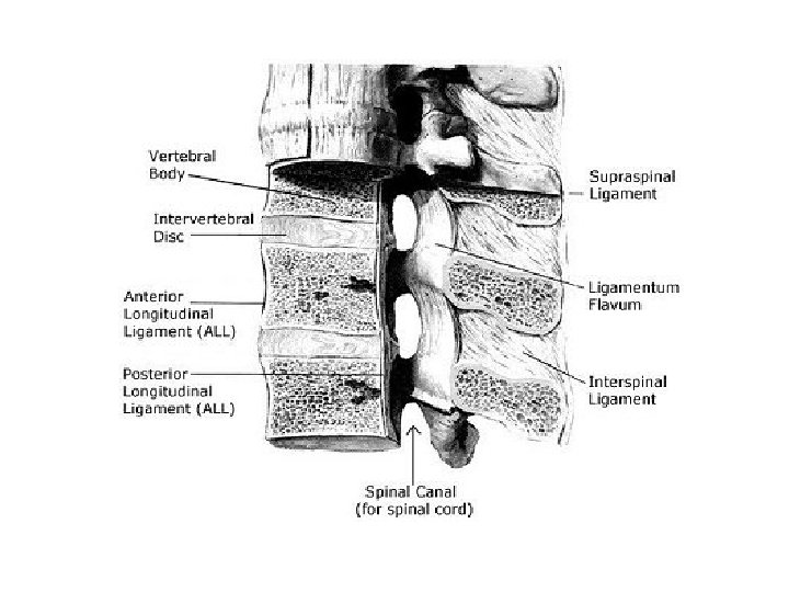
Intervertebral disc • Anterior intervertebral joint • Classification – Symphysis – – Material that connects the bones: •

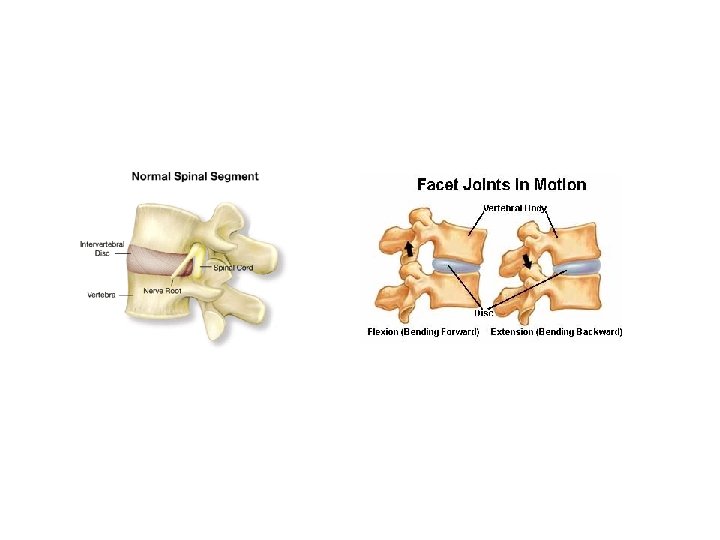
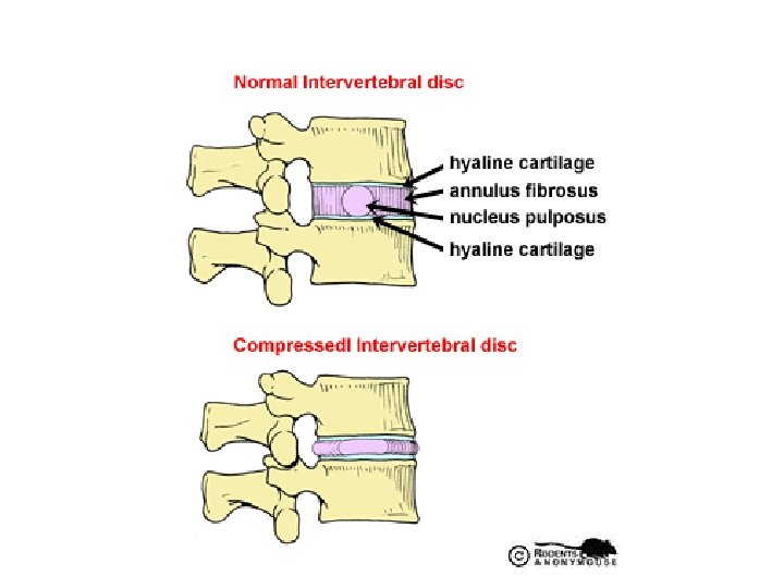
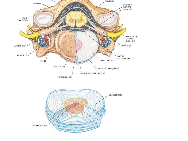
Overall structure of intervertebral disc • Hyaline cartilage on surface of bodies • Disc – made of fibrocartilage – Anulus fibrosis – Nucleus pulposus
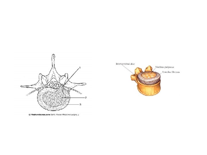

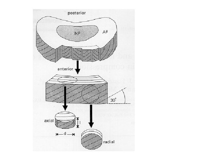
Anulus fibrosus • Fibrous rim of disc • Concentric lamellae of fibrocartilage – Fibers run at an angle – Fibers in one lamellae are perpendicular to fibers in the next lamellae – Lamellae are thinner and less numerous posteriorly than anteriorly or laterally
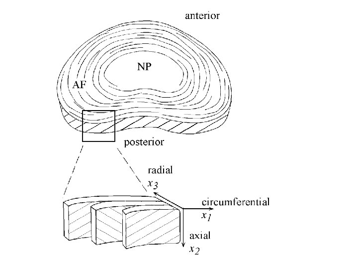
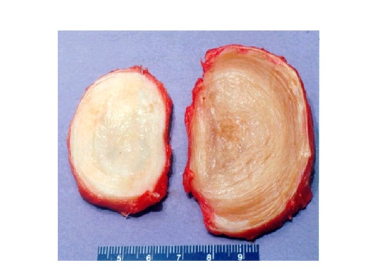

Nucleus pulposus • Central core of disc • Located more posteriorly than central • More cartilaginous than fibrous

Blood supply to nucleus pulposus • Nucleus is avascular • Diffusion from blood vessels in the periphery of the anulus fibrosus

• Nucleus pulposus water content – High water content – Desiccation starts around age 30

Function of nucleus pulposus • Shock absorber for axial forces • Acts like semiliquid ball bearing • Becomes broader when compressed


Disc Injury • If anulus fibrosis is weakened, the nucleus pulposus may rupture and protrude – AF strength? – Ligamentous suppport? • May press on: – Spinal cord – Nerve roots – Cauda equina

Joints of the vertebral arch • Zygapophyseal joints – Synovial joint – Classification • Inferior vertebrae – superior articular facet • Superior vertebrae – inferior articular facet

 • Joint capsule – Attached")
Structure of Zygapophyseal Joints • Cartilage on facets: (type) • Joint capsule – Attached to margins of processes – Longer and looser in the:


Function of zygapophyseal joints • Allow gliding movement • Control flexion, extension and rotation in the cervical and lumbar regions • Share weight-bearing with intervertebral discs

Innervation of zygapophyseal joints • Nerves from medial branches of the posterior primary rami • Figure (page 506)

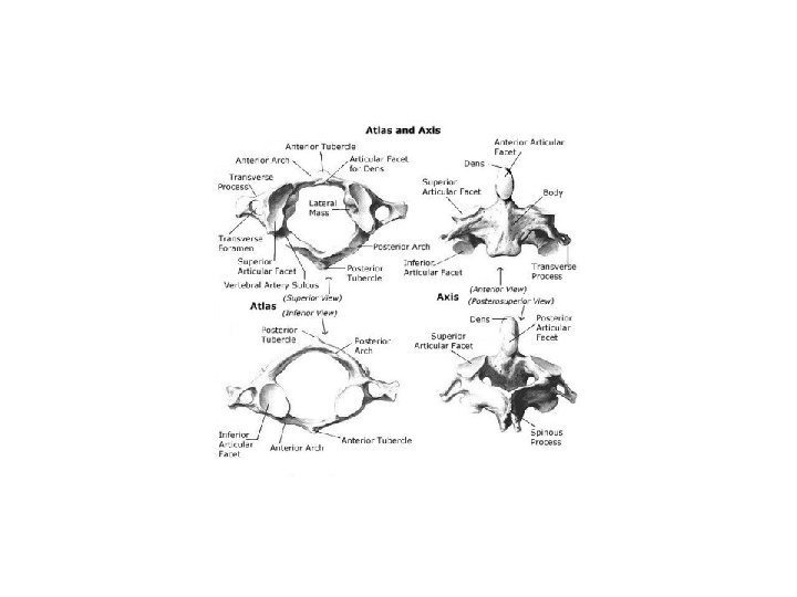
Craniovertebral joints • Atlanto-occipital joint • Atlanto-axial joint
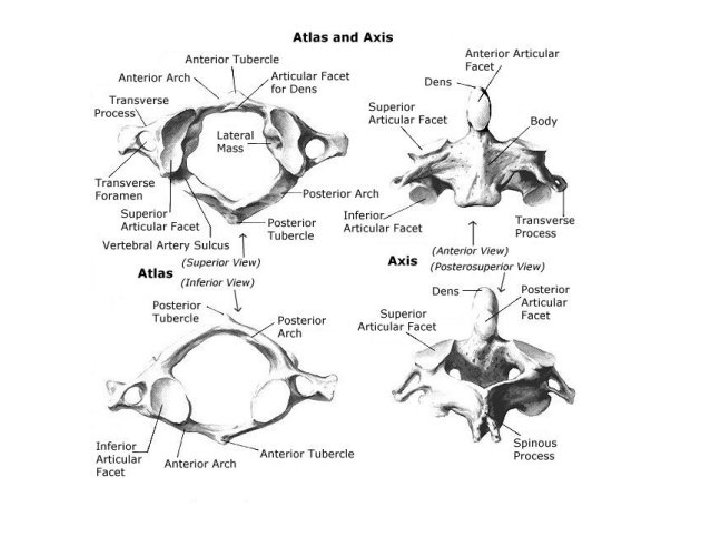
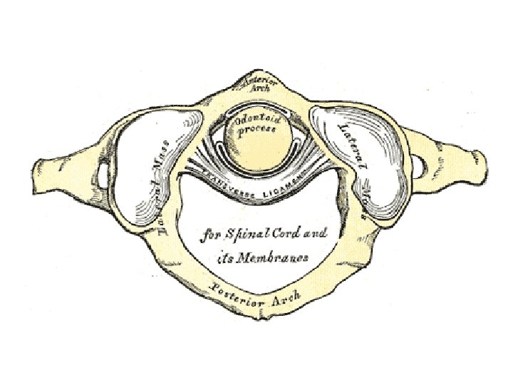

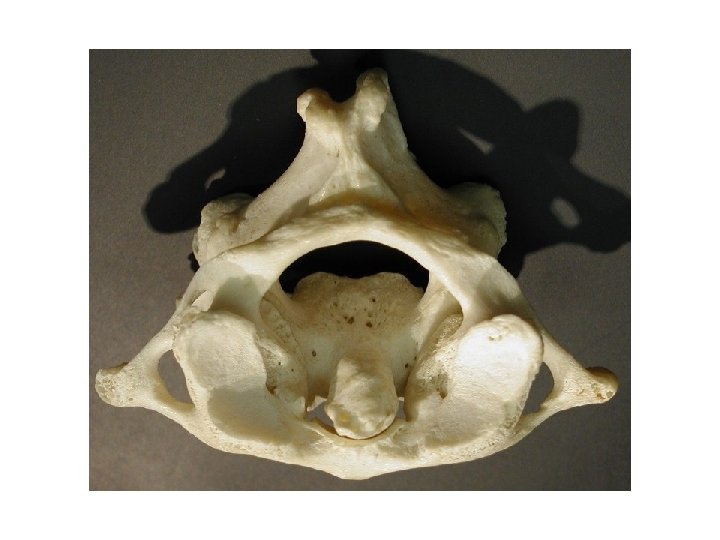
Structures • Atlas – Anterior and posterior arches – Lateral mass – Posterior tubercle – No spinous process – Transverse process • Transverse foramen – Vertebral foramen – Joints • Superior articular facet • Joint with dens and transverse ligament
")
• Axis – Body – Bifid spinous process – Dens (facet for atlas) • Occipital bone – Foramen magnum – Occipital condyles • Anterolateral to foramen magnum

Atlanto-occipital joint • Articulating bones – – • Movements – Primarily: – Slight:

Atlanto-Occipital joint • Classification of joint • How many degrees of movement? • Loose articular capsule with synovial membrane

Atlanto-occipital membranes • Connect C 1 and skull • Anterior and posterior atlanto-occipital membranes • Extend from anterior and posterior arches of atlas to the anterior and posterior margins of the foramen magnum
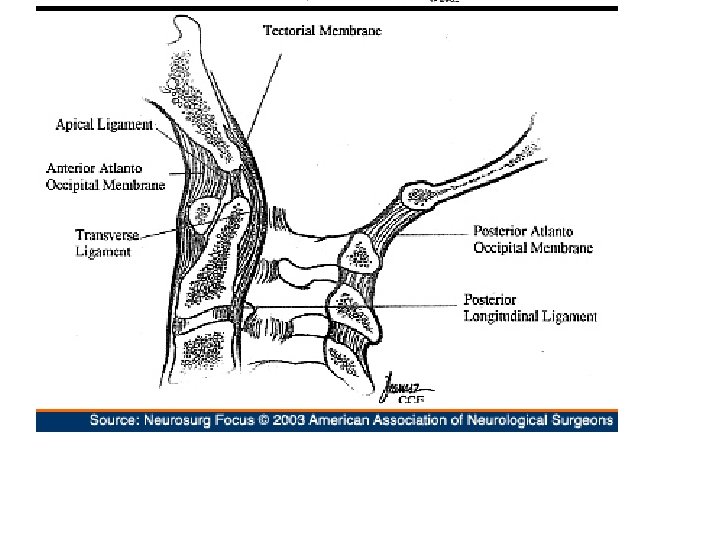

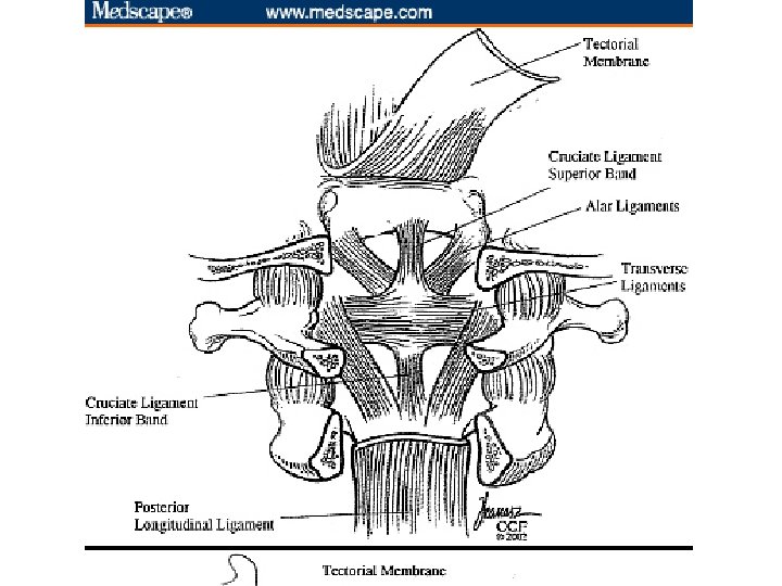
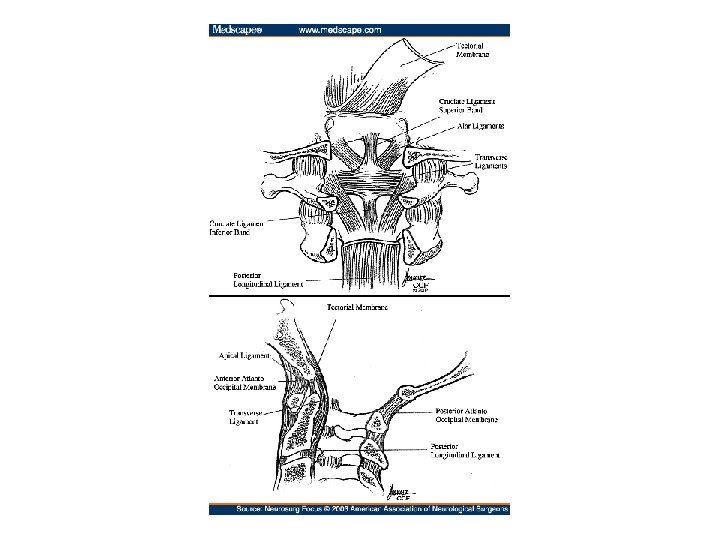
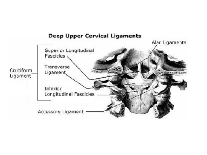
Cruciate ligament • Transverse ligament of the atlas • Superior and inferior longitudinal bands

Transverse Ligament of the Atlas • Strong band • Between tubercles on medial sides of the lateral masses of the atlas • Holds dens in place against anterior arch of atlas • Forms synovial joint between bones

Superior and inferior longitudinal bands of the cruciate ligament • Attach to the occipital bone superiorly and the body of axis inferiorly

Alar ligaments • From dens to lateral margins of foramen magnum • Round cords and as thick as pencils • Check lateral rotation and side-to-side movements of the head • Attach skull to axis


Tectorial membrane • Superior continuation of posterior longitudinal ligament • Attaches to: – Body of axis – Internal surface of occipital bone • Covers the alar and cruciate ligaments

Atlanto-axial joint • Between C 1 and C 2 • 3 joints – Medial joint – 2 lateral joints • Movement: the skull and C 1 rotate as a unit on C 2

Apical ligament of the dens • Tip of dens to internal surface of occipital bone

General movements of the vertebral column • Limits of movement – Intervertebral discs – Resistance from muscles and ligaments in the back – Shape of the zygapophyseal joints and the tension of their respective joint capsules

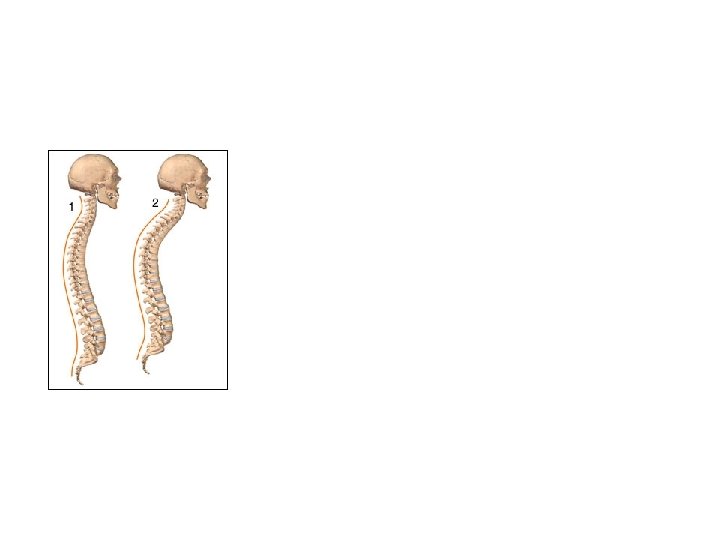
Physiological Curves of the Back • Embryo • Curves change during development

1º curvatures • Develop during fetal period • Concave anteriorly • Which parts of the spine?

2º Curvatures • Present at birth, but not well developed • Concave posteriorly • Regions of the spine • Develop more after birth

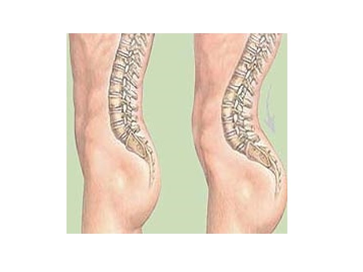
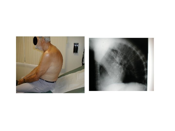
Abnormal curves • Lordosis • Kyphosis • Scoliosis


- Slides: 60