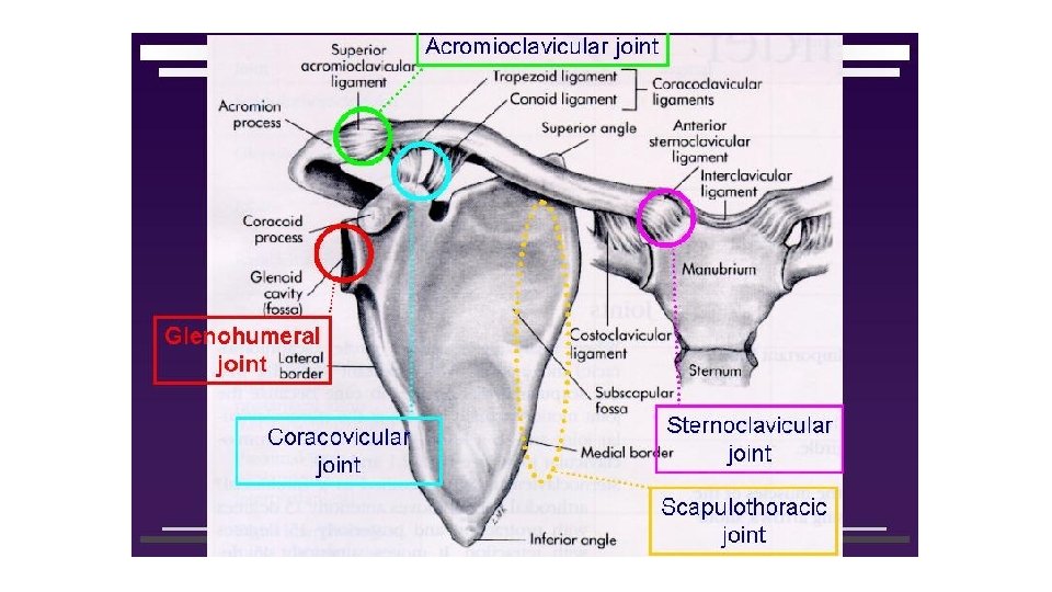
Joints of the shoulder region 3 Joints acromioclavicular


 GIRDLE • - Consists of both the scapula and the")
 JOINT Articulation: Between head of humerus and glenoid cavity of scapula, deepened")


 joint is a ball-and-socket, synovial joint that permits a")


- Slides: 33
Joints of the shoulder region
3 Joints acromio-clavicular sterno-clavicular Shoulder joint
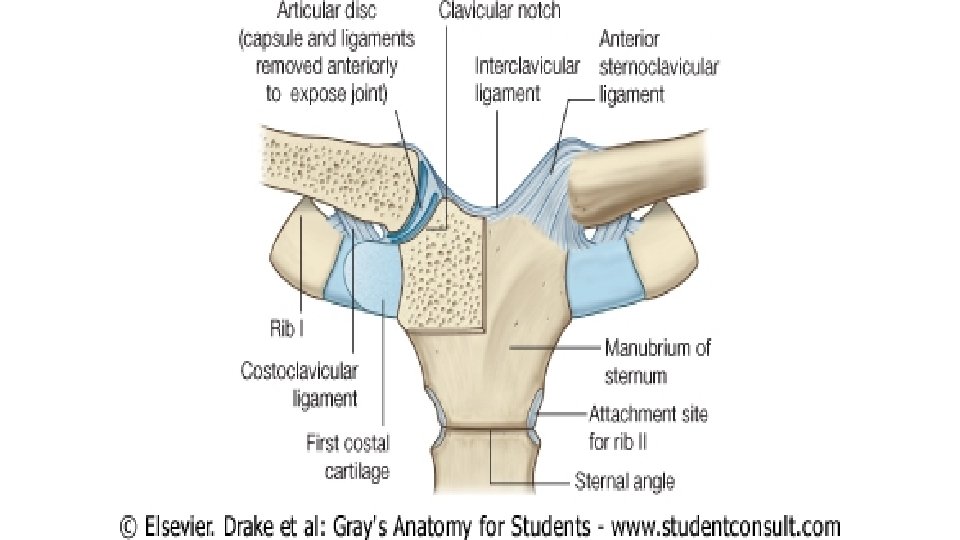
STERNOCLAVICULAR JOINT Articulation: Between sternal end of clavicle, manubrium sterni and first costal cartilage • Type: Synovial joint double-plane type Ligaments: Anterior sternoclavicular ligament Posterior sternoclavicular ligament Accessory ligaments: Costoclavicular ligament Fibrocartilagenous disc: Divide the joint into medial and lateral compartments
Function: 1 -fixation of the medial end of clavicle preventing its dislocation during movements of shoulder girdle. 2 -transmission of any force applied to upper limb to the chest
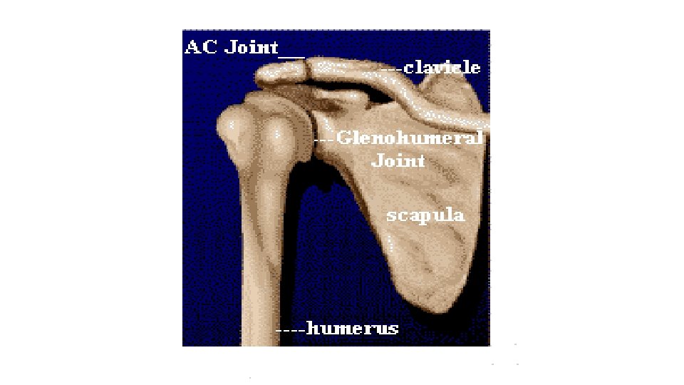
ACROMIOCLAVICULAR JOINT • Articulation: Between acromion of scapula and lateral end of clavicle • Type: Synovial joint, plane type • Capsule: • Surrounds the joint and attached to margins of articular surfaces • Ligaments (and fibrocartilagenous disc): • Superior acromioclavicular ligament • Inferior acromioclavicular ligament • Accessory ligaments: Coracoclavicular ligament • -it consists of 2 parts : conoid &trapezoid
Function: Transmission of most of the weight of the upper limb to the clavicle
• SHOULDER (PECTORAL) GIRDLE • - Consists of both the scapula and the clavicle • - Occur at sternoclavicular and acromioclavicular joints at the same time • Movements of shoulder girdle & muscles doing: • Elevation of scapula: • - Upper part of trapezius - Levator scapula • Depression of scapula: • - Pectoralis minor - Lower part of trapezius • Protraction of scapula (forward movement): • - Serratus anterior - Pectoralis minor • Retraction of scapula(Backward movement): • - Middle part of trapezius - Rhomoideus major • - Rhomboideus minor • Upward rotation of scapula: • - Upper fibers of trapezius - Lower fibers of trapezius • - Lower 5 digitations of serratus anterior • Downward rotation of scapula: • - Levator scapula - Rhomboideus major • - Rhomboideus minor - Pectoralis minor
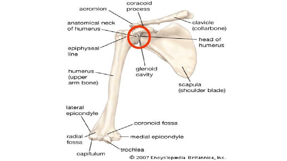
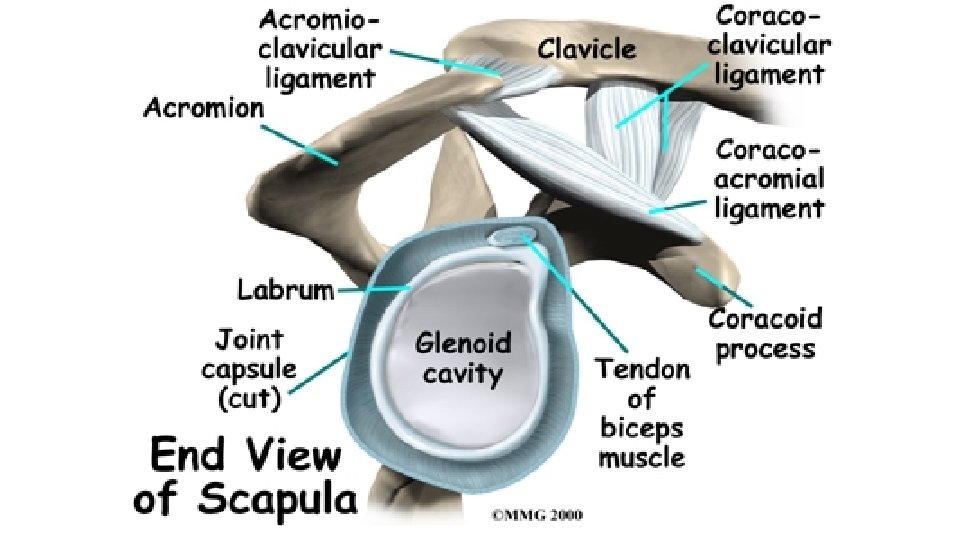
SHOULDER (GLENOHUMERAL) JOINT Articulation: Between head of humerus and glenoid cavity of scapula, deepened by glenoid labrum Type: Synovial joint, ball and socket Capsule: Surrounds the joint, attached medially to margin of glenoid cavity outside labrum, laterally attached to anatomical neck of humerus, strengthened by rotator cuff muscles Ligaments: Glenohumeral ligaments (3) Transverse humeral ligament Coracohumeral ligament Accessory ligaments: Coracoacromial ligament
-Synovial membrane: -Lines the capsule and attached to margins of cartilage covering articular surfaces -It forms a tubular sheath around tendon of long head of biceps
Synovial membrane extends through anterior wall of capsule to form subscapularis bursa (deep to subscapularis)
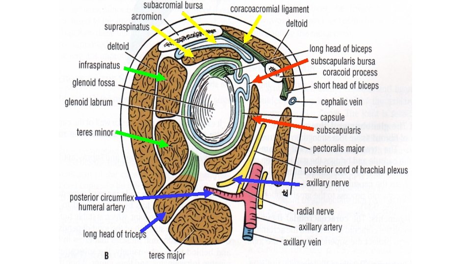
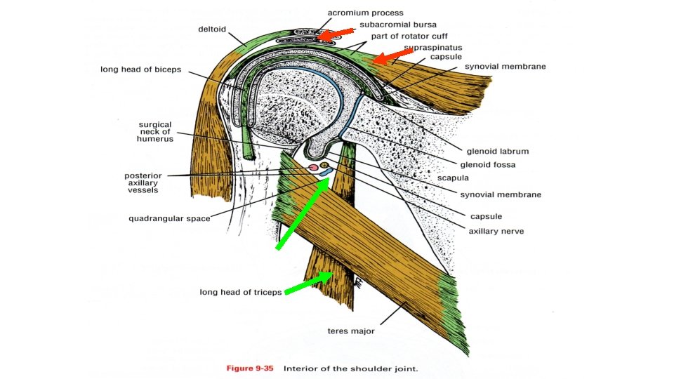
Relations of shoulder joint Anteriorly: - Subscapularis - Subscapular bursa Posteriorly: - Infraspinatus - Teres minor Superiorly: - Supraspinatus - Subacromial bursa - Coracoacromial arch Inferiorly: - Long head of triceps - Quadrangular space Bursae related to shoulder joint: Subscapular bursa Infraspinatus bursa Subacromial bursa Subcutaneous bursa
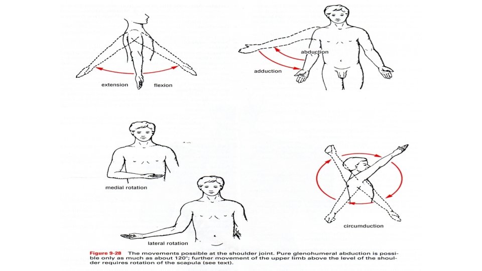
Movements of shoulder joint and muscles doing: Flexion: - Anterior fibers of deltoid -Biceps brachii Extension: - Posterior fibers of deltoid Abduction: -Supraspinatus Adduction: -Pectoralis major -Teres major Lateral rotation: - Infraspinatus Medial rotation: - Subscapularis -Teres major - Pectoralis major - Coracobrachialis - Latissimus dorsi - Teres major - Middle fibers of deltoid - Latissimus dorsi - Teres minor - Posterior fibers of deltoid - Latissimus dorsi - Anterior fibers of deltoid Circumduction: Combination of the above movements
• Abduction is divided into 3 stages: 1 - From 0 to 15: produced by supraspinatus 2 - From 15 to 90: Produced by middle fibres of deltoid 3 - Above 90: Produced through rotation of scapula by: - Upper fibres of trapezius - Lower 5 digitations of serratus anterior
Head of humerus Greater tuberosity Glenoid cavity
Dislocation of the shoulder joint -Occurs due to laxity of the capsule and disproportionate articular surfaces -It is always subglenoid (head slips downwards) -It endangers the axillary nerve which is closely related to lower part of capsule
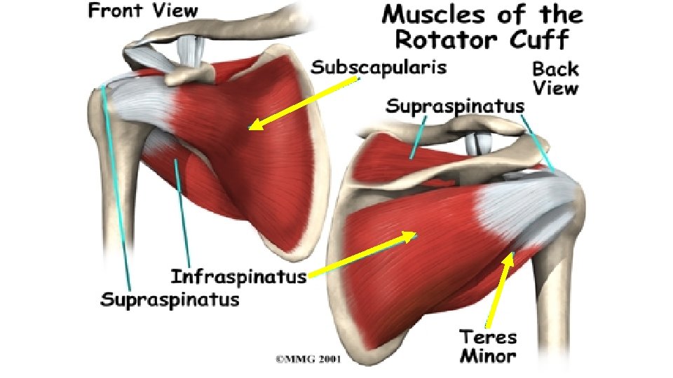
Glenohumeral Joint The glenohumeral (shoulder) joint is a ball-and-socket, synovial joint that permits a wide range of movement; however, its mobility makes the joint relatively unstable The large spherical humeral head articulates with the relatively small and shallow glenoid cavity of the scapula, which is deepened slightly by the ring-like, fibrocartilaginous glenoid labrum (lip). Both articular surfaces are covered with hyaline cartilage. The glenoid cavity accepts little more than a third of the humeral head, which is held in the cavity by the tonus of the musculotendinous rotator cuff (supraspinatus, infraspinatus, teres minor, and subscapularis). 12/28/2021 25
The loose fibrous layer of the joint capsule surrounds the glenohumeral joint and is attached medially to the margin of the glenoid cavity and laterally to the anatomical neck of the humerus. Superiorly, the fibrous layer encloses the proximal attachment of the long head of biceps brachii to the supraglenoid tubercle of the scapula within the joint. The inferior part of the joint capsule, the only part not reinforced by the rotator cuff muscles, is its weakest area. Here, the capsule is particularly lax and lies in folds when the arm is adducted; however, it becomes taut when the arm is abducted. 26
The synovial membrane lines the internal surface of the fibrous capsule and reflects from it onto the humerus as far as the articular margin of its head. The synovial membrane also forms a tubular sheath for the tendon of the long head of the biceps brachii. Anteriorly, there is a communication between the subscapular bursa and the synovial cavity of the joint. 27
LIGAMENTS OF GLENOHUMERAL JOINT The glenohumeral ligaments, evident only on the internal aspect of the capsule, strengthen the anterior aspect of the capsule. The coracohumeral ligament, a strong band that passes from the base of the coracoid process to the anterior aspect of the greater tubercle, strengthens the capsule superiorly. The glenohumeral ligaments are intrinsic ligaments that are part of the fibrous layer of the capsule. The transverse humeral ligament is a broad fibrous band that runs from the greater to the lesser tubercle, bridging over the intertubercular sulcus (groove) and converting the sulcus into a canal for the tendon of the long head of biceps brachii and its synovial sheath. 28
The coraco-acromial arch is an extrinsic, protective structure formed by the smooth inferior aspect of the acromion and coracoid process of the scapula, with the coracoacromial ligament spanning between them. The coraco-acromial arch overlies the head of the humerus, preventing its superior displacement from the glenoid cavity. The arch is so strong that a forceful superior thrust of the humerus will not fracture it; the shaft of the humerus or clavicle fractures first. 29
MOVEMENTS OF GLENOHUMERAL JOINT The glenohumeral joint has more freedom of movement than any other joint in the body. This freedom results from the laxity of its joint capsule and the configuration of the spherical humeral head and shallow glenoid cavity. Glenohumeral joint allows movements around the three axes and permits flexion–extension, abduction–adduction, rotation (medial and lateral) of the humerus, and circumduction. Lateral rotation of the humerus increases the range of abduction. When the arm is abducted without rotation, the greater tubercle contacts the coraco-acromial arch, preventing further abduction. If the arm is then laterally rotated 180 degrees, the tubercles are rotated posteriorly and more articular surface becomes available to continue elevation. Stiffening or fixation of the joints of the pectoral girdle (ankylosis) results in a much more restricted range of movement, even if the glenohumeral joint is normal. 30
BLOOD SUPPLY AND INNERVATION OF GLENOHUMERAL JOINT The glenohumeral joint is supplied by the anterior and posterior circumflex humeral arteries and branches of the suprascapular artery. The suprascapular, axillary, and lateral pectoral nerves supply the glenohumeral joint. BURSAE AROUND GLENOHUMERAL JOINT Several bursae containing capillary films of synovial fluid are located near the joint where tendons rub against bone, ligaments, or other tendons and where skin moves over a bony prominence. Some bursae communicate with the joint cavity; hence, opening a bursa may mean entering the cavity of the joint. The subacromial bursa, sometimes referred to as the subdeltoid bursa, is located between the acromion, coracoacromial ligament, and deltoid superiorly and the supraspinatus tendon and joint capsule of the glenohumeral joint inferiorly. Thus, it facilitates movement of the supraspinatus tendon under the coraco-acromial arch and of the deltoid over the joint capsule and the greater tubercle of the humerus. 31
The subscapular bursa is located between the tendon of the subscapularis and the neck of the scapula. This bursa protects the tendon where it passes inferior to the root of the coracoid process and over the neck of the scapula. It usually communicates with the cavity of the glenohumeral joint through an opening in the fibrous layer of the joint capsule 32
Thanks