JOINTS INTRODUCTION Joints are the regions of the


- Slides: 35

JOINTS

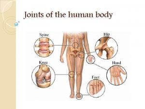
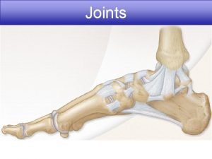
INTRODUCTION • Joints are the regions of the skeleton where - 2 or more bones - bones with cartilage articulate - 2 or more cartilage • Supported by variety of soft tissue structures • Functions: i) to facilitate growth ii) to transmit forces between bones.

Classification of Joints Fibrous Fixed Cartilaginous Slightly movable A. Sutures B. Gomphosis A. Pri. Cart. joints C. Syndesmosis B. Sec. cart. Joints Synovial Freely movable 1. Plane 2. 3. 4. 5. 6. 7. Hinge Pivot Bicondylar Ellipsoid Saddle Ball and socket

CLASSIFICATION 1. Functional classification Immovable (synarthrosis) Cranial sutures in adult Pri cartilaginous jt. in children Slightly movable (amphiarthrosis) Secondary cartilaginous jts Syndesmosis Freely movable (diarthrosis) Synovial jt.

CLASSIFICATION 2. Structural classification Depends on the nature of intervening soft tissue, presence or absence of joint cavity a) Fibrous joint b) Cartilaginous joint c) Synovial joint

FIBROUS JOINT • • • Lacks intervening cart. between 2 bones United by fibrous CT Articulation : -Fixed (ROM restricted/ slight) Lacks joint cavity 3 types: - a) Sutures b) Syndesmosis c) Gomphosis

SUTURE Restricted to skull Synostosis on completion of growth.

SYNDESMOSIS • Fibrous connection between bones • Represented by Interosseous ligament Slender fibrous cord Dense Aponeurotic membrane Eg. Inf tibiofibular jt, post part of sacroiliac jt.

GOMPHOSIS • Peg & socket joints between tooth & its socket

CARTILAGINOUS JOINT 1. Primary Cartilaginous Joint Also called as synchondrosis

CARTILAGINOUS JOINT 2. Secondary Cartilaginous Joints Also called as symphysis

SYNOVIAL JOINT • Most evolved joint. • Freely movable joint. • Possess a joint cavity that consists of synovial fluid.

CHARACTERISTICS OF SYNOVIAL JOINTS 1. Articular cartilage Articular surfaces are covered by thin plates of hyaline cartilage Exceptions: - acromioclavicular sternoclavicular ( atypical synovial joints) Provides smooth friction-free movements & resists compression forces.

2. Fibrous capsule Longitudinal & interlacing bundles of parallel fibers of white collagen. Completely encloses a jt except where it is interrupted by synovial membrane. Stabilizes the jt in such a way that it permits movements but resists dislocation.

3. Synovial membrane Thin highly vascular memb of CT. Pink, smooth and shiny. Lines capsule, covers exposed osseous surfaces , tendon sheaths, bursa but doesn't cover the articular cartilage, intra-articular disc / menisci. Function: produces synovial fluid

4. Synovial fluid Clear or pale yellow, viscous, slightly alkaline at rest. Fluid vol : - < 0. 5 ml in large jt (knee) Composition: Hyaluronic acid, Lubricin, Proteinase and Collagenase. Fxn : - reduce friction, shock absorption, nutrient and waste transportation. 5. Intra-articular menisci, disc and fat pads fibrocartilage, not covered by synovial membrane.

BLOOD SUPPLY • Periarticular arterial plexuses – circulus articularis vasculosus • Articular cartilage: avascular • Fibrous capsule & ligaments: poor blood supply • Synovial membrane: rich blood supply

NERVE SUPPLY • HILTON’S LAW The nerves supplying the joint capsule also supply the muscles regulating the movement of the jt & skin over the joint.

TYPES OF SYNOVIAL JOINT 1. Based on shape of articular surface Articulating surface- Flat Gliding or Sliding Movements Eg. Intercarpal & Intertarsal Intermetacarpal Intermetatarsal Zygapophyseal

TYPES OF SYNOVIAL JOINT Uniaxial Resemble hinge of door Articular surface- pulley shaped Eg. Humero-ulnar Jt. Interphalangeal Jt. Knee & Ankle Jt 2. HINGE JOINT

TYPES OF SYNOVIAL JOINT Biaxial Elliptical convex surface of one bone articulates with elliptical concave surface of other bone Eg. Radio-Carpal Joint Atlanto Occipital Joint Meta-tarso phalangeal Joint Meta-carpophalangeal Joint 3. ELLIPSOIDAL JOINTS

TYPES OF SYNOVIAL JOINT Uniaxial Joint Eg. Superior Radio-ulnar Jt. Median Atlanto-axial Articular surface of one bone is rounded & fits into the concavity of another bone. Further rounded part surrounded by a Ligamentous ring. 4. PIVOT JOINT

TYPES OF SYNOVIAL JOINT Biaxial 5. BICONDYLAR JOINT Round articular surface of one bone fits into socket type articular surface of another bone. Eg. Knee Joint, Temporo-mandibular Joint

TYPES OF SYNOVIAL JOINT Bi-axial Articular surfaces are reciprocally saddle shaped i. e Concavo-convex. Eg. Carpo-metacarpal joint of thumb, Calcaneo-cuboid Joint Sterno-clavicular Joint Incudo malleolar Joint 6. SADDLE JOINT

TYPES OF SYNOVIAL JOINT 7. BALL AND SOCKET JOINT Multi-axial Rounded convex surface of one bone fits into the cup-like socket of another bone. Eg Hip Joint, Shoulder Joint, Incudo-stapedial Joint.

2. Based on plane of movements I. Uniaxial joint : Hinge, Pivot joint II. Biaxial joint: Condylar, Ellipsoid, Saddle joint III. Multiaxial joint: Ball and socket joint.

3. Based on no. of articulating bone I. Simple joint: only 2 bones take part in formation of a joint. II. Compound joint: > 2 bones take part in formation of a joint. III. Complex joint: joint cavity is divided into 2 by the intra-articular disc or meniscus, eg. TM joint, knee joint.

MOVEMENTS OF SYNOVIAL JOINTS 1. TRANSLATION: gliding or sliding movements 2. ANGULATION: change in the angle betn the topographical axes of the articulating bones. 4 types a). Flexion b). Extension c). Abduction d). Adduction 3. ROTARY / CIRCULAR MOVEMENTS a). Axial rotation b). Circumduction

ARTHRITIS • Inflammation of one or more joints, synovial membrane. • > 100 different forms of arthritis. • Symptoms: swollen jt, tender, warm, stiffness limits the movements. • Main complaint: jt pain ( due to inflammation that occurs around the jt, damage to the jt from disease, daily wear and tear of the jt, muscle strains caused by forceful movement) • Most common: osteoarthritis

OSTEOARTHRITIS • Most common form of arthritis • degenerative joint disease • Cause: mechanical stress, overweight, hereditary, developmental deficits • Symptoms: jt pain, tenderness, stiffness, locking and sometimes an effusion. • T/t : -exercise - lifestyle modification - analgesics - jt replacement surgery used to improve quality of life.

OSTEOARTHRITIS

RHEUMATOID ARTHRITIS • Is an autoimmune disease that results in a chronic, systemic inflammatory disorder that may affect many tissues , organs and jts. • Women 2 -3 times more affected than men. • Onset is frequent during middle age. • Pathology: destruction of articular cartilage and ankylosis of the joints. • Commonly involved parts: hands, feet and cervical spine but larger jt can also be involved. • Symptoms: -pain ( lasts for more than 1 hour) -stiffness mainly occurs in the morning -disabling & painful condition can lead to loss of function.

RHEUMATOID ARTHRITIS

RHEUMATOID ARTHRITIS • T/t: - physical therapy -nutritional therapy -analgesia/ anti-inflammatory (NSAIDS) -steroids -DMARDS (disease- modifying anti rheumatic drugs)

 Mikael ferm
 Hát kết hợp bộ gõ cơ thể
 Slidetodoc
 Bổ thể
 Tỉ lệ cơ thể trẻ em
 Gấu đi như thế nào
 Tư thế worm breton
 Chúa yêu trần thế
 Các môn thể thao bắt đầu bằng tiếng bóng
 Thế nào là hệ số cao nhất
 Các châu lục và đại dương trên thế giới
 Công thức tính thế năng
 Trời xanh đây là của chúng ta thể thơ
 Mật thư anh em như thể tay chân
 101012 bằng
 độ dài liên kết
 Các châu lục và đại dương trên thế giới
 Thơ thất ngôn tứ tuyệt đường luật
 Quá trình desamine hóa có thể tạo ra
 Một số thể thơ truyền thống
 Cái miệng bé xinh thế chỉ nói điều hay thôi
 Vẽ hình chiếu vuông góc của vật thể sau
 Biện pháp chống mỏi cơ
 đặc điểm cơ thể của người tối cổ
 V cc
V cc Vẽ hình chiếu đứng bằng cạnh của vật thể
 Phối cảnh
 Thẻ vin
 đại từ thay thế
 điện thế nghỉ
 Tư thế ngồi viết
 Diễn thế sinh thái là
 Dạng đột biến một nhiễm là
 So nguyen to
 Tư thế ngồi viết
 Lời thề hippocrates


